|
When you have finished your scanning and have found the area of the smear
where cells are optimally spread, turn to the 100X oil immersion objective
and begin your differential count and morphology assessment. The high
dry (40X) objective should never be used for this purpose since the
magnification is too low to see adequately all the features of the cells.
Begin the differential count by moving back and forth across the 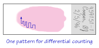 smear in a pattern that avoids covering the same territory. Identify
each leukocyte that is encountered until 100 cells have been counted and
sorted by type. As you see each cell, record it in the proper category
on the counter (using either a mechanical differential counter or the
program on your computer in the dry lab). The percentage of each cell
type that results is termed the relative differential count.
smear in a pattern that avoids covering the same territory. Identify
each leukocyte that is encountered until 100 cells have been counted and
sorted by type. As you see each cell, record it in the proper category
on the counter (using either a mechanical differential counter or the
program on your computer in the dry lab). The percentage of each cell
type that results is termed the relative differential count.
The absolute differential is derived by multiplying the percentage of each cell type by the WBC to get the number of each type of leukocyte /”l. If nucleated red cells (nRBC) are encountered, they should be recorded separately from the leukocytes and reported as the number of nRBC/100 WBC. This number is then used to correct the apparent WBC (which is actually just a count of nucleated cells) so that it represents the number of leukocytes/”l. To help you learn to identify leukocytes and assess blood cell morphology, this module contains sections that display photomicrographs of the different kinds of leukocytes and some of the species variations. You can gain access to these reference pictures by returning to the introduction page and clicking on the appropriate button. |
Last Updated:Thursday, February 08, 1996