Platelet counts can be done manually or using automated cell counters. Since many laboratories use instruments that count platelets, red cells and leukocytes concurrently, a platelet count is a routinely reported result on many samples of dog and horse blood. However, platelet clumping will lower (and in some instances invalidate) the platelet count when determined by any method. This occurs especially in samples of cat and cow blood (see image below). In these instances, an accurate platelet count cannot be provided, but for most purposes, significant changes in platelet number can be detected during the microscopic examination of the stained blood smear (see section on platelet estimation). In such samples, close attention should be paid to the platelet smear estimate part of our hemogram. Platelet clumping is usually Manual platelet counts Impedance-based platelet
counts Flow cytometry-based
platelet counts
Platelets are identified by their size (< 30 fL) and refractive index (n = 1.35 to n = 1.40). The platelet cytogram on the left is a graphical representation of how the Advia counts platelets. Low light scatter is plotted against the X axis and high light scatter is plotted against the Y axis (B). Platelets are detected in the region labeled 1. Large platelets (section 2) are identified on the basis of size (> 20 fL) and refractive index (which distinguishes them from red cells). In species with very small red cells, e.g. goats, some red blood cells may be counted as platelets. The analyzer also detects platelet clumps and flags their presence. When this occurs, a comment is appended to the platelet count or smear estimate (if a count is not provided) demonstrating the presence of clumps.
Last Updated: June 2000 |
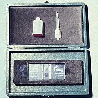 Unopette® system for platelet counting with Newbauer hemocytometer.
 due
to a sample collection problem and can be minimized by collecting blood
from a large peripheral vein (cephalic or jugular), such that blood
flows smoothly into the vacutainer or syringe, and using a 22 or 23
g needle (in a dog or cat). The blood should be mixed with the anticoagulant
as soon as possible after collection, by gentle rotation or inversion.
Platelet clumping increases with time, so platelet counts should be
done as soon as possible after collection to maintain accuracy.
due
to a sample collection problem and can be minimized by collecting blood
from a large peripheral vein (cephalic or jugular), such that blood
flows smoothly into the vacutainer or syringe, and using a 22 or 23
g needle (in a dog or cat). The blood should be mixed with the anticoagulant
as soon as possible after collection, by gentle rotation or inversion.
Platelet clumping increases with time, so platelet counts should be
done as soon as possible after collection to maintain accuracy.