|
In vitro aging of blood cells in the specimen tube
causes changes in the appearance of the cells on stained blood films. Eventually, cells will
break down altogether, rendering the specimen useless for analysis.
When analysis will be delayed (e.g., lab closed or when sending a sample to a reference lab), smears should be made and sent along with the EDTA tube. Air-dried unfixed smears hold up very well unless subjected to flies or moisture. Ensure that the slides are not in direct contact with "cool"packs, as the slide can freeze or become moist, thus rupturing the cells. 
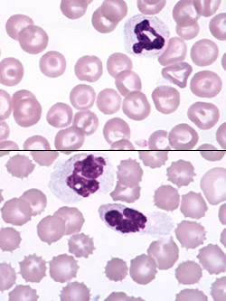
Shown at right are two smears from the same sample, the upper one made while the blood was fresh, the lower made after overnight storage of blood at refrigerator temperature. The aged blood smear illustrates echinocyte formation and swelling of neutrophil nuclei. Age-related changes that occur are: 1) Red blood cells: Crenation (echinocyte formation), lysis, hemoglobin crystallization. 2) White blood cells: Swellling and smoothing of the nuclear chromatin (mimicking band neutrophil formation), pyknosis and karyhorrhexis of nuclei, cell smudging, and prominence of Dohle bodies (mimicking toxic change). 3) Platelets: Clumping, degranulation (the latter makes platelets difficult to see and enumerate). 
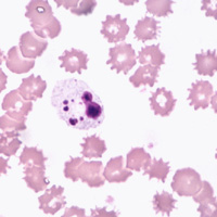
Shown at left is a smear from a sample which had been in EDTA for 48 hours at room temperature. Extreme crenation of the erythrocytes is evident, which could easily mask or render suspect significant pathologic shape abnormalities. The leukocyte nucleus has undergone pyknosis and karyorrhexis, making certain identification impossible. |