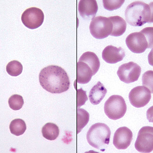
BSE may be seen as a feature of regenerative anemia, especially in ruminants (but also in dogs and cats). In this setting, most of the stippling is seen in young red cells (i.e., polychromatophilic or normochromic macrocytes). BSE can also occur in the absence of anemia in animals with lead poisoning. In this situation, stippling results from the poisoning of the 5' nucleotidase normally responsible for degrading RNA, and hence may be seen in older, smaller red cells as well as nucleated red blood cells. Increased numbers of nRBC (erythroblastosis) usually accompanies the stippling in this condition (as does polychromasia). |