|
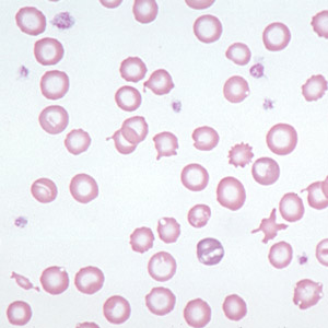
Blood film from a dog with chronic blood loss resulting in
iron deficiency anemia. Note that, in addition to the hypochromic cells (with increased areas
of central pallor), a variety of other shape abnormalities are present (keratocytes,
schistocytes).
|
Hypochromasia is a term with two common usages. As a
descriptor of erythroid cells as seen on a blood film, it refers to the appearance of
increased central pallor with a thin rim of cytoplasm. For all practical purposes, true
hypochromasia in common domestic animal species occurs only in the context of chronic
iron deficiency anemia. It is recognized most frequently in dogs.
The other application of this term is to denote an MCHC below reference range. Hypochromasia
in this sense does not necessarily correlate with the appearance of increased central
pallor in a smear. In developing iron deficiency anemia, the appearance of hypochromasia
in smears precedes the subnormal MCHC.
Hypochromasia in advanced Fe lack is accompanied by red cell shape abnormalities
suggesting fragmentation (schistocytes, keratocytes, eccentrocytes). This may result from diminished
deformability of the iron deficient cells.
|