|
Epithelial cells from the urinary tract used to be defined as "cells from the upper, middle
or lower tracts", according to their morphology.
However, except for the male urethra and the female trigone area and urethra, the excretory
system is lined with an epithelium of several layers (transitional or urothelial epithelium).
The characteristics of these layers are common throughout, so that the morphological differences
depend on the layers from which they come and not on the part of the tract where exfoliation
took place.
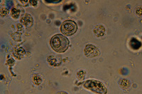 (400x) (64K, jpg) The deep basal cells are small, rounded or polyhedral, mostly mononuclear, the nucleus having rather coarsely granular chromatin and relatively little cytoplasm.
 The cells of the middle layers vary considerably in size and form: they may be rounded, raquet-shaped, oval, polyhedral, etc. |
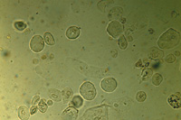
The cells from the surface layers are large, flat, umbrella-shaped, oval or rhomboid.
Quite often they have several nuclei and the chromatin network is quite well preserved.
A few of these cells, particularly those from the "surface layers", can be found even in the
urine of healthy people; a definite increase, especially of those from the middle and deep
layers, is a sign that normal exfoliation has been accentuated, mostly as a result of
irritant factors. This finding is difficult to interpret. It is usually, but not necessarily
seen in acute inflammation or during relapse, and is of only limited interest in chronic
pyelonephritis.
The trigone area and almost all of the female urethra, and the last 0,5-1 cm of the male
urethra are lined with squamous epithelial cells.
These cells have a rather smaller nucleus and a lower nucleus to cytoplasm ratio than
the transitional epithelial cells.
By using the Sternheimer-Malbin staining technique the nuclei of the epithelial cells
appear mauve and the cytoplasm purple.
With Papanicolaou stain the nuclei take on a deep purple shade and the cytoplasm becomes
red or blue-grey.
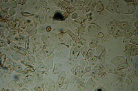 Keratinized squamous cells, clearly eosinophilic, are often a sign of prolonged irritation of the bladder wall; they are clearly visible with Papanicolaou stain. Cells from the vulva may have similar characteristics. |