On the basis of this patients clinical presentation and history, a likely diagnosis of pyoderma gangrenosum can be made. However, you must keep in mind that as many as 50% of cases can be idiopathic. A skin biopsy is necessary to exclude fungi and mycobacterial infections that can look just like this. The diagnosis is based on exclusion of other etiologies of leg ulcers (infections, neoplasms, other inflammatory disorders, gammopathies). Pyoderma gangrenosum is a spontaneous ulcerative condition of the skin and is associated with a variety of disorders. This includes inflammatory bowel disease, chronic hepatitis, rheumatoid arthritis and myeloproliferative disorders. The lesions have a characteristic presentation that begins with a small pustule, which becomes undermined and develops a heaped-up border. Treatment consists of systemic corticosteroids with refractory cases being treated with Dapsone or Cyclosporine.
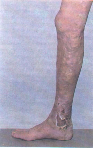
Venous insufficiency tends to occur over the medial malleoli and is associated with irregular borders without the characteristic purulent appearance of pyoderam gangrenosum.
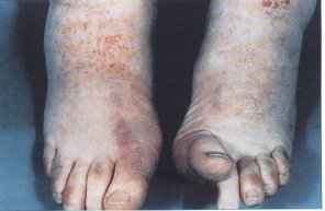
Vasculitic lesions are usually characterized by multiple ulcers that begin as purpuric papules. This particular patient had polyarteritis nodosa with a commonly associated mononeuritis not allowing him to flex his right big toe.
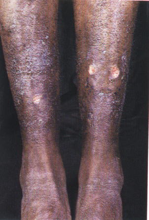
Arterial insufficiency ulcers, have a punched-out appearance, are very painful and tend to occur distally. This particular patient had sickle-cell anemia and developed these arterial ulcers over the shin.
References:
![]()