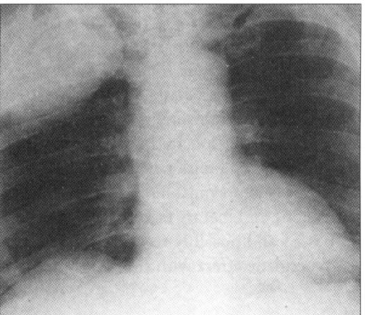
The classic triad of Horner's Syndrome consist of ipsilateral miosis, ptosis and anhidrosis. However the anhidrosis is often absent or difficult to appreciate. The majority of cases are idiopathic, but Horner's syndrome may be caused by a neoplasm impinging on the sympathetic chain or sympathetic cervical ganglia. Damage to the sympathetic contribution to the third cranial nerve results in paresis of the iris dilator muscle. Given this patient's history of smoking and lack of any other focal neurological abnormalities on examination the next step would be getting a chest x-ray looking for an apical tumor (Pancoast's tumor). Pain in the shoulder, radiating down the upper inner arm, can be the first sign of a Pancoast tumor.
Given that he has no other neurological deficits an MRI would be a low-yield, expensive test. Always keep in the back of your mind that posterior cerebral artery aneurysms can present with a dilated pupil. In that case, a lumbar puncture would confirm the presence of bloody CSF and lead you into getting an MRA or cerebral angiogram.
The ultrasound of the carotids would be prudent if the patient presented with carotid artery disease related neurological deficts. Remember that you can divide "strokes" into two broad categories: 1) anterior circulation = carotid artery disease or 2) posterior circulation = basilar artery disease.
Anterior circulation typically involve sensory and motor deficits (i.e. numbness and weakness of a limb). Posterior circulation involves cerebellar deficits (i.e. dizziness, nausea, ataxia and cranial nerve deficits).
A fundoscopic exam would be prudent in any patient with "eye" findings. However, most patients with dilated pupils secondary to retinal disease will complain of vision changes. This is because, to cause significant afferent pupillary defects (retinal cause to a dilated eye) a significant part of the retina would have to be destroyed.
Diabetics can get isolated cranial nerve findings due to nerve ischemia. The most common is a lateral gaze palsy from involvement of the 6th cranial nerve.
This patient had this chest x-ray:
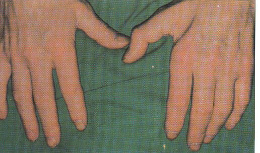
Other signs of a Pancoast tumor may arise from compression of the brachial plexus:
References:
![]()