|
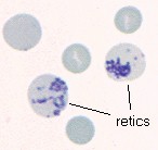
Reticulocytes ("retics") are young, anucleate erythrocytes. They are released to
the blood in increased numbers as
a response to anemia caused by
hemolysis or blood loss in most species
(horses are a notable exception).
Evaluation of the reticulocyte response is an initial step in characterizing anemia in species other than the horse. If the number of retics is increased in an anemic animal, it is a sign that the anemia is "regenerative" in character. Regenerative anemias are caused by loss (bleeding) or destruction (hemolysis) of erythrocytes from the peripheral circulation. Anemias without increased reticulocytes are termed "non-regenerative", and are due to conditions which decrease the production of erythrocytes by the marrow.
Last Updated: June 2000 |
 Reticulocytes in canine IHA