
|
|
CRYSTALS Contents of This Section (All links are to subsections within this file.) Except for the cystine crystals and a few others, the majority of crystals found in the urinary sediment are of limited clinical value. It is tempting to associate crystals with a risk of urolithiasis, but the majority of patients with a crystalluria do not have and will not develop kidney stones. Many benign situations can provoke crystal formation. In the majority of cases, the crystals found in urine are not present in the freshly voided specimen. Alkalization and refrigeration are promoters of crystals formation. The interpretation of a persistent crystalluria must be done according to the clinic. Drug crystals are sometimes found in urine. In most cases, these findings are of little clinical value except, if the sediment's picture indicates a possible renal obstruction. Crystal casts are pathognomonic of this situation. Some think that giving much time to the identification of unusual crystals is not worthwhile. Crystals related to urolithiasis are, except for cystine, usual and easy to identify. Calcium is found in 80 to 95% of kidney stones, mostly as oxalate or phosphate crystals. Many stones are not homogeneous. Some have a nucleus of a different composition from the surrounding matrix. The following table shows stones composition listed by occurrence.
In many cases, the presence of crystals is a pest to the microscopic examination. The elimination of these crystals can be made by gently heating the specimen at 37įC. To attain complete dissolution, it is preferable to heat the whole specimen. Once decanted, it is often impossible to dissolve the bulk in the small remaining volume. It is possible to dissolve the obscuring crystals by adjusting the pH. Phosphates can be dissolved by adding a drop or two of 2% acetic acid. Amorphous urates can be dissolved by adding an alkali like a 2% ammonia solution. But heating is by far a preferred method. It is not wise to solve a urate problem by creating a phosphate precipitation. About crystalluria—It is impossible to dissolve the quantity of calcium, phosphate, and oxalate eliminated in a 24-hours urine specimen into 1 to 2 liters of water. It is therefore necessary to conclude that substances inhibiting the crystallization are present. Known inhibitors of urinary crystallization are pyrophosphate, citrate, magnesium, and certain macromolecules. The Tamm-Horsfall protein is believed to be an important calcium oxalate inhibitor. This role is thought to be due to the sialic acid residue of the protein. While the fully sialated protein is an inhibitor, the sialic residues lacking protein is a crystallization promoter. Urine is a supersaturated solution of calcium, phosphate and oxalate in equilibria. Crystal formation can be caused:
Some crystals are found exclusively in acid urine, others are found exclusively in alkaline urine. Amorphous crystals are often identified on the basis of the urine pH. In an acid specimen, urates are reported, in an alkaline specimen, amorphous phospate are reported. This simplification should be used with care. Amorphous phosphates and triple phosphates are sometimes observed in slightly acid specimens. (pH 6,5)
Specific clinical conditions that explain urolithiase formation can also explain a persistent crystalluria. Hypercalciuria—An increased urinary calcium elimination can result in a crystalluria, mostly as calcium oxalates. The superior limit for the calciuria is 75 mmol/d under a 250 mmol/d diet. Hypercalciuria can be caused by:
Hyperoxaluria—The calcium oxalate is probably the crystal that one meets the most frequently in a urinary sediment. In the majority of cases, the presence of these crystals is without any clinical meaning. According to Conyers, only 10 to 15% of the urinary oxalate is directly related to the diet. The majority of the urinary oxalates is produced by the metabolism (glyoxilic acid cycle). It seems that even light hyperoxaluria is, after the decreased urinary volume, the most significant factor in the recurrent calcium oxalate urolithiasis. In some cases, the crystallization of the calcium oxalate is massive and catastrophic. A typical example of the oxalate clinical catastrophe is the cases of ethylene glycol poisoning. In this situation, one can find oxalate crystals in the patient's tissues. The toxicity syndrome affects organs like the liver, the kidney, and the brain and is accompanied by a metabolic acidosis. Naturally, the oxalate crystalluria is massive, and is predominated by the ovoid crystals (Whewellite) forming microlithes. Calcium oxalate casts are highly significant. These imply that the oxalate crystals were already formed when the urine was at it's maximum dilution. Conyers has reported other substances that can lead to oxalosis. Some of these substances are use as glucose substitutes in parental alimentation. Other causes of hyperoxaluria are:
Hyperuricosuria and calcium oxalates—An increased elimination of urate is most frequently caused by a high purine diet. Overproduction, can also be a cause of hyperuricosuria. With a urinary pH greater than 5,5, amorphous urates will be the major crystal form. Below 5.5, uric acid crystals are observed. It is not rare to observe calcium oxalate crystals with amorphous urate in the same urinary sediment. Urate crystals seem to have an enhancer effect on the calcium oxalate crystal formation. A possible explanation is that urates and oxalates are competing for the litho-inhibitor macromolecules. Hypocitraturia—The chelating effect of citrate is known to reduce the saturation in calcium salt. Also, the soluble chelating calcium complex seems to have an inhibiting effect on crystal formation. One can therefore expect an increase in crystals formation, and even urolithiasis in conditions leading to hypocitraturia. Hypocitraturia is seen in conditions like in:
Many bacteria infecting the urinary tract reduce the citrate concentration. 5% of the hypocitraturia are of unknown causes. Uric acid—Approximately 66 to 75% of the uric acid is eliminated by the urine. The quantity to eliminate depends mostly on the diet (meat). In increased uricosuria, values > 4,5 mmol/d are observed. Uric acid crystals are formed when the urinary pH is <5,5 since the pK of uric acid is 5,5. Uric acid crystalluria is mainly due to a poor dilution volume at an acid pH, or due to an overproduction. In the majority of cases, this finding is of little clinical value and represents a pinpoint situation. Acid pH. Some conditions, like chronic diarrhea, can be responsible for uric acid urolithiasis. Many of these patients will also have calcium stones. Overproduction. Uric acid stones are seen in cases of gout, myeloproliferative syndrome, glycogenosis and neoplasms. Cystinuria—Cystine crystals are found in urine of almost exclusively patients with a genetic disease giving an impairment with the tubular reabsorption of the basic aminoacid: lysine, arginine, ornithine and cystine. This disease is called cystinuria. For a few patients with cystinuria, stones will develop. The urolithiasis is highly dependent of the urinary pH and water intake. Cystine is less soluble at a pH lower than 5,0 (saturation 300 mg/l); saturation is of 500 mg/l at a pH of 7,4. Infection—Infection with urea splitting bacteria (ex: proteus species) leads to a production of ammonia an alkalinization of the urine. The produced ammonia generates magnesium ammonium phosphate crystals, also called triple phosphates. The mineralogical name of triple phosphate is Struvite. Triple phosphates are usually found with amorphous phosphates, owing to their low solubility at alkaline pH. Solid substances are divided in two large groups, amorphous
substances and crystalline substances. Crystals have defined
geometrical shapes while amorphous substances have not. More,
crystals have a precise melting point, while amorphous substances
have a melting point that spreads over an interval of temperature.
In crystallography, one speaks of planes, axes, and angles
to describe their shape. Crystals, while preserving their primary
shape, have a very variable size, but the ratios and angles
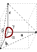
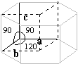
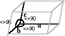
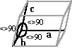
between the faces and between the sides, are constant. A characteristic of crystals is that their form is predictable from the elementary structure. There are 230 possible geometrical forms that are grouped in 32 classes, based upon the arrangement of elements of symmetry. These elements of symmetry are axes of symmetry, planes of symmetry, the center of symmetry. These 32 classes are further regrouped in 6 crystalline systems. Crystallographic constants of these crystalline systems are described by a system of coordinate, 3 axes (a, b, c), and by the angles formed by these axes between them (a, b, g).
Crystals of the cubic system are said to be isotropic, since these have the same properties in all direction. One of these properties is the refractive index. Crystals of the other systems are said to be anisotropic, having two (birefringent) or even three refractive indexes. Anisotropic crystals subdivide into two groups: uniaxial (two refractive indexes) and biaxial (three refractive indexes). Crystals of the tetragonal system and the hexagonal system are uniaxial while the orthorombic, monoclinic and the triclinic are biaxial. Some anisotropic crystals form interference patterns when viewed under polarized light. Uric acid are polychromatic, and cholesterol ester in a liquid crystal state generates a maltese cross pattern. Birefringency is a crystal behavior. The following table shows the birefringent behavior of some crystals found in urine.
Urine is a complex medium which influences the crystallization process. The same substance can crystallize into different shapes depending on the urine composition. Crystals found in urine are often truncated and eroded. Spherical crystals are frequent. Slow crystallization tends to give larger crystals. In the urinary sediment, one can find two forms of calcium oxalate crystals. The most frequent form is the di-hydrated calcium oxalate. The mineralogical name of the calcium oxalate 2(H2O) is Weddellite. The second form is the mono-hydrated calcium oxalate whose mineralogical name is Whewellite. The two forms have different crystallographic characteristics. It seems that the calcium/magnesium ratio plays an important role in the formation of the calcium oxalate crystals. Crystals of calcium oxalate are found mainly in an acidic urine, but these can also be seen in slightly alkaline specimens.
Weddelite crystals are poorly birefringent and do not show any interference pattern under polarized light. Weddelite crystals are usually of little clinical value. Many specimens develop weddelite crystals on standing.
Uric acid crystals usually have a characteristic yellow color. The intensity of the color depends on the thickness of the crystal, thus very thin plates seem colorless, while the massive crystals have a color that tends to be brown. Under polarized light, uric acid shows a polarization color, and with thicker crystals, a series of concentric black lines. The color variation seen under polarized light is quite typical of uric acid. With rare exceptions, uric acid crystals are of little clinical value and represent a punctual situation.
These crystals often adhere to mucus threads and casts. This property sometimes generates structures that mimic the dirty brown cast. Polarized light examination can easily discriminate these urates pseudocasts. Amorphous urates are of little clinical value.
The main cause of this crystalluria is the alkaline pH that decreases the solubility of the calcium phosphate and entails a precipitation of the former. The alkaline pH can be caused by the diet (vegetarian, rich in phosphates..) but can also represent a pathological situation. Usually, the presence of these crystals is non significant. The distinction between amorphous urates and amorphous phosphates is often made on the urinary pH basis. With a simple examination of the centrifuge pellet, the precipitate of calcium phosphate is white, while that amorphous urate is pink.
The primary factor to the triple phosphate crystals formation is the ammonia concentration. Alkalanisation of a urine specimen with ammonia generates triple phosphates while alkalanisation with sodium hydroxide does not. A normal freshly voided specimen contains little free ammonia; this substance is mainly generated by urea splitting bacteria. Triple phosphates are usualy associated with bacterial growth. With a first-morning fresh specimen, triple phosphates can indicate urinary tract infection. Otherwise, triple phosphates are of little clinical value. Several crystals are found only rarely in the urine. Often these crystals are difficult to differentiate from some particular shapes of a "usual" crystals. Before considering the possibility of an exotic crystal, it is necessary to exhaust the possibilities of a rare shape of a usual one. Some rare crystals are found mainly in acidic specimens, while others are found in alkaline media. Again. this represents only a tendency.
Cystine crystals are a clinically significant finding. A confirmatory test is described elsewhere. Leucine and tyrosine—Crystals of the amino acids leucine and tyrosine are very rarely seen in a urinary sediment. These crystals can be observed in some hereditary diseases like tyrosinosis and the "Maple syrup disease", but these conditions are very rare. The majority of cases where one finds these crystals are in patients with a serious hepatic problem, often in a terminal stage. In these cases, a concurrent presence of leucine and tyrosine is observed.
Calcium phosphate crystals are found with triple phosphates and their clinical meaning is identical.
The presence of iatrogenic crystals is a rare event. Except for the Indinavir crystal, a light crystalluria due to a medication or from a contrast agent for X-ray imagery has no clinical significance. On the other hand, an abundant crystalluria associated with a hematuria, a marked cellularity, and oliguria could indicate an obstructive renal disease. The presence of casts with crystalline inclusions clearly indicates that the crystallization is intratubular. The majority of these drugs crystallize in an acid media often around a pH of 5,0. Drugs of the sulfamides group (sulfamethoxazole, acetylsulfadiazine, sulfadiazine) are the most frequently observed.
The crystals are birefringent under polarized light with a color dispersion as seen with uric acid. Indinavir crystal, unlike uric acid, are seen at a neutral (6,5 7,5) while uric acid crystals are seen at a more acidic pH (5,0 - 5,5) The aim of these tests is to confirm the identity of crystals. These tests are rarely used, and only if indicated by a clinical situation. Many confirmation tests can easily be done using routine reagents (uric acid, cholesterol, phosphates, calcium...). However, some tests need special reagents rarely used for other reasons. In these cases, reagents will probably have to be freshly made. To be sure that the reaction is due to crystals, it is wise to do two tests. The first test is done with the centrifuged supernatant and the second one is done with the dispersed sediment. The color difference between the two tests is then due to solid matters. A good strategy is often necessary with mixed crystalluria. Since the sediment's volumes are normally small, it is necessary to have a good idea on the identity of the crystals. Before making tests to identify rare crystals, the possibility of a rare form of a usual crystal should be considered and excluded. From: Ringsrud KM, Linnť JJ: Urinalysis and Body Fluids Reagents Ammonia 10%
Sodium cyanide 5%
Sodium nitroprusside 5%
Procedure
NB: Similar reagents are included in kits for urinary calculus identification. Hemosiderin can be demonstrated by the Roux reaction ( Prussian blue). This stain reacts with the iron depot of a cytospun specimen. These depots can be free (amorphous mass), within macrophages, within tubular cells, and embedded in casts. Reagents Potassium ferricyanide 2%
HCl 1%
Working reagent
Procedure
From: Ringsrud KM, Linnť JJ: Urinalysis and Body Fluids Reagents HCl 10%
Sodium nitrite 0,1%
Ammonium sulfamate 0,5%
Diazo reagent 0,1%
Procedure
NB: Similar reagents can be found in some LAP kits. |
|||||||||||||||||||||||||||||||||||||||||||||||||||||||||||||||||||||||||||||||||||||||||||||||||||||||||||


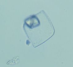
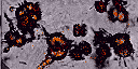
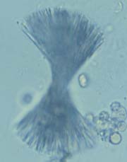
 The
weddelite or calcium oxalate di-hydrate crystallizes in the
tetragonal system. The classic crystal shape is the eight-face
bi-pyramid. In bright field microscopy, the weddelite crystals
are recognized easily by their shape that reminds a mail envelope.
More complex shapes of weddelite are possible. The dumbbell
shape is not rare. The former has no precise angles or sides.
This form is, in reality, an microcrystalline agglomerate that
takes the shape of a biconcave disc.
The
weddelite or calcium oxalate di-hydrate crystallizes in the
tetragonal system. The classic crystal shape is the eight-face
bi-pyramid. In bright field microscopy, the weddelite crystals
are recognized easily by their shape that reminds a mail envelope.
More complex shapes of weddelite are possible. The dumbbell
shape is not rare. The former has no precise angles or sides.
This form is, in reality, an microcrystalline agglomerate that
takes the shape of a biconcave disc.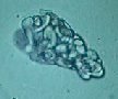
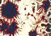
 The
whewellite crystal is a rare form of crystallization of calcium
oxalate. In theory, the whewellite, or calcium oxalate mono-hydrate
crystallizes in a monoclinic leave shape, but in the majority
of cases, the former precipitates as an oval egg shape. The
dumbbell structure is often erroneously associated to this
form of oxalate. X-ray analysis have shown that the dumbbell
structure can also represent weddelite crystals. Contrarily
to the weddelite, the whewellite is found in situations of
massive calcium oxalate precipitation. According to Berg, the
abundance of oxalates formed of ovoid structures strongly agglutinated,
twin structures, and microliths, is an indication of a pathological
massive precipitation. Urines of patients with a calcium oxalate
urolithiase have a tendency to have a sediment with some of
the preceding characteristics.
The
whewellite crystal is a rare form of crystallization of calcium
oxalate. In theory, the whewellite, or calcium oxalate mono-hydrate
crystallizes in a monoclinic leave shape, but in the majority
of cases, the former precipitates as an oval egg shape. The
dumbbell structure is often erroneously associated to this
form of oxalate. X-ray analysis have shown that the dumbbell
structure can also represent weddelite crystals. Contrarily
to the weddelite, the whewellite is found in situations of
massive calcium oxalate precipitation. According to Berg, the
abundance of oxalates formed of ovoid structures strongly agglutinated,
twin structures, and microliths, is an indication of a pathological
massive precipitation. Urines of patients with a calcium oxalate
urolithiase have a tendency to have a sediment with some of
the preceding characteristics.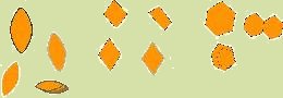
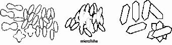
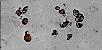
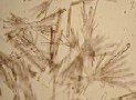
 Uric
acid crystallizes in the orthorombic system. Uric acid crystals
can appear under several shapes. The classic crystals are thin
rhombus shaped plates with more or less eroded tops. The other
forms are the hexagonal plate, the needle and the rosette.
Uric
acid crystallizes in the orthorombic system. Uric acid crystals
can appear under several shapes. The classic crystals are thin
rhombus shaped plates with more or less eroded tops. The other
forms are the hexagonal plate, the needle and the rosette. The
amorphous urates seen in urine specimens and are, most of the
time, the result of refrigeration. A pink pellet after centrifugation
is characteristic of amorphous urates. Under the microscope,
amorphous urates appear as yellow-brown mass of small rounded
particles.
The
amorphous urates seen in urine specimens and are, most of the
time, the result of refrigeration. A pink pellet after centrifugation
is characteristic of amorphous urates. Under the microscope,
amorphous urates appear as yellow-brown mass of small rounded
particles.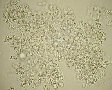 Amorphous
phosphates is the name given to a granular precipitate containing
calcium and phosphate in an alkaline urine. Calcium phosphate
crystals, regrouped under the term apatite, have mineralogical
names that differ according to their chemical composition.
The CaH2PO4*(2H2O) is called Brushite, the calcium hydroxyl
phosphate is called hydroxyl-apatite, the calcium bicarbonate
phosphate is called Dahlite or carbonate-apatite.
Amorphous
phosphates is the name given to a granular precipitate containing
calcium and phosphate in an alkaline urine. Calcium phosphate
crystals, regrouped under the term apatite, have mineralogical
names that differ according to their chemical composition.
The CaH2PO4*(2H2O) is called Brushite, the calcium hydroxyl
phosphate is called hydroxyl-apatite, the calcium bicarbonate
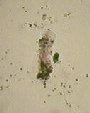
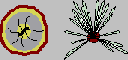
phosphate is called Dahlite or carbonate-apatite. Triple
phosphates are found in urines whose pH is superior to 6,5.
The former crystallize in the orthorombic system. The crystal
is slightly birefringent and often shows a polarization color.
The classic shape is the pyramid, that reminds a coffin lid.
The crystalluria is usually polymorphous. The rosette-shaped
crystal shown in the picture bank has been observed in a specimen
with a pH of 6,5.
Triple
phosphates are found in urines whose pH is superior to 6,5.
The former crystallize in the orthorombic system. The crystal
is slightly birefringent and often shows a polarization color.
The classic shape is the pyramid, that reminds a coffin lid.
The crystalluria is usually polymorphous. The rosette-shaped
crystal shown in the picture bank has been observed in a specimen
with a pH of 6,5.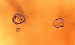


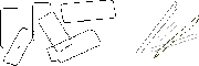

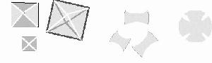
 Indinavir,
a protease inhibitor (antiviral agent) widely used to treat
patients with HIV infection, has been associated with nephrolithiasis.
Indinavir is insoluble at physiological pH so that, 20% of
the person receiving Indinavir have characteristic crystal
in their urine. The crystal are seen as plate-like rectangles
exibiting rosette formation
Indinavir,
a protease inhibitor (antiviral agent) widely used to treat
patients with HIV infection, has been associated with nephrolithiasis.
Indinavir is insoluble at physiological pH so that, 20% of
the person receiving Indinavir have characteristic crystal
in their urine. The crystal are seen as plate-like rectangles
exibiting rosette formation