|
|
MICROORGANISMS AND OTHER ELEMENTS This section is dedicated to all those elements of the urinary sediment that cannot be classified as cells, casts or crystals. We therefore discuss elements as varied as bacteria, mucus and artifacts. In some cases, we mention the possibility of a misidentification with another element of the sediment. Contents of This Section (All links are to subsections within this file.)
Bacteria associated with urinary tract infection are mostly bacillus (E. Coli) but other shapes cannot be ruled out. Bacteria-coated urothelial cells are frequent in cystitis. This situation is different from Clue Cells, which are vaginal squamous cells coated with a coccobacillus (Gardnerella Vaginalis), forming a crust over the cell. Under the microscope, these latter cells have a granular aspect with a blunted border.
Other forms of yeast can be seen in the urine specimens. With a wet uncolored sediment, some of these forms are difficult to differentiate from red blood cells or some other elements. However, yeast contains DNA that can be demonstrated with an usual stain like the Sedistain(tm). This dye preparation stains yeast cells in blue. In a number of specimens, the presence of yeast is the result of a contamination with vaginal secretion. Yeasts are often observed in specimens that contain sugar. It is important to be careful with these specimens because a yeast infection is a frequent finding with diabetic patients. Yeast containing casts have a very high clinical value; these are pathognomonic of pyelonephritis.
Identification of the living cell is quite easy owing to its spectacular motility. Identification of immobile cells is less obvious. Special stain can be use for Trichomonas. These stains can be found in the parasitology literature Other parasites can be seen in the urine sediment. But these situations are rare and are mostly seen in exposed population. Identification of these parasites should be transferred to the parasitology department. This laboratory section has the tools and the expertise to make a proper identification. Some cellular manifestations of viral infection can be seen in the urine sediment. To see these manifestations, the specimen must be stained. In some cases, phase contrast microscopy can be used but, a good staining procedure on a cytospin specimen gives a preparation that is easier to read. Identification of infected cells is within the scope of the cytology department. Suspected specimens should be transferred to the cytology lab.
Some labs do not report spermatozoa. The problem with such a policy is that the labs rarely know all the elements to make a truly wise decision. Most cases are due to a normal human "activity" that should stay personal. But some cases, not necessarily known to the lab, are a result of reprehensible activity. Reporting the sperm cells found in a young girl's specimen is an easy decision but, cases of abuse are not always so obvious. We think that the lab should report all cases of spermatozoa and let the clinician decide what to do with the result. Mucus is a frequent finding of the urinary sediment. The exact function of mucus is unknown. Some think that this substance is a protection against bacterial infection. This action is done by coating the bacterial's pilis, essential to colonization of the lower urinary tract wall. The mucus coated bacteria are eliminated through miction. Mucus can also protect the lower urinary tract against irritating chemical agents.
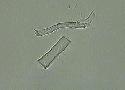
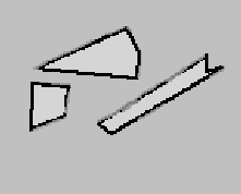
In the majority of cases, presence of mucus threads is a benign situation. An irritating factor could stimulate mucus secretion. The number of contaminating elements found in the urine sediment is surprising. Some of these artifacts are unavoidable, such as glass fragments, bubbles. Other artifacts are accidental, such as fibers and hair.
|
|||||||||||||||||||||
 The
urinary infection is the most frequently observed anomaly in
urinary microscopy. The presence of many white blood cells
and bacteria is characteristic of this situation. On the other
hand, urine specimens for routine examination are not usually
obtained through a sterile technique with the result that old
specimens can have a lot of bacteria with only a few leukocytes.
The presence of numerous squamous cells can indicate an external
genital source for the bacteria. In these two situations, a
positive nitrite result may indicate a urinary tract infection
but a safe diagnosis is made only by a positive culture obtained
with a midstream specimen.
The
urinary infection is the most frequently observed anomaly in
urinary microscopy. The presence of many white blood cells
and bacteria is characteristic of this situation. On the other
hand, urine specimens for routine examination are not usually
obtained through a sterile technique with the result that old
specimens can have a lot of bacteria with only a few leukocytes.
The presence of numerous squamous cells can indicate an external
genital source for the bacteria. In these two situations, a
positive nitrite result may indicate a urinary tract infection
but a safe diagnosis is made only by a positive culture obtained
with a midstream specimen.
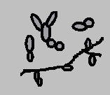 Like
bacteria, the presence of yeast in the urine sediment may indicate
an infection. A frequently seen yeast in urine is Candida.
Identification of this organism is relatively easy because
of its usual club shape. In the majority of cases, only the
isolated cells are seen but, in some cases, budding pseudohyphea
may be observed.
Like
bacteria, the presence of yeast in the urine sediment may indicate
an infection. A frequently seen yeast in urine is Candida.
Identification of this organism is relatively easy because
of its usual club shape. In the majority of cases, only the
isolated cells are seen but, in some cases, budding pseudohyphea
may be observed. The
parasite that is the more frequently seen in urine is Trichomonas.
Usually, this cell comes from genital secretions contaminating
the specimen. But Trichomonas should be mentioned because cases
of vesical and prostate colonisation by this organism have
been reported in the literature.
The
parasite that is the more frequently seen in urine is Trichomonas.
Usually, this cell comes from genital secretions contaminating
the specimen. But Trichomonas should be mentioned because cases
of vesical and prostate colonisation by this organism have
been reported in the literature. Urinary
spermatozoa is a contamination arising from sexual activity.
With a male subject, these represent a residual drainage while
with a female, these have a vaginal contamination source.
Urinary
spermatozoa is a contamination arising from sexual activity.
With a male subject, these represent a residual drainage while
with a female, these have a vaginal contamination source. Mucus
forming cells are found scattered all over the urinary tract
from the ascending section of the loop of Henle to the bladder.
Consequently, mucus can originate from the kidney or from the
lower urinary tract. Mucus originating from the kidney is made
of Tamm-Horsfall protein. This explains the frequent association
of mucus threads and casts. In elderly patients, mucus is a
frequent finding and seems to originate from the lower urinary
tract.
Mucus
forming cells are found scattered all over the urinary tract
from the ascending section of the loop of Henle to the bladder.
Consequently, mucus can originate from the kidney or from the
lower urinary tract. Mucus originating from the kidney is made
of Tamm-Horsfall protein. This explains the frequent association
of mucus threads and casts. In elderly patients, mucus is a
frequent finding and seems to originate from the lower urinary
tract.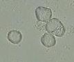 Since
the advent of systematic use of latex gloves by the hospital's
staff, the presence of starch crystals or sometimes talc has
become very frequent. The crystals are birefringent with a
maltese cross interference pattern when seen under crossed
polarized filter. The aspect of these crystals under bright
field allows one to easily distinguish them from the birefringent
fatty droplets.
Since
the advent of systematic use of latex gloves by the hospital's
staff, the presence of starch crystals or sometimes talc has
become very frequent. The crystals are birefringent with a
maltese cross interference pattern when seen under crossed
polarized filter. The aspect of these crystals under bright
field allows one to easily distinguish them from the birefringent
fatty droplets.