Ed Friedlander, M.D., Pathologist
scalpel_blade@yahoo.com


Cyberfriends: The help you're looking for is probably here.
Welcome to Ed's Pathology Notes, placed here originally for the convenience of medical students at my school. You need to check the accuracy of any information, from any source, against other credible sources. I cannot diagnose or treat over the web, I cannot comment on the health care you have already received, and these notes cannot substitute for your own doctor's care. I am good at helping people find resources and answers. If you need me, send me an E-mail at scalpel_blade@yahoo.com Your confidentiality is completely respected.
 DoctorGeorge.com is a larger, full-time service.
There is also a fee site at myphysicians.com,
and another at www.afraidtoask.com.
DoctorGeorge.com is a larger, full-time service.
There is also a fee site at myphysicians.com,
and another at www.afraidtoask.com.
Translate this page automatically
 |
With one of four large boxes of "Pathguy" replies. |
 I'm still doing my best to answer
everybody.
Sometimes I get backlogged,
sometimes my E-mail crashes, and sometimes my
literature search software crashes. If you've not heard
from me in a week, post me again. I send my most
challenging questions to the medical student pathology
interest group, minus the name, but with your E-mail
where you can receive a reply.
I'm still doing my best to answer
everybody.
Sometimes I get backlogged,
sometimes my E-mail crashes, and sometimes my
literature search software crashes. If you've not heard
from me in a week, post me again. I send my most
challenging questions to the medical student pathology
interest group, minus the name, but with your E-mail
where you can receive a reply.
Numbers in {curly braces} are from the magnificent Slice of Life videodisk. No medical student should be without access to this wonderful resource. Someday you may be able to access these pictures directly from this page.
Also:
Medmark Pathology -- massive listing of pathology sites
Freely have you received, freely give. -- Matthew 10:8. My
site receives an enormous amount of traffic, and I'm
handling about 200 requests for information weekly, all
as a public service.
Pathology's modern founder,
Rudolf
Virchow M.D., left a legacy
of realism and social conscience for the discipline. I am
a mainstream Christian, a man of science, and a proponent of
common sense and common kindness. I am an outspoken enemy
of all the make-believe and bunk that interfere with
peoples' health, reasonable freedom, and happiness. I
talk and write straight, and without apology.
Throughout these notes, I am speaking only
for myself, and not for any employer, organization,
or associate.
Special thanks to my friend and colleague,
Charles Wheeler M.D.,
pathologist and former Kansas City mayor. Thanks also
to the real Patch
Adams M.D., who wrote me encouragement when we were both
beginning our unusual medical careers.
If you're a private individual who's
enjoyed this site, and want to say, "Thank you, Ed!", then
what I'd like best is a contribution to the Episcopalian home for
abandoned, neglected, and abused kids in Nevada:
My home page
Especially if you're looking for
information on a disease with a name
that you know, here are a couple of
great places for you to go right now
and use Medline, which will
allow you to find every relevant
current scientific publication.
You owe it to yourself to learn to
use this invaluable internet resource.
Not only will you find some information
immediately, but you'll have references
to journal articles which you can obtain
by interlibrary loan, plus the names of
the world's foremost experts and their
institutions.
Alternative (complementary) medicine has made real progress since my
generally-unfavorable 1983 review linked below. If you are
interested in complementary medicine, then I would urge you
to visit my new
Alternative Medicine page.
If you are looking for something on complementary
medicine, please go first to
the American
Association of Naturopathic Physicians.
And for your enjoyment... here are some of my old pathology
exams
for medical school undergraduates.
I cannot examine every claim that my correspondents
share with me. Sometimes the independent thinkers
prove to be correct, and paradigms shift as a result.
You also know that extraordinary claims require
extraordinary evidence. When a discovery proves to
square with the observable world, scientists make
reputations by confirming it, and corporations
are soon making profits from it. When a
decades-old claim by a "persecuted genius"
finds no acceptance from mainstream science,
it probably failed some basic experimental tests designed
to eliminate self-deception. If you ask me about
something like this, I will simply invite you to
do some tests yourself, perhaps as a high-school
science project. Who knows? Perhaps
it'll be you who makes the next great discovery!
Our world is full of people who have found peace, fulfillment, and friendship
by suspending their own reasoning and
simply accepting a single authority that seems wise and good.
I've learned that they leave the movements when, and only when, they
discover they have been maliciously deceived.
In the meantime, nothing that I can say or do will
convince such people that I am a decent human being. I no longer
answer my crank mail.
This site is my hobby, and I presently have no sponsor.
This page was last updated February 6, 2006.
During the ten years my site has been online, it's proved to be
one of the most popular of all internet sites for undergraduate
physician and allied-health education. It is so well-known
that I'm not worried about borrowers.
I never refuse requests from colleagues for permission to
adapt or duplicate it for their own courses... and many do.
So, fellow-teachers,
help yourselves. Don't sell it for a profit, don't use it for a bad purpose,
and at some time in your course, mention me as author and KCUMB as my institution. Drop me a note about
your successes. And special
thanks to everyone who's helped and encouraged me, and especially the
people at KCUMB
for making it possible, and my teaching assistants over the years.
Whatever you're looking for on the web, I hope you find it,
here or elsewhere. Health and friendship!
 I am presently adding clickable links to
images in these notes. Let me know about good online
sources in addition to these:
I am presently adding clickable links to
images in these notes. Let me know about good online
sources in addition to these:
Pathology Education Instructional Resource -- U. of Alabama; includes a digital library
Houston Pathology -- loads of great pictures for student doctors
Pathopic -- Swiss site; great resource for the truly hard-core
Syracuse -- pathology cases
Walter Reed -- surgical cases
Alabama's Interactive Pathology Lab
"Companion to Big Robbins" -- very little here yet
Alberta
Pathology Images --hard-core!
Cornell
Image Collection -- great site
Bristol Biomedical
Image Archive
EMBBS Clinical
Photo Library
Chilean Image Bank -- General Pathology -- en Español
Chilean Image Bank -- Systemic Pathology -- en Español
Connecticut
Virtual Pathology Museum
Australian
Interactive Pathology Museum
Semmelweis U.,
Budapest -- enormous pathology photo collection
Iowa Skin
Pathology
Loyola
Dermatology
History of Medicine -- National Library of Medicine
KU
Pathology Home
Page -- friends of mine
The Medical Algorithms Project -- not so much pathology, but worth a visit
National Museum of Health & Medicine -- Armed Forces Institute of Pathology
Telmeds -- brilliant site by the medical students of Panama (Spanish language)
U of
Iowa Dermatology Images
U Wash
Cytogenetics Image Gallery
Urbana
Atlas of Pathology -- great site
Visible
Human Project at NLM
WebPath:
Internet Pathology
Laboratory -- great site My team:
Ed Lulo's Pathology Gallery
Bryan Lee's Pathology Museum
Dino Laporte: Pathology Museum
Tom Demark: Pathology Museum
Dan Hammoudi's Site
Claude Roofian's Site
Pathology Handout -- Korean student-generated site; I am pleased to permit their use of my cartoons
Estimating the Time of Death -- computer program right on a webpage
Pathology Field Guide -- recognizing anatomic lesions, no pictures

St.
Jude's Ranch for Children
I've spent time there and they are good. Write "Thanks
Ed" on your check.
PO Box 60100
Boulder City, NV 89006--0100
More of my notes
My medical students
Clinical
Queries -- PubMed from the National Institutes of Health.
Take your questions here first.
HealthWorld
Yahoo! Medline lists other sites that may work well for you

We comply with the
HONcode standard for health trust worthy
information:
verify
here. 

![]()
The student will describe the specificity of the common stains used in histopathology.
The student will correctly define the following terms as used in pathology, supply them (given a definition), and mention their significance:
amyloid
hemosiderosis
dystrophic calcification
jaundice
fatty change
lipophage
fatty ingrowth
metastatic calcification
ferruginous body
viral inclusion
hemochromatosis
xanthoma
Given a photomicrograph or glass slide, plus any clinical or special-stain information that may be necessary, the student will recognize:
alcoholic hyaline
glycogen (liver cell nuclei)
cholesterol crystals ("needles")
metastatic calcification
dystrophic calcification
stromal infiltration of fat
fatty change in the liver
tophus of gout
Gaucher cells
viral inclusion (herpes)
 The student will explain the origins of each of the important tissue pigments (bilirubin, carbon,
hemosiderin, lipofuscin, melanin), and recognize each in tissue sections (given appropriate
supplementary information when necessary).
The student will explain the origins of each of the important tissue pigments (bilirubin, carbon,
hemosiderin, lipofuscin, melanin), and recognize each in tissue sections (given appropriate
supplementary information when necessary).
The student will recognize the following "hyaline" substances, given the appropriate setting:
amyloid
basement membrane
hyalinized collagen (keloid)
fibrin
Mallory's hyaline (prekeratin)
fibrinoid (necrosis)
Russell body (immunoglobulin)
The student will recognize why liver cells accumulate fat during alcohol abuse, and list the classic causes of fatty change in the liver and heart respectively.
The student will describe and account for the accumulation of glycogen in cells in patients with diabetes, storage disease, and on IVs.
The student will recognize the major mechanisms of jaundice.
Given a yellow patient, the student will correctly distinguish carotenemia, jaundice, and uremia.
The student will describe typical sites and settings for dystrophic and metastatic calcification, myxoid change, and mitochondrial aberrations.
QUIZBANK
Necrosis #'s 55-65, 69-70, 72
Minerals and pigments (all)
LEARN FIRST
Hematoxylin stains nucleic acids, bacteria, and calcium blue. Eosin stains arginine and lysine (i.e., protein molecules) pink. PAS stains insoluble carbohydrates magenta; d-PAS stain is used to prove something PAS-positive is, or is not, glycogen.
Acid-fast stains selectively stain mycobacteria.
Fatty change is too much fat in business cells which shouldn't ordinarily accumulate it; it's a sign that the cell is sick. Fatty ingrowth is extra fat cells in an organ where they don't usually belong.
Hemosiderin is iron storage pigment. You distinguish it using the Prussian Blue stain.
Melanin is the familiar skin pigment. You distinguish it because it loses its color on being exposed to hair bleach.
Bilirubin pigment generally occurs with bile plugs or bile lakes in the liver.
Lipofuscin is an inert, wear-and-tear pigment. You distinguish it by its location, or by process of elimination.
Dystrophic calcification results from disease at the site of calcification. Metastatic calcification results from disease remote from the site of calcification that has caused elevated blood calcium or phosphate.
"Hyaline" is a generic term for amorphous masses protein (usually a single protein).
Amyloid is beta-pleated anything. You identify it using Congo Red staining.
"Fibrinoid necrosis" of a vessel means the endothelium has been badly damaged, and fibrin and
other proteins have precipitated in the wall.
 Amyloidosis
Lots of apple green birefringence
Wash U, St. Louis
INTRODUCTION
This unit will teach you to recognize some things that pop up again and again when we look at disease. You know much of this material already. We want you to start feeling comfortable with gross and microscopic views of disease processes.
You will already have heard about some of these pathologic changes. This unit will emphasize how they look.
STAINS FOR MEDICAL STUDENTS
|
|
Tissue stains stain whatever is left in the embedded tissue after processing. (Standard processing of formalin-fixed tissue removes small non-protein molecules, especially lipids.)
Clinicians need to know only a little about stains used by pathologists in establishing diagnosis. The following will come in handy in understanding pathology slides and reports:
Hematoxylin and eosin (H&E) is the standard tissue stain.
The pedagogical explanation ("hematoxylin is basic and stains acids, eosin is acidic and stains bases") isn't exactly so.
Hematoxylin (blue-purple) is a metal chelator. We usually use it with aluminum ions, which link it to fixed phosphate groups. Hematoxylin stains nucleic acids, calcium salts, and bacteria blue-purple. (* Hematoxylin comes from an exotic jungle tree.)
Eosin (red) binds to free amino groups and thus stains arginine and lysine pink. (* Eosin, which is fluorescent, is a major component of many colors of lipstick. It's also the red dye on pistachio nuts.)
Air, water, fat, and carbohydrate end up unstained.
The Romanowsky stain family, including Wright and Giemsa, use a similar color scheme, though with somewhat different dyes and some nice metachromasia. You'll encounter these stains when we study blood and bone-marrow smears.
Periodic acid-Schiff (PAS) is a stain based on the familiar * periodic acid (H+IO4-) oxidation (cis-diols to aldehydes) and * Schiff-base reactions.
Anything with a cis-dihydroxy group, i.e., insoluble sugar compounds such as ...
... gets stained magenta.
Diastase-PAS ("d-PAS") is the PAS reaction performed on tissue previously digested by diastase, which removes glycogen.
The pathologist can buy diastase, or can spit on the slide before staining. If something is PAS-positive and d-PAS negative, you know it is glycogen.
Reticulin stains the delicate fibers that surround small blood vessels and hold together liver, spleen, and lymph nodes.
Prussian Blue uses a special solution of * ferrocyanide.
By the familiar reaction (college chemistry, laundry bluing, old-fashioned blueprints), it stains all exposed ferric ions very blue.
Congo Red is a special dye that fits tightly into beta-pleated proteins of all sorts.
Beta-pleated proteins in humans are abnormal and are called amyloids. Congo Red stains all amyloids brick red, and everything else pink. Because of the way beta-pleated sheets line up Congo Red, amyloids also acquire an apple green birefringence when stained with Congo Red and then examined under polarized light.
|
{10880} congo red stain of amyloid, kidney
|
Sudan / Oil Red O are oil-based stains that can only be used on thin slices ("frozen sections") of
tissue from which fat has not been removed.
These stain only fat (usually black or bright red). The principle is of course hydrophobic bonding.
{17413} fat stain, atherosclerosis, oil red O
Mucicarmine is a special dye that stains only epithelial mucin (usually red). The actual chemistry
remains mysterious.
* Alcian blue stains certain mucoid substances (hyaluronic acid, sulfomucin,
maybe carboxymucin
depending on the recipe).
Trichrome uses familiar aniline dyes to stain collagen (type I, also basement membranes) blue or
green and everything else some other color. The stain depends on the special way collagen is woven.
Acid-fast stains (ZN, auramine O,
others) stain certain waxes a permanent red (or some other color). This
shows up mycobacteria (TB bugs) and certain other rare substances.
Argentaffin stains test the ability of cell structures to bind and reduce silver, while argyrophil
stains demonstrate all sites of silver binding, whether or not reduction occurs. (Everything that is
argentaffin is argyrophil, but not vice-versa.)
Methenamine silver is the most sensitive and specific common
stain for fungi and pneumocystis. It stains them black.
Elastic stains (Verhoeff, Van Gieson) selectively stain elastic fibers (typically black).
Metachromatic stains take advantage of molecule-stacking. A single dye will impart a variety of
hues to different structures. The most important metachromatic dyes are those use to stain blood and
bone marrow smears (the various azures); * Bismark brown imparts a metachromatic yellow on pap smears.
* Light green stains RNA green.
* Orange G stains disulfide bonds orange.
* Papanicolaou's stain is used for cytology (i.e., smears of cells on glass slides, "Pap" smears).
It contains hematoxylin, eosin, light green, orange G, and sometimes Bismark brown.
* Methyl green pyronine stains RNA red and everything else green.
Immunostaining (immunofluorescence, immunoperoxidase) uses monoclonal antibodies to
demonstrate specific antigens (i.e., specific proteins) in tissues.
Nucleic acid probes are now being
introduced to stain particular genes and their RNA's. For example, a
cancer cell that contains mRNA for albumin must be of hepatocyte origin.
FATTY CHANGE ("fatty metamorphosis", "fatty degeneration", "steatosis"): accumulation of
excess neutral fat in vacuoles within non-adipocytes
If there's one big fat vacuole, it's "macrovesicular". If there's many little fat vacuoles, it's
"microvesicular".
Fatty change of injured cells occurs classically in the liver and the heart.
There are at least six mechanisms by which the liver cell accumulates fat during disease, any or all
of which may be operating in a given situation.
1. Too much free fat coming to the liver
2. Too much fatty acid synthesis by the liver
3. Impaired fatty acid oxidation by the liver
4. Excess esterification of fatty acid to triglycerides by the liver
5. Too little apoprotein synthesis by the liver
6. Failure of lipoprotein secretion by the liver.
At first the fat accumulates in the rough endoplasmic reticulum, but soon fat globules occur that are
not membrane-bound.
You will care for many patients with fatty liver.
Fatty liver develops during heavy drinking, and all six mechanisms are known to contribute here.
Other causes of heavy-duty fatty liver include kwashiorkor (why?), Reye's syndrome, poisoning by
phosphorus, carbon tetrachloride, * non-alcoholic steatohepatitis, * outdated tetracycline, pregnancy (rare and mysterious), * the
bad kind of galactosemia, and * following ileal bypass for weight reduction.
{46294} fatty liver in kwashiorkor
As you'd expect (why?), liver hypoxia from any cause will produce mild fatty change. Also note
that both ischemia and toxic injury are worst in the centers of the lobules, since this is where the
oxygen supply is poorest (why?)
* Among the viruses, only hepatitis C produces much fatty change. Nobody knows why.
After a boozy weekend, a person can have several hundred grams of excess fat in the liver. People
dying on benders often have livers weighing more than 4000 gm (normal is 1500 gm or so). These
livers hurt (stretched Glisson's capsule), can be palpated below the costal arch, and sections float.
By itself, the fat is probably harmless enough, but its presence is a marker for injury.
Patients with fatty livers do occasionally "die of it". A blow to a drinker's abdomen can disrupt
enough hepatocytes to cause fatal fat embolization (lung, brain). Or the patient may die of
hypoglycemia (not due to the fat, but to the sick liver's not being able to buffer a falling blood
glucose.)
{08357} fatty liver, gross (we would confirm
our impression microscopically)
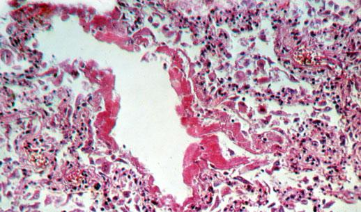
Fatty change in the heart is seen in two classic situations, both fortunately rare today:
(1)
It most often reflects poor oxygenation (i.e., chronic severe anemia). It is distributed away from the
vessels, and produces a "tiger-stripe" or "thrush-breast" heart.
(2)
The heart damaged by diphtheria exotoxin is uniformly flabby and often fatty. (The old idea
that diphtheria toxin block fatty acid burning by inhibiting the carnitine shuttle
has been replaced by the finding that the protein is a nonspecific
and very potent inhibitor of protein synthesis.).
Notice that the injured, fat-laden cell may not be permanently damaged or killed. And remember
cells can and do die without undergoing fatty changes.
Accumulation of fat in phagocytic cells is a common theme in pathology. The fat is usually made
up largely of cholesterol esters.
Atherosclerosis, still the #1 disease in our country, results when phagocytic cells in the intimal layers of
large arteries become engorged with cholesterol and its esters. The phagocytes themselves
tend to die off and leave the cholesterol to crystallize.
{11051} early atherosclerosis ("fatty streaks", all of you have these already)
Lipophages are scavenger macrophages that have devoured fat. This is common wherever lipid-rich
tissues (belly fat, brain, others) have been injured, or where alveoli cannot drain (surfactant).
The cytoplasm of these cells typically looks "foamy" ("foam cells", etc.)
{05955} foam cells, wall of gall bladder (these are laden with cholesterol)
* Likewise, fixed phagocytes the kidney turn into "foam cells" when sick people pass lipoproteins
through their glomerular basement membranes.
"Tumors" (nodular hyperplasias, really) composed of these cells are called xanthomas ("xanthos"
means yellow). These often (but not always) suggest some problem with blood lipids.
{09741} xanthelasma
Fatty ingrowth ("stromal infiltration of fat", * "lipomatosis") is totally different from fatty change. It
is metaplasia of an organ's capillary pericytes into mature adipocytes.
This is a common finding in lymph nodes, in the pancreas, and in the right ventricle and atria of the
heart. We're seeing it now in the muscles of people on the new anti-HIV medicines.
Usually it has no effect on organ function.
* You probably know that all adipocytes are modified pericytes.
* I learned to call this "fatty infiltration". Textbooks use "fatty infiltration" as a not-recommended
synonym for fatty change.
* The most important appearance of fatty ingrowth in medical pathology is as a component of most
muscular dystrophies. You'll make the diagnosis on other criteria, however.
* Fatty ingrowth in skeletal muscle is a hallmark of the
HIV-insulin resistance syndrome, seen in patients on long-term antiretroviral
therapy (J. Clin. Endo. Metab. 89: 2171, 2004.)
GLYCOGEN ACCUMULATION
Glycogen ordinarily is present in the livers of people in the fed state, and is abundant if the patient
has an IV line infusing glucose ("dextrose", "D5", etc.).
In hyperglycemia,
it is common to see glycogen "in" hepatic nuclei (really, in a deep cup-shaped
depression in the side of the nucleus; the nucleus looks clear), pancreatic beta cells, and (if control is
really poor) in the proximal tubular epithelial cells (why?). These accumulations are probably
harmless.
You'll study glycogen storage diseases later.
{17421} glycogen "in" hepatocyte nuclei
The various glycogen storage diseases result from inborn errors of metabolism.
ACCUMULATIONS OF COMPLEX LIPIDS AND CARBOHYDRATES
These typically result from inborn errors of metabolism. Typically the substance is stored in
lysosomes. Eventually enough accumulates to compromise organ function.
Now's a good time to memorize what accumulates in what disease:
Gaucher's disease is common, and produces huge, pink-staining, glucocerebroside-laden, "crumpled
tissue paper" (* old-timers say "watered-silk") macrophages in the bone marrow and elsewhere.
Before we leave the subject of inborn errors of metabolism, remember that in gout, uric acid
accumulates in nodules in the tissues. These are called "tophi" (singular "tophus").
IATROGENIC ACCUMULATIONS
Lipogranulomas in the spleen and celiac lymph nodes are thought to have several causes, including
oral mineral oil.
* Enjoy reading about "sclerosing lipogranuloma of the penis" in J. Urol. 133: 1046, 1985. One of the great
pathology mysteries solved.
Lymphangiogram contrast medium is an oil
that stays in lymph nodes for years.
Argyria results from silver salts
being permanently deposited in the
skin (J. Clin. Path. 47: 556, 1994).
With improved industry safety standards, the usual cause today is quack medicines.
Two faddists make their baby horribly sick: J. Ped. Gastro. 33: 439, 2001.
Thorium dioxide was, believe it or not, once used to make liver x-rays. It stays around forever,
giving off radiation.
PIGMENTS
{12216} carotenemia
Insoluble pigments inside cells are typically stored in phagolysosomes.
{37887} jailhouse tattoo
Carbon settles in macrophages, where it remains indefinitely. Carbon in the lungs and nearby lymph
nodes is called "anthracosis". It is inert though ugly. ("If you keep smoking, kid, your lungs will
get blacker and blacker!")
{17438} carbon in the lung, gross
Other mineral dusts include silica (colorless, very harmful) and iron oxide ("rusty lung", not very
harmful).
* NOTE: Professional tattooers use metal salts. Lots of people are allergic to the red mercuric
sulfide.
Lipofuscin (* "lipochrome"; "fuscus" is Latin for brown)
This is another brown pigment that is now known to be the un-digestible residue of subcellular
membranes whose unsaturated lipids have been scrambled ("polymerized", "peroxidated", etc.) by
free radicals. (See, for example, Br. J. Surg. 81: 1300, 1994).
Lipofuscin is the "cellular clinker" or "wear-and-tear pigment". It is considered harmless, and * does
not stain with lipid dyes.
* Well, maybe it's not harmless. Lipofuscin's an important component of the cores of extracellular
Alzheimer's lesions (Am. J. Path. 140: 1389, 1992; Am. J. Path. 152: 983, 1998).
* Not all lipofuscins are clinkers. Even in youth, the interstitial cells of the testis, and the epithelial cells of the epididymis and
seminal vesicles are packed with lipofuscin, and there is lipofuscin at the poles of
the cardiac nuclei even in babies. And earwax pigment is a lipofuscin.
"Ceroid" is lipofuscin that has become acid-fast and autofluorescent for some reason. It is of no
special significance. (* Future pathologists: the best acid-fast stain for this purpose is the Fite.)
* Hamazaki-Wesenberg bodies are giant lysosomes / residual bodies
loaded with lipofuscin. Look for them in lymph nodes especially near sarcoid granulomas.
They are a minor mystery of medicine.
{17348} lipofuscin (EM and H&E)
* The chemistry of lipofuscin formation is just getting worked out. Recipe: Bioch. Biophys. Acta
1290: 319, 1996.
Lipofuscin becomes more abundant during normal aging, or following atrophy.
"Brown atrophy" is simply atrophy where the lipofuscin is visible grossly.
{17454} hypertrophy vs. brown atrophy
A good place to find lipofuscin is at the poles of the nuclei of cardiac muscle cells from elderly
people. By age 90, the heart may contain 30% lipofuscin by weight.
{17443} lipofuscin (H&E, EM)
* In other species, the faster the rate of basal metabolism, the faster lipofuscin accumulates (J. Ger.
47: B-126, 1992).
Melanosis coli pigment is a curious pigment found within macrophages in the mucosa of the colon.
This imparts a tortoise-shell appearance to the colons of certain people.
It is typical of people who like over-the-counter anthraquinone ("cascara", etc.) laxatives. However,
this isn't the whole story (Dis. Col. Rect. 32: 235, 1988). The pigment apparently results from
apoptosis of the colonic epithelial cells, and they are then digested by those macrophages, with
indigestible cell fragments becoming the pigment.
You may see it in people with inflammatory bowel syndrome who do not use
laxatives (from all the damaged cells, J. Clin. Gastro. 26: 167, 1998).
Read all about it: Am. J. Path. 131: 465, 1988.
Even the chemical nature of melanosis coli pigment remains unknown. It reportedly stains as iron,
melanin, and lipofuscin.
{49202}
melanosis coli
Tobacco pigment is common in alveolar macrophages in heavy smokers. It is a fine, powdery
mix of brown pigments, sometimes including iron. Don't confuse it was carbon, which is black.
Melanins are widely distributed in the animal kingdom, and the melanin in octopus ink is much like
ours. Neuromelanin in the brain is much like skin melanin.
Natural selection: Races from near the equator are protected
from skin cancer and hypervitaminosis D by dark
pigmentation. Races from high latitudes are protected from rickets by light pigmentation.
The most important single skin color gene is MC1R, which binds
melanocyte-stimulating hormone and which determines both depth of
pigment and how much will be pheomelanin (Nat. Genet. 11: 238, 1995,
big dosage effect obviously).
That this is the origin of most red-headedness is now amply confirmed
(J. Inv. Derm. 117: 1314, 2001; Hum. Mol. Genet. 9: 2531, 2000).
Most human redheads have zero function at this locus.
* Cloning the mouse redhead gene perhaps homologous to a rufous albinism (red-hair, red skin in
Africans; Nature 361: 72, 1993). People lacking pro-opiomelanocortin
also have red hair, along with obesity and congenital hypocortisolism (Hum. Molec. Genet. 11: 1997, 2002;
Ann. N.Y. Acad. Sci. 994:
233, 2003).
Albinos cannot make melanin, and usually have genetic defects of tyrosine metabolism. To think
about: Why are children with phenylketonuria more fair-complected than their parents?
Melanin is characteristically seen in melanocytes and their tumors (common "moles", malignant
melanomas)
Most (not all) melanomas contain at least a little melanin, and finding melanin production by tumor
cells proves a cancer is a melanoma.
To prove a pigment is melanin, the pathologist applies a little hydrogen peroxide (hair bleach) to the
section. If it's melanin, it bleaches. * There's also a Fontana stain, etc.
Abnormalities of melanin occur in other settings. You will learn them soon enough.
Diseases with increased ACTH ("MSH" -- think especially of primary adrenocortical insufficiency -- why?)
cause
hyperpigmentation.
Ferric ion blocks breakdown of melanin, a fact that explains the dark pigmentation in the skin of
hemochromatosis patients and over dermatofibromas.
Melanin in the urine indicates extensive malignant melanoma.
Tryptophan metabolites provide the yellow color to lipid (adipose tissue, adrenal cortex, tissue
necrosis, atherosclerosis; they do not cross the blood-brain barrier, or we'd talk about "yellow
matter"). Their importance, if any, is unknown.
The stainable form of iron is hemosiderin, a complex mixture of proteins and ferric ions. It is faintly
visible as shiny golden granules in unstained tissue sections.
The best way to demonstrate hemosiderin is using acid ferrocyanide, which forms a striking blue
complex with stainable ferric ion ("Prussian blue").
In hepatocytes, hemosiderin tends to locate near the bile
canaliculi.
Note that ferrous iron in heme groups (hemoglobin, myoglobin, cytochromes) does not stain.
Neither does the ferric iron stored as ferritin, since the apoferritin protein shields the iron atoms.
Normally there is some stainable hemosiderin in marrow, spleen, liver.
Lack of stainable iron of course indicates systemic iron deficiency.
Localized accumulations of iron ("local hemosiderosis") reflect longstanding congestion (lungs, leg
veins), repeated minor injury (shrapnel fragments, sports, etc.).
Too much iron in the whole body ("generalized hemosiderosis") has several causes that are worth
learning now:
In a few cases, the disease has been detected by sufferers' tripping airport metal detectors.
* The oral iron chelators (alpha-ketohydroxypyridines, L1, etc.) were orphan drugs introduced
in the early 1990's that have
helped people with transfusional hemosiderosis. You'll
learn about them soon.
{34364} hemosiderin at site of hemorrhage in a malignant brain tumor
A special case of iron deposition is the ferruginous body of asbestosis -- iron-calcium salts encrusted
on an asbestos fiber in the lungs.
{36189} ferruginous body in asbestosis
Copper pigment
Copper penny and blue copper-salt colors. Deposited in the liver and/or basal ganglia in Wilson's
disease, an important diagnosis not to miss.
* Minocycline, an antibiotic, is acted upon by the peroxidases
in the thyroid and turns the gland black. For keeps.
Homogentisic acid polymer ("alkapton"):
Patients with the hereditary arthritis syndrome "alkaptonuria" accumulate this substance, which
breaks down into black pigment, in their cartilages (nose, ears), joints, sweat. The accumulation
itself is called "ochronosis". Histopathology: Am. J. Clin. Path. 90: 95, 1988.
Genetics: Nat. Gen. 14: 19, 1996.
{18252}
ochronosis (black cartilage in the ear)
Hemozoin: This is a ferric iron pigment that looks like hemosiderin when unstained, but that
does not exhibit the Prussian Blue reaction because the iron is sequestered by protein.
It consists of polymerized heme with each iron atom joined to a carboxyl group on the next
porphyrin unit (Proc. Nat. Acad. Sci. 88(2): 325, 1991).
It is seen in RE cells in malaria; the plasmodia protect themselves from free iron-heme complex by
converting it into this substance. Once it's been deposited, it apparently autocatalyzes its own
production (Nature 374: 269, 1995). Some of the antimalarials work by preventing the bugs from
producing this stuff.
* Malaria kills as many or more people as AIDS.
Almost every one of the deaths could be prevented fairly easily.
The ongoing stupidity is a prime example of Virchow's dictum that
its overriding public health problem is its politicians.
See Br. Med. J. 328: 1033 & 1086 & 1378, 2004; Lancet 363: 237, 2004;
lots more.
* Hematin, sometimes deposited in RE cells in other cases of heavy hemolysis, is ferriprotoporphyrin
IX hydroxide.
{12220} jaundice
This is the non-iron-containing, yellow-orange pigment that results from breakdown of porphyrin
rings (mostly hemoglobin).
Bilirubin by itself is insoluble in water and is carried on albumin to the liver, where hepatocytes
conjugate it with glucuronic acid and pour it into the bile.
Elevated levels of bilirubin in the blood mean jaundice. Mechanisms:
You may see bile plugs (bile in distended canaliculi; big ones that ruptured are "bile lakes") or
intracellular bilirubin in the liver in obstructive jaundice or primary cancer of hepatocytes.
{24559} liver that is green from biliary obstruction
Of course, if you find a cancer cell is making bile, you know the cancer arose from a hepatocyte.
* Trivia: Two proteins, ligandin and protein Z, process bilirubin in the hepatocyte. Phenobarbital
increases ligandin and speeds processing of bilirubin. Once it was fashionable to treat everyone with
Gilbert's "disease" with phenobarbital, thereby turning healthy adults with a slightly abnormal lab
value into chronically and iatrogenically sick adults with a normal lab value.
CALCIFICATION: A subject of interest to most physicians, not just radiologists.
Calcium salts (hydroxides, phosphate-hydroxides) are deposited. Regardless of cause, calcium salts
stain dark blue on H&E. (* If there is any doubt, special stains like the Von Kossa of Alizarin red
demonstrate it is
calcium.)
Dystrophic calcification
This is calcification that takes place locally, in the presence of normal overall calcium-phosphorus
metabolism.
The calcifications may be of any size.
"Dystrophic" means "seeking out the bad". While a necrotic cell whose mitochondria calcified may
provide a nidus for stone-building, some texts suggest that only dead things calcify. This is simply
not true.
* In extracellular calcification, calcium salt is complexed to coagulation factors II, VII, IX, and X,
the ones that contain gamma-carboxy glutamic acid (and therefore require vitamin K, etc.)
In intracellular calcification, the first organelle to calcify is usually the mitochondrion. Of course,
that's the end of the cell....
Several normal structures tend to calcify during adult life.
This includes the pineal gland, the cartilages in the airways, the media of large arteries
("Monckeberg's") and the mitral valve annulus. These are probably harmless.
Around 1% of adults develop calcifications in their otherwise-normal sinuses of Valsalva, causing
deadly aortic valve stenosis.
{03560} calcified aortic valve, x-ray
Little bits of calcium help mammographers recognize breast cancer.
Advanced atherosclerotic plaques undergo calcification, but this is not the principal problem in
atherosclerosis.
* Nobody knows why most dystrophic calcifications happen. One
suspect is certain nanobacteria that thrive on the hydroxyapatite surfaces
that they build. Tetracycline-sensitive -- let's wait and see.
PNAS 95: 7896 & 8274, 1998.
* "Chelation therapy" (infusions of EDTA) is a perennial health
fraud that claims to "cure atherosclerosis by
removing calcium from the walls of vessels".
Patients feel their fingers tingle
during the infusion, and they are told that this is "proof that the circulation was being restored." Explain.
Dystrophic calcification is characteristic of other diseases as well. The reasons are generally
obscure.
Malformed or damaged cardiac valves tend to calcify, especially congenitally bicuspid aortic valves
(another common cause of aortic valve stenosis).
Caseous granulomas (tuberculosis, histoplasmosis, others) often calcify.
Scars (surgical, myocardial) often calcify.
The fingertip pulp calcifies in scleroderma and CREST syndrome.
Certain tumors contain "psammoma bodies", little spherules of basement membrane that calcify.
(Think of thyroid cancer, ovarian cancer, meningioma, somatostatinoma). Little spherical
calcifications inside giant cells in granulomas are called "Schaumann bodies" or "conchoid
bodies". More about these things later.
{35552} Schaumann bodies in giant cells of granulomas (berylliosis case)
* Uterine fibroids (smooth muscle tumors) often calcify. (This is an ancient finding: Arch. Path.
107: 91, 1983.)
* Calcification of the pinna of the ear occurs for some reason in some cases of longstanding
adrenocortical insufficiency (Addison's disease; there are other causes too).
If a fetus dies and calcifies, it may be retained for years as a "lithopedion" ("stone child" -- * read
Michael Bishop's famous non-supernatural horror story, "Within the Walls of Tyre").
A special case of dystrophic calcification is precipitation of calcium stearate in pancreatitis-associated fat necrosis.
If the celiac plexus is involved, this produces one of medicine's most intractable
pain syndromes.
Metastatic calcification:
"Metastatic" means "another place". Here the serum calcium and/or phosphate ion concentration is
already elevated for some reason. Healthy tissues calcify.
* High blood calcium is usually due to cancer destroying bone, high hPTH levels (parathyroid
adenomas and hyperplasias, squamous cell carcinoma of the lung, rarely others), sarcoid, vitamin D
abuse, milk and antacid abuse.
High blood phosphate is almost always due to kidney failure or massive tumor lysis.
* Virchow first explained the mechanism, relating metastatic calcification of
lung and stomach to demineralization of the bones and kidney failure.
Metastatic calcification occurs predictably in the alveolar walls, the gastric fundic epithelium (near
parietal cells), the basement membranes of certain renal tubules, and the walls of small blood
vessels.
Note that all but the last are sites of pH gradients. The calcium precipitates first where there is
excess hydroxyl ions.
In very severe lung or kidney involvement, respiratory insufficiency (* "pumice lung") or renal
tubular failure can occur. But usually metastatic calcification is harmless evidence of serious
disease elsewhere.
* Future pathologists: A few calcifications around the thin limb of Henle's loop is "normal" and
does not imply a calcium problem.
{39670}calcification, metastatic, in the lung;
* Modern-era pathologists: calcification of collagen is diagnostic of sustained electrical
injury (happens at the cathode during electrical torture; J. Clin. Path. 53: 569, 2000;
Nature 301: 75, 1983).
Any kind of calcification can ossify, i.e., produce bone and even bone marrow. (It's commonplace to
see bone, with active marrow, in airway sections of elderly patients, in calcified atherosclerotic
plaques, and so forth.)
HYALINE: Any substance (intracellular or extracellular) that stains a homogeneous (say "homo-JEAN-yuss") pink on
routine H&E stains.
{17485} treatise on hyaline
When patient are losing lots of protein trough their glomeruli, it's common to see eosinophilic
droplets in the proximal tubular epithelial cells. This is a good autopsy marker, especially if the
patient was not well worked-up during life.
{46308} hyaline droplets in proximal tubules
Russell bodies are round accumulations of monoclonal immunoglobulin that are (or used to be)
inside constipated plasma cells. Nobody knows how or why they form.
Viral inclusions are crystalloids of virus components within infected cells. The most famous are
herpes inclusions -- look in the cell nucleus, since they will give you the diagnosis.
Mallory's alcoholic hyaline is scrambled prekeratin intermediate filaments plus ubiquitin, typically
in liver cells. This usually reflects weeks of heavy drinking ("alcoholic hepatitis" -- * there are other
rare causes). The stuff is eosinophilic and flocculent (i.e., it looks like pink cottage cheese).
{09103} Mallory's alcoholic hyaline, electron micrograph
(upper right)
Alpha-1 protease inhibitor ("antitrypsin") globules look like multi-sized cherries within
hepatocytes that are unable to secrete this product (inborn error, regenerating cells).
* Biotin causes eosinophilic inclusions and optically-clear
nuclei in the endometrial glands in pregnancy: Lancet 364: 532, 2004.
* Giant mitochondria, a feature of alcoholic liver disease. If you can spot these,
you have exceeded undergraduate pathology expectations. These are called "Yokoo bodies",
having been discovered by one of my teachers.
Collagen can hyalinize, especially in keloids
/ hypertrophic scars and other abnormal fibrous proliferations.
{17646} keloid, gross
Excess basement membrane and other proteins "hyalinizes" the body's small arteries in high blood
pressure and diabetes.
Of course this narrows the lumens. * Complement components also bind to the structural
components of the vessel, etc., etc.
Thickening and excesses of basement membrane are a major theme in renal glomerular disease,
and in diabetes mellitus.
Much more about this later.
{17472} hyaline arteriolar sclerosis (right, one on left is normal)
Amyloid, mentioned above, is another extracellular accumulation that always has a hyaline
appearance.
{13613} amyloid, H&E
{11427} fibrin in a premature baby's lung ("hyaline membranes")
Fibrinoid is a special "material" seen in the walls of blood vessels that are dead but still contain
flowing blood. A mix of plasma proteins and dead cell debris solidifies in the media and stains
intensely pink.
If solid "hyaline" looks inflamed, it's usually fibrin or "fibrinoid".
* The centers of rheumatoid nodules may be filled with "fibrinoid", and it is also characteristically
seen in the myocardium in rheumatic fever.
* Radiation injury to vessels appears as hyaline-fibrinoid in vessel walls. Early, there may be some
inflammation and/or necrosis. Later, the vessels look more subdued, but the lumen continues to
narrow throughout the patient's life-span.
{01917} radiation injury to vessels; the hyaline /
fibrinoid is pink
* Future pathologists only: The Splendore-Hoeppli phenomenon of little pink-staining club-shaped
things around actinomyces colonies, fungus masses,
schistosome eggs, and occasionally other organic-based "foreign bodies".
* Future pathologists only: Spironolactone bodies are found in
the mineralocorticoid-producing cells of the adrenal gland
in people treated with this Rx. They are hyaline, laminated
things, up to 15 microns, derived from S.E.R.
In kidney disease, entire glomeruli "hyalinize". Depending on the disease, these dead glomeruli
are replaced by basement membrane-mesangial matrix, collagen, and/or plasma proteins.
MYXOID CHANGE
Increased ground substance.
* The comb and wattles of a rooster is the familiar example from normal comparative physiology.
There is increased ground substance throughout much of the body in hypothyroidism (myxedema).
* (Curiously) many patients with Graves' disease (which usually produces hyperthyroidism) have
localized accumulation of ground substance on their shins ("pretibial myxedema"; it may be severe
enough to compress the lymphatics and give superimposed elephantiasis. See Lancet 341: 403,
1993).
If there is associated damage to the connective tissue fibers, we use the term myxomatous
degeneration ("myxoid degeneration").
The two real-life examples are "cystic medial necrosis" of the aorta (prelude to a lethal tearing called
"aortic dissection") and Barlow's floppy mitral valve (a semi-disease that if you really search for it
affects a few percent of
humankind.)
* Myxoid change of the intima narrows the renal arteries in scleroderma and Balkan nephropathy,
eventually causing kidney failure.
Accumulation of epithelial mucin, as large pools, may be seen in several diseases, most notably
"colloid cancers". More about them later.
ABNORMAL MITOCHONDRIA
Selective damage of the mitochondria (i.e., extreme swelling seen on electron microscopy) is the
essential lesion of true Reye's syndrome.
Parking-lot crystals inside mitochondria are a hallmark of the genetic diseases of mitochondria, in
which a portion of their genetic code is faulty. Abnormal creatine kinase is being synthesized and
accumulating.
* Most of these diseases show up primarily in muscle (why?), and the sick mitochondria accumulating
around the edges of the worse-involved fibers create a "ragged red" appearance. More about this
later.
Hürthle cells (oncocytes, Ashkenazy cells, oxyphil cells) have their cytoplasm so packed with
mitochondria that there is little room for anything else. You already know these from parathyroid
and armpit (apocrine cell) histology. We'll see them at other sites as disease markers.
We've already mentioned the giant mitochondria of the alcoholic's liver.
Amyloidosis
Pittsburgh Illustrated Case
{38782} sudan stain, fatty liver 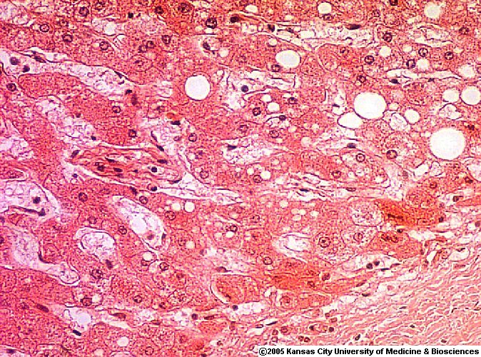 Liver
Liver
Fatty change
Dave Barber MD, KCUMB
* Immunostaining, and studying how antigens can be lost and recovered,
has clarified the mechanism of formalin fixation. Recoverable antigens
involve cross-linking of a tyrosine and an amino-sidechain (usually arginine).
Am. J. Clin. Path. 121: 190, 2004.
If the fat is periportal, think of malnutrition / total parenteral nutrition / AIDS wasting.
{40039} Reye's syndrome, liver; microvesicular fatty change
{40040} Reye's syndrome, liver; microvesicular fatty change; oil red O
Something clear in an H&E-stained cell? Is it...
{08366} fatty liver, micro
{08829} fatty liver, micro
{37589} fatty liver, micro
You'll see this in brain necrosis and in xanthomas (hyperlipidemia,
"strawberry gallbladder", idiopathic).
{11648} early atherosclerosis, gross (natural-color and "oil red O stain")
{08108} foam cells, wall of gall bladder (ditto)
{01453} microglia (macrophages) eating up necrotic myelin lipid following a stroke
{38332} eruptive xanthomas
{24886} xanthoma histology (* the thing that looks like a flower is a "floret" giant cell)
 Cholesterol polyps
Gall bladder xanthomas!
Pittsburgh Pathology Cases
{17422} the real picture of a "glycogen nucleus"
{46306} glycogen in the proximal tubule in poorly-controlled diabetes
Gaucher cells
WebPath Photo
 Soluble pigments do not appear in tissue sections. They include carotene (carrot gluttons, most
yellow on palms and soles, take a history; your lecturer likes his carrots; NEJM 346: 821, 2002),
bilirubin (except in bile plugs), and urochrome (kidney
failure, check the patient's lab results).
Soluble pigments do not appear in tissue sections. They include carotene (carrot gluttons, most
yellow on palms and soles, take a history; your lecturer likes his carrots; NEJM 346: 821, 2002),
bilirubin (except in bile plugs), and urochrome (kidney
failure, check the patient's lab results).
 Carbon particles enter our bodies in smoke and soot
or as the pigment in jailhouse tattoos (Lancet
338: 380, 1991).
Carbon particles enter our bodies in smoke and soot
or as the pigment in jailhouse tattoos (Lancet
338: 380, 1991).
{17439} interesting tattoos
{38249} more tattoos
{10943} carbon in macrophages from an excised tattoo
 Smoker's Lung
Most anthracotic lungs are
MUCH blacker. AFIP
* Future pathologists: "Blue scars" result from wounds sustained in a coal
mine. The dust becomes trapped in the fresh wound for life.
{17437} carbon in the lung, histology
{36157} carbon in macrophages, papanicolaou stain
{26047} carbon in macrophages, papanicolaou stain
Carbon pigment, lung
Mild by today's standards.
WebPath Photo
{18720} hypertrophy vs. brown atrophy
Melanosis coli
There is also a colon cancer
Urbana Atlas of Pathology
Melanoma
Cancer cell with melanin pigment
Urbana Atlas of Pathology
 From Greek "melos", black. This is the principal pigment of human skin. It is a complex largely
polymerized 5,6-dihydroxyindole and other tyrosine metabolites.
From Greek "melos", black. This is the principal pigment of human skin. It is a complex largely
polymerized 5,6-dihydroxyindole and other tyrosine metabolites.
 Most people make mostly eumelanin, redheads make mostly pheomelanin (this stuff doesn't protect
from sunlight but actually generates more free radical when light-exposed). Actually most
mammals (including most people) make both; you can tell the granules apart under an electron
microscope (eumelanin granules are ellipsoid, pheomelanin granules are spherical). Dark-skinned
and most light-skinned make equal amounts of tyrosinase (the rate-limiter), but it works much better
in dark-skinned people.
Most people make mostly eumelanin, redheads make mostly pheomelanin (this stuff doesn't protect
from sunlight but actually generates more free radical when light-exposed). Actually most
mammals (including most people) make both; you can tell the granules apart under an electron
microscope (eumelanin granules are ellipsoid, pheomelanin granules are spherical). Dark-skinned
and most light-skinned make equal amounts of tyrosinase (the rate-limiter), but it works much better
in dark-skinned people.
* Organic chemists: The red is mostly trichochrome c(1a)
(J. Org. Chem. 66: 6958, 2001).
Trichosiderin, supposedly an iron-rich pigment in red hair,
is an artifact from extraction.
 {53602} albino
{18250} phenylketonuria patient
{18253} phenylketonuria patient
 Suntanning is physiologic (and does not keep out cancer-causing rays). Ingestion or application of
psoralens (celery juice, limes, etc.) makes the skin sensitive to sunlight.
Suntanning is physiologic (and does not keep out cancer-causing rays). Ingestion or application of
psoralens (celery juice, limes, etc.) makes the skin sensitive to sunlight.
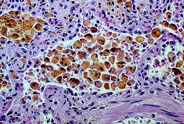
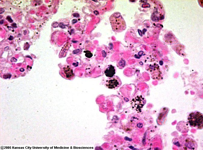
Hemosiderin
In lung macrophages
WebPath Photo
 Excess
hemosiderin eventually causes organ injury by generating free oxygen radicals. This leads to
organ failure, called "hemochromatosis". ("Hemochromatosis is generalized hemosiderosis
that has made you sick.") This is a major, under-diagnosed, treatable disease in the U.S.
Excess
hemosiderin eventually causes organ injury by generating free oxygen radicals. This leads to
organ failure, called "hemochromatosis". ("Hemochromatosis is generalized hemosiderosis
that has made you sick.") This is a major, under-diagnosed, treatable disease in the U.S.
{37592} hemosiderin, Prussian blue stain; there is also black carbon
{38491} hemosiderin-laden macrophages in the lung ("heart failure cells")
 Heart Failure Cells
Hemosiderin-laden macrophages in the lung
KU Collection
 Pulmonary macrophages
Mixed pigment -- iron, carbon, tobacco
Dave Barber MD, KCUMB
{39787} bile plug
{06461} calcified aortic valve, gross
{45702} calcified carotid artery (the proximal portion of the common carotid, and the proximal
portions of both internal and external carotid arteries are visible; look carefully!)
{15856} dystrophic calcification in heart muscle (myocarditis patient)
Breast cancer with microcalcification
WebPath Photo
Severe atherosclerosis with calcium
WebPath photo
* The most grisly example of metastatic calcification
is the severe autosomal recessive disease familial tumoral calcinosis.
Gene GALNT3: Nat. Genet. 36: 579, 2004.
{08099} calcification, metastatic, in the lung
{17440} hyaline droplets in proximal tubules
Dideoxyinosine therapy for HIV is another
cause of Mallory bodies.
Am. J. Clin. Path. 108: 280, 1997.
{17418}
Mallory's alcoholic hyaline, H&E and electron micrograph
* Certain alleles of alpha1-antitrypsin produce globbies
in end-stage livers regardless of cause:
Am. J. Clin. Path. 107: 692, 1997.
Alpha-1 antitrypsin globules
WebPath Photo
{17647} keloid, histology
{17648} keloid, histology
{46351} keloid, histology
Keloids (hypertrophic scars?)
Actor Benjamin Bratt
UCSF Skin-In-Cinema
Amyloid
Congo red
WebPath Photo
 Hyaline membrane disease
Fibrin membranes in lung
KU Collection
Fibrin
On the epicardium
WebPath Photo
Usually fibrinoid necrosis results from type III immune injury
(immune complex precipitation and complement activation).
High physical pressure ("malignant hypertension"), toxemia of pregnancy,
and some infections
can do the same.
Oncocytoma
Mitochondrion-rich tumor
VCU Pathology
Hürthle cell carcinoma
Thyroid
Pittsburgh Pathology Cases


| Visitors to www.pathguy.com reset Jan. 30, 2005: |
Ed says, "This world would be a sorry place if
people like me who call ourselves Christians
didn't try to act as good as
other
good people
."
Prayer Request
 Teaching Pathology
PathMax -- Shawn E. Cowper MD's
pathology education links
Ed's Autopsy Page
Notes for Good Lecturers
Small Group Teaching
Socratic
Teaching
Preventing "F"'s
Classroom Control
"I Hate Histology!"
Ed's Physiology Challenge
Pathology Identification
Keys ("Kansas City Field Guide to Pathology")
Ed's Basic Science
Trivia Quiz -- have a chuckle!
Rudolf
Virchow on Pathology Education -- humor
Curriculum Position Paper -- humor
The Pathology Blues
 Ed's Pathology Review for USMLE I
![]()
![]()
 | Pathological Chess |
 |
Taser Video 83.4 MB 7:26 min |

 Lipids
 Stains and molecular markers
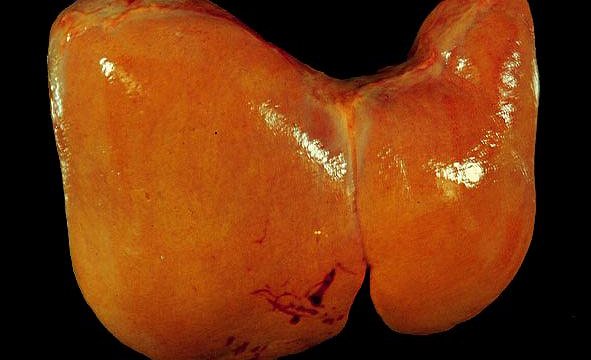 Fatty liver
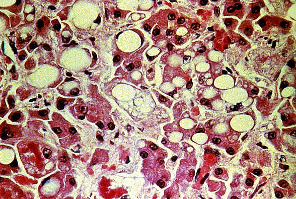 Fatty liver
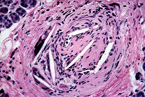 Cholesterol emboli
 Juvenile xanthogranuloma
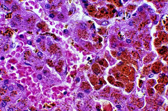 Hemochromatosis
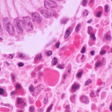 Russell Body
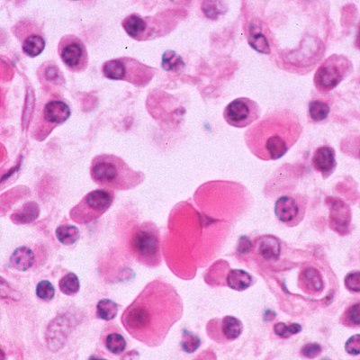 Russell Bodies
 Keloid
)window.location='http://edcenter.med.cornell.edu/Pathology_Images/295.gif') Fibrinoid in an arterial wall
Fibrinoid in an arterial wall