Ed Friedlander, M.D., Pathologist
scalpel_blade@yahoo.com


Cyberfriends: The help you're looking for is probably here.
Welcome to Ed's Pathology Notes, placed here originally for the convenience of medical students at my school. You need to check the accuracy of any information, from any source, against other credible sources. I cannot diagnose or treat over the web, I cannot comment on the health care you have already received, and these notes cannot substitute for your own doctor's care. I am good at helping people find resources and answers. If you need me, send me an E-mail at scalpel_blade@yahoo.com Your confidentiality is completely respected.
 DoctorGeorge.com is a larger, full-time service.
There is also a fee site at myphysicians.com,
and another at www.afraidtoask.com.
DoctorGeorge.com is a larger, full-time service.
There is also a fee site at myphysicians.com,
and another at www.afraidtoask.com.
Translate this page automatically
 |
With one of four large boxes of "Pathguy" replies. |
 I'm still doing my best to answer
everybody.
Sometimes I get backlogged,
sometimes my E-mail crashes, and sometimes my
literature search software crashes. If you've not heard
from me in a week, post me again. I send my most
challenging questions to the medical student pathology
interest group, minus the name, but with your E-mail
where you can receive a reply.
I'm still doing my best to answer
everybody.
Sometimes I get backlogged,
sometimes my E-mail crashes, and sometimes my
literature search software crashes. If you've not heard
from me in a week, post me again. I send my most
challenging questions to the medical student pathology
interest group, minus the name, but with your E-mail
where you can receive a reply.
Numbers in {curly braces} are from the magnificent Slice of Life videodisk. No medical student should be without access to this wonderful resource. Someday you may be able to access these pictures directly from this page.
Pathology Education Instructional Resource -- U. of Alabama; includes a digital library
Also:
Medmark Pathology -- massive listing of pathology sites
Freely have you received, freely give. -- Matthew 10:8. My
site receives an enormous amount of traffic, and I'm
handling about 200 requests for information weekly, all
as a public service.
Pathology's modern founder,
Rudolf
Virchow M.D., left a legacy
of realism and social conscience for the discipline. I am
a mainstream Christian, a man of science, and a proponent of
common sense and common kindness. I am an outspoken enemy
of all the make-believe and bunk which interfere with
peoples' health, reasonable freedom, and happiness. I
talk and write straight, and without apology.
Throughout these notes, I am speaking only
for myself, and not for any employer, organization,
or associate.
Special thanks to my friend and colleague,
Charles Wheeler M.D.,
pathologist and former Kansas City mayor. Thanks also
to the real Patch
Adams M.D., who wrote me encouragement when we were both
beginning our unusual medical careers.
If you're a private individual who's
enjoyed this site, and want to say, "Thank you, Ed!", then
what I'd like best is a contribution to the Episcopalian home for
abandoned, neglected, and abused kids in Nevada:
My home page
Especially if you're looking for
information on a disease with a name
that you know, here are a couple of
great places for you to go right now
and use Medline, which will
allow you to find every relevant
current scientific publication.
You owe it to yourself to learn to
use this invaluable internet resource.
Not only will you find some information
immediately, but you'll have references
to journal articles which you can obtain
by interlibrary loan, plus the names of
the world's foremost experts and their
institutions.
Alternative (complementary) medicine has made real progress since my
generally-unfavorable 1983 review linked below. If you are
interested in complementary medicine, then I would urge you
to visit my new
Alternative Medicine page.
If you are looking for something on complementary
medicine, please go first to
the American
Association of Naturopathic Physicians.
And for your enjoyment... here are some of my old pathology
exams
for medical school undergraduates.
I cannot examine every claim which my correspondents
share with me. Sometimes the independent thinkers
prove to be correct, and paradigms shift as a result.
You also know that extraordinary claims require
extraordinary evidence. When a discovery proves to
square with the observable world, scientists make
reputations by confirming it, and corporations
are soon making profits from it. When a
decades-old claim by a "persecuted genius"
finds no acceptance from mainstream science,
it probably failed some basic experimental tests designed
to eliminate self-deception. If you ask me about
something like this, I will simply invite you to
do some tests yourself, perhaps as a high-school
science project. Who knows? Perhaps
it'll be you who makes the next great discovery!
Our world is full of people who have found peace, fulfillment, and friendship
by suspending their own reasoning and
simply accepting a single authority which seems wise and good.
I've learned that they leave the movements when, and only when, they
discover they have been maliciously deceived.
In the meantime, nothing that I can say or do will
convince such people that I am a decent human being. I no longer
answer my crank mail.
This site is my hobby, and I presently have no sponsor.
This page was last updated February 6, 2006.
During the ten years my site has been online, it's proved to be
one of the most popular of all internet sites for undergraduate
physician and allied-health education. It is so well-known
that I'm not worried about borrowers.
I never refuse requests from colleagues for permission to
adapt or duplicate it for their own courses... and many do.
So, fellow-teachers,
help yourselves. Don't sell it for a profit, don't use it for a bad purpose,
and at some time in your course, mention me as author and KCUMB as my institution. Drop me a note about
your successes. And special
thanks to everyone who's helped and encouraged me, and especially the
people at KCUMB
for making it possible, and my teaching assistants over the years.
Whatever you're looking for on the web, I hope you find it,
here or elsewhere. Health and friendship!
 I am presently adding clickable links to
images in these notes. Let me know about good online
sources in addition to these:
I am presently adding clickable links to
images in these notes. Let me know about good online
sources in addition to these:
Online Health Resources
Houston Pathology -- loads of great pictures for student doctors
Pathopic -- Swiss site; great resource for the truly hard-core
Syracuse -- pathology cases
Walter Reed -- surgical cases
Alabama's Interactive Pathology Lab
"Companion to Big Robbins" -- very little here yet
Alberta
Pathology Images --hard-core!
Cornell
Image Collection -- great site
Bristol Biomedical
Image Archive
EMBBS Clinical
Photo Library
Chilean Image Bank -- General Pathology -- en Español
Chilean Image Bank -- Systemic Pathology -- en Español
Connecticut
Virtual Pathology Museum
Australian
Interactive Pathology Museum
Semmelweis U.,
Budapest -- enormous pathology photo collection
Iowa Skin
Pathology
Loyola
Dermatology
History of Medicine -- National Library of Medicine
KU
Pathology Home
Page -- friends of mine
The Medical Algorithms Project -- not so much pathology, but worth a visit
National Museum of Health & Medicine -- Armed Forces Institute of Pathology
Telmeds -- brilliant site by the medical students of Panama (Spanish language)
U of
Iowa Dermatology Images
U Wash
Cytogenetics Image Gallery
Urbana
Atlas of Pathology -- great site
Visible
Human Project at NLM
WebPath:
Internet Pathology
Laboratory -- great site My team:
Ed Lulo's Pathology Gallery
Bryan Lee's Pathology Museum
Dino Laporte: Pathology Museum
Tom Demark: Pathology Museum
Dan Hammoudi's Site
Claude Roofian's Site
Pathology Handout -- Korean student-generated site; I am pleased to permit their use of my cartoons
Estimating the Time of Death -- computer program right on a webpage
Pathology Field Guide -- recognizing anatomic lesions, no pictures

St.
Jude's Ranch for Children
I've spent time there and they are good. Write "Thanks
Ed" on your check.
PO Box 60100
Boulder City, NV 89006--0100
More of my notes
My medical students
Clinical
Queries -- PubMed from the National Institutes of Health.
Take your questions here first.
HealthWorld
Yahoo! Medline lists other sites which may work well for you

We comply with the
HONcode standard for health trust worthy
information:
verify
here. 


![]()
Define the following terms:
activation (of oncogene)
activation (of carcinogen)
Ames test
amplification
carcinogenesis
chromosomal instability
clonal selection
complete carcinogen
fusion gene
growth fraction
immortalization
immune surveillance
inducer (genotoxic carcinogen)
Knudson two-hit model
labeling index
microsatellite instability
monoclonality
oncogene
procarcinogen
promoter (non-genotoxic carcinogen)
proto-oncogene
retrovirus
transduction
transformation
tumor progression
tumor marker
ultimate carcinogen
Recognize the ways in which the growth properties of cancer cells differ from normal. Explain the Nowell multi-step clonal evolution model for tumorigenesis, and cite evidence that it is true.
Briefly describe how the classic transforming viruses caused cancer in experimental animals. Explain why cancers produced by a particular transforming virus in a particular experiment will, as a rule, be antigenically identical.
Tell the features common to most or all genuine chemical carcinogens. Explan why cancers produced by a particular chemical carcinogen in a particular experiment will, as a rule, be antigenically dissimilar.
Give the evidence for radiation carcinogenesis in humans, and the resulting tumors.
Discuss oncogenes in some detail. Explain how we believe certain mutations make ras oncogenic. Explain the importance of myc oncogenes in certain cancers, and the two ways myc is activated.
Describe tumor-suppressor genes (anti-oncogenes) in considerable detail, and explain why tumors lose heterozygosity at their loci. Explain the Knudson two-hit model.
Recognize the important tumor viruses for humans. Tell how they differ from the transforming viruses of the classic animal experiments. Tell how they effect their damage.
Recognize the major known and suspected human chemical carcinogens and the tumors they produce. Describe the Delaney Clause and its repeal.
Evaluate media and government claims about "things that cause cancer" intelligently and honestly.
Critique the following statement, overheard in a supermarket check-out line: "Cancers are continually forming in our bodies, but our immune system destroys them. The natural way to cure cancer is by strengthening the immune system."
Give the overall cure rate for newly-diagnosed cancers in the US today. Identify cancers that are increasing and decreasing in the U.S., and suggest reasons why. Identify cancers that are common in some countries and rare in others. Suggest reasons why.
Distinguish "benign" and "malignant" tumors. Explain how certain benign tumors cause serious disease.
Explain the various mechanisms by which cancer causes pain, disability, and death.
Explain how paraneoplastic syndromes happen (tumor products, tumor immunity). Given the name of a paraneoplastic syndrome, tell its effect on the patient.
Explain the concept of "tumor markers", substances produced by the tumor that appear in the bloodstream and assist diagnosis. Explain what is meant by an "oncofetal antigen".
Describe the common tumor-suppressor gene deletion syndromes ("autosomal dominant tumor susceptibility syndromes") in principle.
Recognize each of the following tumor-family syndromes by physical signs:
Recognize cancer quackery and its methods. Recognize why a scientific physician must not "keep an open mind" toward obvious untruths, or "debate / dialogue" with these people.
Appreciate the devastating impact of a cancer diagnosis to a patient, and the need for intelligent, humane care of the whole person.
RECOMMENDED READING: The "Neoplasia" chapters in Big or Baby Robbins. or R&F. All are pretty good. I've followed the sequence in Big Robbins.
QUIZBANK
Cell growth #'s 1-21, 31-147
|
|
LEARN FIRST
If you have gotten this far, you should already know how to recognize benign and malignant tumors grossly and microscopically. Please ask for help if this is still a problem.
Tumors are overgrowths, clones within clones, of cells bearing cumulative genetic injuries, each of which confers growth advantages over the neighbors ("NOWELL'S LAW"). Tumor cells typically have failure of division control, failure of senescence ("immortalization"), and failure of proper apoptosis. (At least some of these are already gone bad in the seemingly-normal cells from which tumors arise). We understand much more than we did a few years ago about the nature of these injuries, and how they produce the neoplastic phenotype.
Genetic lesions are somewhat stereotyped for individual tumors. Risk factors are known for various kinds of genetic injury, and various kinds of cancer.
Oncogenes are slightly altered (activated) versions of normal genes involved in cell division (proto-oncogenes). Activation may occur by point mutation, translocation, or increased copy number (amplification). Oncogenes tell the cell to divide when it shouldn't. This message overrides normal instructions from the gene's normal counterpart.
Tumor suppressor genes (anti-oncogenes) are normal genes that tell cells when not to divide. Usually the malignant phenotype is expressed only when both copies are damaged or missing.
The Knudson two-hit model explains the tremendously high prevalence of certain tumors in people unfortunate enough to have inherited one defective copy of a tumor suppressor gene.
Viral carcinogenesis is probably important in only a few common human tumors. Viruses cause cancer in humans by (1) binding and inactivating the products of normal tumor-suppressor genes, and/or (2) increasing cell turnover, allowing selection of abnormal clones, and/or (3) scrambling the genome. Cancer of the cervix and hepatocellular carcinoma are the most important cancers caused by known infectious agents.
Truly benign tumors are those in which the genome has not and will probably not become destabilized.
People with cancer do not die of the disease itself, but of secondary effects of the tumor or its treatment.
INTRODUCTION
This will be a whirlwind tour of cancer biology. Don't worry, you will see most of this material again and again.
There is more erroneous information circulating about cancer than about any other disease (even AIDS and mental illness). Some of this is the result of disinformation campaigns by charlatans. Most, however, results from our failure as physicians simply to explain things to our patients.
The essential "cause of cancer", once mysterious, is now clear. Both benign and malignant tumors are clonal overgrowths of cells bearing multiple genetic injuries. The visible tumor is the result of the overgrowth of clones within clones within clones. This is as well-established as anything in science. We are now simply filling in the details. Cancer is the great acquired genetic disease of humankind. Understand this.
Caring for a cancer patient requires a combination of great knowledge (of cancer biology, of therapeutics, of community resources, of the human heart), total integrity, and real compassion and love. When the time comes, I know you'll all be ready.
For a review of the common cancers (genes, screens): Disease-A-Month, Oct 1997 (still good). For an update on cancer genetics, see Nat. Med. 10: 849, 2004 and CA 55(1):45-54, Jan.-Feb. 2005.
ATTRIBUTES OF CANCER CELLS
Carcinogenesis is a generic term for a series of events leading up to expression of full malignant potential. Transformation is the term for this process as applied to cells themselves.
Much of the material in "Big Robbins" in loc. is primarily of research interest. Worth remembering:
Tumors are clonal overgrowths, generally monoclonal (G6PD marker studies). By the time a tumor is visible, the changes have been underway for a long time (at least months, usually years). The really bad areas represent clones which have arisen from clones, bearing cumulative genetic injuries. There is now overwhelming evidence that each successive mutation confers an unfair growth advantage the cell line which bears it. (This is the famous Nowell multi-step clonal evolution model of tumorigenesis model, first articulated Science 194: 23, 1976; it should probably now be called "Nowell's Law", and is by your lecturer.)
If the tumor results from only a few mutations of large effect, the genome will probably remain stable. Otherwise, the genome will be destabilized, and the cells will eventually acquire new abilities.
As a destabilized (i.e., malignant) tumor grows, non-disjunction creates cells with extra chromosomes (the deprived cells, of course, die off), and many (but not all) cancers become aneuploid.
Tumor progression refers both to the growth and distant spread of cancer, and to the way the front-line cells become more aggressive and more resistant to therapy (i.e., by the emergence, and selection for, nasty subclones; "multiple-steps").
It is fundamentally wrong to think of cancer just as "cells growing more rapidly than other cells". Rather, they are less subject to normal controls, and are growing faster than they are dying off.
Growth fraction is the percent of cells making nucleic acid at a given time. The monoclonal antibody Ki-67 stains proliferating cells, and is now standard to determine growth fraction; this is now emerging as a guide to therapy.
"Increased numbers of mitotic figures seen in cancer" represent mitoses that got stuck because of the cell disorganization. A mitotic figure can perhaps stick around for weeks.
The ability to invade and spread:
Benign tumors, with basically stable genomes, simply expand or perhaps reach a stable size. Malignant tumors, however, eventually gain the ability to invade the surrounding tissues. The mechanisms of invasion and metastasis involve binding to laminin of basement membranes, and destruction of type IV collagen (the early work Cancer 73: 22, 1994, Nature 370: 14, 1994) in basement membrane. Until this can happen, a would-be carcinoma cannot get through a basement membrane. Unfortunately, when a single cell acquires these abilities, it has a tremendous growth advantage over its neighbors, and the tumor will progress accordingly.
Please read, at your convenience, the speculative material about metastasis, vascular homing, and so forth in "Big Robbins". It is likely to be important on your licensure exam and in future therapy. Update Clin. Ortho. 451-S: S19, 2003.
* These are continually being updated, and now we can guess where the tumor will go based on microassay arrays: Nat. Med. 9: 999, 2003. And there are genes whose products seem to suppress growth of cells which may have reached secondary sites, without effecting the growth of the primary (Science 268: 884, 1995; J. Urol. 169: 1122, 2003; KAI1, CD44, MAPK, others; these do not include endostatin or angiostatin). For some reason, nuclear factor κB seems to turn on the pro-metastatic factors and turn off the anti-metastatic factors. Blocking it may be a target for antimetastatic drugs (Cancer Res. 60: 6557, 2000).
This work is now finding some clinical relevance. For example, genetic profiling of breast cancers predicts fairly well whether they will metastasize (NEJM 351: 2817, 2004; Lancet 365: 671, 2005).
Of course, telomerase is required to allow cancer cells to remain immortal. And so the successful clones in a cancer mutate so as to express telomerase. It's a target for anti-cancer therapy today: Nat. Med. 5: 1164, 1999; Anticancer Res. 20: 4419, 2000. And at least in some initial studies for exfoliative cytology, only cancer cells stain positive for telomerase (Cancer 90: 117, 2000). Is this the dream of a pathologist's "stain for malignancy"?
And there's more... Benign or malignant, a tumor must be able to induce its own new blood supply. After decades of searching for "the" factor that the neoplastic cells must learn to elaborate, we've finally come up with VEGF ("vascular endothelial growth factor"; Nature 367: 576, 1994).
In questionable cases, production of VEGF confirms that a cancer is actually invading (Am. J. Path. 156: 159, 2000; Am. J. Path. 155: 1967, 1999). VEGF can be blocked (* monoclonal antibody "avastin"), or its receptor blocked, and the growth of many (but by no means all) cancers arrested nicely (PNAS 98: 8829, 1998)
* When an altered VEGF receptor is administered, it not only soaks up the VEGF in the blood that's contributing to angiogenesis -- it intercalates itself into the membranes of the endothelial cells and turns the regular VEGF response itself off (PNAS 95: 8795, 1998. Stay tuned.
* Also watch Tie2/Tek, an endothelial tyrosine kinase that promotes vessel growth in cancers and can be blocked by gene therapy in animals: PNAS 95: 8829, 1998.
We are prognosticating cancer progression by the extent of neovascularization (microvessel density, which has been one of the most-studied subjects in pathology over the past ten years. Brain Cancer 77: 362, 1996; lung: J. Thor. Card. Surg. 115: 652, 1998; and Ann. Thor. Surg. 61: 470, 1996; cervix Am. J. Ob. Gyn. 178: 314, 1998; larynx Am. J. Surg. 174: 523, 1997; prostate Cancer 78: 345, 1996; stomach Surg. 131(S1): S48, 2002; cartilage tumors Clin. Orth. 397: 76, 2002; almost everyplace Anticancer Res 21(6B): 4373, 2001; other head & neck did not show a correlation Arch. Ot. 124: 80, 1998), using Factor VIII antigen (why?) as the marker for the new vessels.
Angiostatin, the angiogenesis inhibitor which is a breakdown product of plasminogen, raises the possibility of therapy with the substance and/or the gene (J. Clin. Invest. 101: 1055, 1998; Blood 101: 1857, 2003). Endostatin, a bit of collagen XVIII, is also a potent angiogenesis inhibitor.
* A group at Harvard discovered both little molecules; here's a possible future Nobel prize.
* Other targets include basic fibroblast growth factor (bFGF) and its receptor (Am. J. Surg. 174: 540, 1997), mitogen-activated protein kinase (MAPK: Nat. Med. 5: 736, 1999), and the primitive fibronectins laid down in new growths. All are possible novel remedies (Cancer 80(S-12): 2378, 1997).
* For some reason, plasminogen activator inhibitor (PAI1) also seems to be required for invasion and metastasis. Paradoxical -- definitely stay tuned. Nat. Med. 4: 923, 1998.
Effective metastasis is probably to a site where fibrin has been laid down. The successful cancer cells themselves accomplish this (Am. J. Pat. 152: 399, 1998), and this too will probably be targeted by new therapies.
Altered growth properties in tissue culture: "Cancer cells can be characterized as antisocial, fairly autonomous units that appear to be indifferent to the constraints and regulatory signals imposed on normal cells" (Big Robbins). They exhibit:
Relatively unregulated proliferation -- a feature of cells from both benign and malignant tumors. ("High labeling index" is a measure of the number of cells in S-phase, i.e., those that will label with tritiated thymidine).
Failure to mature: i.e., they never assume postmitotic forms.
Transplantability: i.e., they grow easily in culture or syngenic hosts or athymic ("nude") mice.
Immortality: i.e., the culture won't die out after around fifty generations, like cells from healthy tissues do (i.e., the "Hayflick phenomenon" does not take place because the cells can re-grow their telomeres and no longer obey the "do-not-divide" signals even if they are shortened.)
Loss of contact inhibition: i.e., cultured cells continue dividing and actually pile up, instead of stopping once they have formed a nice monolayer
* I know a student is faking when my question, "What makes this cell look malignant?" gets answered: "I can see it has lost contact inhibition."
Loss of serum and anchorage requirement: i.e., cancer cells will grow suspended in fluid, and in relatively low concentrations of (presumably growth-factor rich) serum
Loss of density-dependent growth inhibition: i.e., you can grow a lot more of them on just a little medium, presumably because they are less dependent on growth factors, less subject to various inhibitory influences, etc.
Morphologic changes on biopsy
These were mostly described in the previous lecture. Very rarely does a tumor which is anatomically "benign" ever metastasize.
Karyotypic changes
Many cancers (as well as benign tumors, even the banal lipoma; Int. J. Cancer 48: 194, 1991) have trademark chromosomal abnormalities (deletions, translocations) that are characteristic for that particular tumor. It seems likely that we will eventually discover one or more such changes ("the genetic fingerprint") for each common cancer. * Update on chromosomal aberrations in solid tumors: Nat. Genet. 34: 369, 2003.
Good to know (because there is a reason for each):
Microassay technology has made it possible to do elaborate
genetic profiling on tumors, examining the levels of expression of
thousands of genes at the same time. Antigenic changes
This is a huge subject which has contributed nothing to patient care.
Rules:
1. All tumors evoked by a specific oncogenic retrovirus (in one organ in one species) tend to have the
same tumor-specific antigens (Nowell's law;
laboratory retroviruses carry extremely potent
oncogenes sufficient to transform by themselves).
2. Tumors induced by a specific chemical are all pretty much different antigenically
(Nowell's law,
the background of other mutations is different in each case).
Metabolic changes
Another old much-studied subject which ended up showing
that "cancer is not other, it is us."
Cancer cells do the same biochemical pathways as do normal cells.
* Old ideas about cancer focused on increased aerobic metabolism (Warburg hypothesis),
accumulation of polyamines, persistent trophoblast,
etc., etc. None of these worked, and today only the quacks talk about them.
* The laetrile fraud generated an elaborate literature, focusing on "cancer as a chymotrypsin-and-vitamin-B17 deficiency
disease". Laetrile was supposedly taken up by all cells, killing only cancer
cells because they could not detoxify its cyanide. This was just a bold,
cynical lie. In fact laetrile does not
even enter human cells, and humans do not possess the enzyme that supposedly activated it.
* Cell surface and membrane changes:
Still another historic field that yielded nothing useful.
Typical changes include greatly increased lectin agglutinability, loss of
such adaptations as microvilli and pseudopods, increased turnover of plasma membrane, and so
forth.
NOTE: Despite the "many differences between cancer cells and normal cells", the similarities still
predominate. As of this writing, there is still no known antigen unique to any cancer (Nature 369:
357, 1994, still holds). On rotations, you'll see the still-disappointing results of
chemotherapy ("drugs that are
more toxic to cancer cells than normal cells") for the most common cancers. "Cancer is not 'other', it
is 'us'" (to paraphrase Virchow, of course). "To fully understand cancer, we will need to understand all of life."
NOTE: mdr-1 (MRP1) is a resistance gene for chemotherapy, turned on
in many cancer cells. It's a pump which pumps the medicines out of the cells.
NOTE: One apparent problem with Nowell's law as a unified theory for cancer
is that certain cancers are common in children (acute lymphoblastic
leukemias, neuroblastoma, brain tumors, Wilms' tumor, retinoblastoma),
but not in adults.
Cancers that are distinctive for teens and
young adults arise in tissues that have just recently
been growing (i.e., Hodgkin's in reactive lymph nodes, osteosarcomas
in the knees of tall teens, testicular
cancer in the germinal epithelium).
Children's cancers are mostly very undifferentiated,
and it seems reasonable to think that they arise from cells
that were programmed to die as the child got older.
Even though they've gone malignant, the death program is still
activated when the time comes.
 Ring chromosome 22
Brain tumor also
Pittsburgh Pathology Cases
* This is not going to put anatomic pathologists out of business
any time soon, but it's the wave of the future.
Early work: Science 286: 5311, 1999.
It is now under intensive study
both for diagnosis and
initial prognosis
(impressive results: J. Clin. Invest. 113: 913, 2004; review Am. J. Path. 164: 9, 2004).
Pancreas: Am. J. Path. 164: 903, 2004. Watch for lots more.
Future pathologists: How to dissect
out the stromal cells Am. J. Clin. Path. 117: 109, 2002.
* I am much less optimistic about microarray assays for
predicting the response
to chemotherapy,
since the malignant genome changes rapidly for most of the common cancers.
But see Cancer 97(S8): 2076, 2003.
These
differ from the common adults cancers in several ways:
If this seems hard to believe, remember that young
children have easy-to-see "neuroblasts" in their adrenals,
and that it is
commonplace to find scarring typical of
a self-cured childhood-type neuroblastoma
at the autopsy of an older adult.
CHEMICAL CARCINOGENESIS (JAMA 266: 681, 1991; Science 250: 1644, 1990; Science 251: 10 & 387, 1991; little has changed since and this is no longer "cutting edge"; update for the truly-hardcore Mut. Res. 489: 17, 2001)
Classic carcinogenesis experiments disclosed that key steps are often the induction and promotion of cancer by chemicals. Chemical carcinogens and human health.
The historic Delaney Clause from the 1950's forbade the presence of any "cancer-producing chemical" in any concentration in U.S. food. The selective enforcement of this unrealistic (obsolete, frankly silly nowadays) ideal was highly political and kept lawyers busy: Nature 358: 181, 1992.
Now is a good time to learn the following associations:
Soot: Cancer of the scrotum ("chimney sweep's cancer" -- * discovered by Percival Pott)
Cancer chemoRx: Acute leukemia (* the bad ones include cyclophosphamide, chlorambucil, busulfan, melphalan, others -- the alkylating agents)
Cyclophosphamide: Transitional epithelial (mostly bladder) cancers
Other alkylaters: Many cancers (remember nitrogen mustard, bischloromethyl ether, benzyl chloride)
 Polycyclic hydrocarbons::
Tobacco smoking-related cancers (lung, larynx, mouth, throat, esophagus, pancreas, bladder, kidney
-- * remember 3-methylcholanthrene, benz(a)anthracene and benzo(a)pyrene).
Polycyclic hydrocarbons::
Tobacco smoking-related cancers (lung, larynx, mouth, throat, esophagus, pancreas, bladder, kidney
-- * remember 3-methylcholanthrene, benz(a)anthracene and benzo(a)pyrene).
Azo dyes: Bladder cancer (dye factory workers, ?? red-M&M eaters, etc., etc. -- remember "butter yellow" in margarine, "scarlet red" in maraschino cherries, and beta-naphthylamine). Azo dye workers still have a tremendous increase in urothelial cancer but not cancer of other organs: Cancer 76: 1445, 1995; also J. Occup. Env. Med. 42: 762, 2000.
Aflatoxin: Eaters of moldy grain and peanuts (hepatocellular carcinoma, endemic in Africa; the mold is aspergillus species)
Betel nut: Mouth and throat cancer (addictive substance chewed in India and elsewhere; despite older reports the betel nut itself is probably a carcinogen: Lancet Onc. 4: 587, 2003).
* Maté : Uruguayan herbal concoction; with black tobacco, takes blame for Uruguayan epidemic of esophageal and bladder cancer (Cancer 67: 536, 1991; Cancer Ep. 12: 508, 2003.)
* Pickled/salted fish: Balmed for Chinese nasopharyngeal cancer (Lancet 339: 1314, 1992, Int. J. Cancer 77: 228, 1998); the risk is probably real but it is not super-strong like most of the others: Int. J. Cancer 85: 358, 2000.
Pickled vegetables: Chinese esophageal cancer (Lancet 339: 1314, 1992, others; this is now a robust finding)
* Safrole: Sassafras (stomach cancer? liver cancer? other cancers?; a free-radical generator Science News 114: 109, 1993) and binds to DNA (CMAJ 162: 359, 2000). Despite warnings from the FDA, politics still allows the tea to be peddled extensively as a "complementary holistic remedy".
Aristolochia: A "holistic" herbal remedy which has caused end-stage renal disease in hundreds of people, many of whom went on to develop transitional cell kidney and bladder cancers.
Vinyl chloride: Angiosarcoma of the liver (factory workers)
Chromium, nickel: Lung cancer (factory workers -- scramble chromosomes and somehow enhance the effectiveness of real mutagens: Tox. Let. 127: 63, 2002). Cr+6 is the worst known mutagen among the metals.
Cadmium: Lung cancer (strong link) and prostate cancer (weaker; battery factory workers)
Asbestos: Lung cancer, mesothelioma (scrambles chromosomes)
|
 |
Arsenic: Arsenic given as a medication for syphilis was famous for causing skin cancers (amplifies genes -- see Science 241: 79, 1988). Some arsenic occurs naturally in ground water, and this has been a major concern recently. Especially, bladder cancers might have something to do with arsenic in drinking water (Am. J. Epidem. 153: 411, 2001).
By contrast with the questionable risk in the US, there is no question that the epidemic of arsenic exposure in Bangladesh due to deep wells is increasing cancer rates there (Am. J. Pub. Health 94: 741, 2004).
PCB's: Polychlorinated biphenyls (pollutants, perennially accused of causing human cancers; hard evidence, i.e., increased cancer in people who actually WORK with the stuff and are heavily exposed, is conspicuous by its absence, which is apparently why only the World Health Organization is still writing about them: J. Tox. 40: 457, 2002)
Saccharin: Bladder cancer (in huge doses given to animals, but epidemiologically not a significant risk to human users). Saccharin was banned in the 1970's for political reasons.
Cyclamates: Same story as saccharin.
Human feces: Several known carcinogens, including those derived from bile salts (* try and ban feces, Senator Delaney!)
Benzene: Leukemias and related problems
Phenacetin: Transitional epithelial (mostly bladder) cancers
Anabolic steroids: Liver cancer (this particular risk is relatively small; more about "roids" later)
Estrogen: Endometrial hyperplasias and carcinomas
Ferric ion: Liver cancer (hemochromatosis patients); perhaps many other cancers ("free radical generator")
* Herbicides: Chlorophenol herbicides occasionally show small statistical links to soft tissue sarcomas: Epidemiology 10: 788, 1999; Am. J. Epidem. 145: 1061, 1997; the numbers aren't very impressive; this has been tossed around for years.
Some environmental carcinogens are direct-acting ("activation-independent"), and exert their effect directly. However, the majority (procarcinogens) require metabolic conversion (activation) to produce carcinogenic molecules (ultimate carcinogens).
Famous direct-acting carcinogens include the alkylating agents (cancer chemotherapeutic agents) and a few acylators. Some heavy metals actually depolymerize DNA.
All the others, including polycyclic hydrocarbons (smoke), aromatic amines, amides, and azo dyes, natural plant products, and nitrosamines all require activation to ultimate carcinogens. Often (but not always) the carcinogen is activated by the hepatic P-450 mixed function oxidase system.
Probably all chemicals that really induce cancer are mutagens ("genotoxic carcinogens"). You don't want any more exposure to these than absolutely necessary.
The non-mutagens ("non-genotoxic carcinogens") act by promoting cell division ("promoters"); these are clearly dose-dependent and the effect is reversible when the promoter is eliminated. These are a lot less dangerous and include lots of common substances -- hence the absurdity of banning "all traces of anything that causes cancer".
A rule that works most of the time is that the actual carcinogen either damages DNA directly (the alkylating and acylating agents) or is a potent electrophile (* the epoxide ultimate carcinogens derived from polycyclic hydrocarbons, vinyl chloride, and aflatoxins; the N-hydroxylated dye metabolites; the alkyldiazonium ions derived from nitrosamines, etc., etc. etc.: best review article is still Cancer 47: 2327, 1981.)
Review of how environmental carcinogens produce mutations: JAMA 266: 681, 1991 (still good).
When (not "if") you, the physician are asked about media and government claims that something causes cancer, please bear in mind that the relationships that have held up have been striking, apply to animals too, and make sense biologically.
Where the link has proved genuine:

The most dubious "carcinogen" in the public eye in the 1990's was high-tension electric lines. Only in deeply flawed "epidemiologic studies" have "statistical risks" been identified. There is no theoretical mechanism, no one has been able to induce cancer in animals this way, fields orders of magnitude higher have no apparent effect on bio-molecules or cells, and the "electromagnetic field exposure" from the body's own beating heart is far greater. The newer epidemiologic studies haven't shown an effect, either (Br. Med. J. 307: 895, 1993). See also JAMA 265: 1438, 1991; Cancer 68: 455, 1991; Pediatrics 88: 630, 1991; Br. Med. J. 313: 1047, 1996; NEJM 337: 1, 1997. The major article claiming a link was finally branded a fake by Br. Med. J. 319: 337, 1999. The business seems to have ended. (The "cellular phone" business is even sillier. Ask a tort lawyer.)
In June 2005 there was a pronouncement by the EPA that they were gravely concerned about the safety of teflon, one of the most inert substances in existence, because a chemical used in its manufacture was a carcinogen. My search of the NIH database did not show a single publicating supporting this claim.
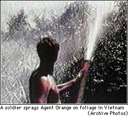 Agent Orange was contaminated by the experimental carcinogen
2,3,7,8-tetrachlorodibenzoparadioxin (TCDD), which remains present
in measurable quantities in some veterans even now
(Am. J. Ind. Med. 30: 647, 1996).
Despite the decision by politicians to
compensate Vietnam veterans with lymphoma (the son of
Admiral Zumwaldt, who ordered the spraying of agent orange, got lymphoma...) and (1993)
tobacco-induced lung cancer (I'm not making this up), any link between agent orange (dioxin) and a plethora
of alleged health problems (cancers, birth defects) remains very soft.
So is evidence that most of our
soldiers were even exposed. See JAMA 265: 898, 1991; Am. J. Pub. Health 81: 289 & 344, 1991;
Arch. Env. Health 53: 199, 1998 (Air Force; no chloracne
or noted increase in common acne
in veterans who sprayed it during Operation Ranch Hand);
Am. J. Epidem. 148: 786, 1998 (no increased mortality;
no increase in total cancer);
J. Occ. Env. Med. 39: 740, 1997 (VA
study finds lung cancer claim fails totally);
Arch. Env. Health
51: 368, 1996 (gestational trophoblastic disease claim fails);
Epidemiology 7: 454, 1996; Ann. Epidem. 5: 414, 1995
(VA; Hodgkin's claim fails completely);
Epidemiology 6: 17, 1995 (claims of more stillbirths and birth
defects fails completely). No link to prostate cancer: J. Urol. 166: 100, 2001.
No link to trophoblastic disease in the Vietnamese people: Arch. Env. Health 41: 368, 1996.
People heavily exposed in industry have only a slight increase in overall cancer risk,
even assuming that the effect isn't due to confounding variables
(Occ. Env. Med. 53: 606, 1996; Env. Health Perspect. 106 S2: 663, 1998);
one epidemiologist actually showed how to juggle the statistics, including
studies the EPA chose to ignore, to claim dioxin protects against
cancer (sort of like broccoli sprouts, I guess; Reg. Tox. Pharm. 26: 134, 1997.)
Nevertheless,
in 1994, the Environmental Protection Agency issued a report concluding that
dioxin as among the "greatest threat[s] to public health", i.e.,
was a grave danger which could be the cause
of 1.3 out of every 100 American cancer deaths. Of course, its own Science Advisory Board
refused to accept this preposterous idea, both in 1995 and after the EPA's 2000
revision (Tox. Sci. 64: 7, 2001 points out even the EPA is not
allowed to divide by zero;
also Reg. Tox. 36: 211, 2002, which considers among other strange things
the EPA's willingness to believe in "U-shaped dose-response curves").
In 1997 the government decided to compensate Vietnam vets whose children
have neural tube defects; again this is probably politics rather than science.
Of course, the Hanoi government claims a tremendous
increase in birth defects "caused by Agent Orange";
there was a conference in 2001 (Nature 413: 442, 2001) which
produced the expected agreement for joint study (Nature 416:
252, 2002).
Agent Orange was contaminated by the experimental carcinogen
2,3,7,8-tetrachlorodibenzoparadioxin (TCDD), which remains present
in measurable quantities in some veterans even now
(Am. J. Ind. Med. 30: 647, 1996).
Despite the decision by politicians to
compensate Vietnam veterans with lymphoma (the son of
Admiral Zumwaldt, who ordered the spraying of agent orange, got lymphoma...) and (1993)
tobacco-induced lung cancer (I'm not making this up), any link between agent orange (dioxin) and a plethora
of alleged health problems (cancers, birth defects) remains very soft.
So is evidence that most of our
soldiers were even exposed. See JAMA 265: 898, 1991; Am. J. Pub. Health 81: 289 & 344, 1991;
Arch. Env. Health 53: 199, 1998 (Air Force; no chloracne
or noted increase in common acne
in veterans who sprayed it during Operation Ranch Hand);
Am. J. Epidem. 148: 786, 1998 (no increased mortality;
no increase in total cancer);
J. Occ. Env. Med. 39: 740, 1997 (VA
study finds lung cancer claim fails totally);
Arch. Env. Health
51: 368, 1996 (gestational trophoblastic disease claim fails);
Epidemiology 7: 454, 1996; Ann. Epidem. 5: 414, 1995
(VA; Hodgkin's claim fails completely);
Epidemiology 6: 17, 1995 (claims of more stillbirths and birth
defects fails completely). No link to prostate cancer: J. Urol. 166: 100, 2001.
No link to trophoblastic disease in the Vietnamese people: Arch. Env. Health 41: 368, 1996.
People heavily exposed in industry have only a slight increase in overall cancer risk,
even assuming that the effect isn't due to confounding variables
(Occ. Env. Med. 53: 606, 1996; Env. Health Perspect. 106 S2: 663, 1998);
one epidemiologist actually showed how to juggle the statistics, including
studies the EPA chose to ignore, to claim dioxin protects against
cancer (sort of like broccoli sprouts, I guess; Reg. Tox. Pharm. 26: 134, 1997.)
Nevertheless,
in 1994, the Environmental Protection Agency issued a report concluding that
dioxin as among the "greatest threat[s] to public health", i.e.,
was a grave danger which could be the cause
of 1.3 out of every 100 American cancer deaths. Of course, its own Science Advisory Board
refused to accept this preposterous idea, both in 1995 and after the EPA's 2000
revision (Tox. Sci. 64: 7, 2001 points out even the EPA is not
allowed to divide by zero;
also Reg. Tox. 36: 211, 2002, which considers among other strange things
the EPA's willingness to believe in "U-shaped dose-response curves").
In 1997 the government decided to compensate Vietnam vets whose children
have neural tube defects; again this is probably politics rather than science.
Of course, the Hanoi government claims a tremendous
increase in birth defects "caused by Agent Orange";
there was a conference in 2001 (Nature 413: 442, 2001) which
produced the expected agreement for joint study (Nature 416:
252, 2002).
|
 |
* In 1998, there was an E-mail flap about sodium lauryl sulfate (the famous saponifier) causing cancer. Readers were exhorted to shun shampoos, toothpastes, and so forth that contained sodium lauryl sulfate. The truth... Some twit read a couple of articles about mixtures containing sodium lauryl sulfate causing a variety of cancers in laboratory animals. What the author didn't realize was that sodium lauryl sulfate was used as the vehicle to deliver the real carcinogens. Gee whiz!!
* The antiperspirants and breast cancer business is just a lie. The business about tampon manufacturers putting cancer-causing asbestos in tampons to make women bleed more is yet another bald-faced lie. The fact that these persist as internet and contemporary folklore shows how willing people are to believe ugly, obvious untruths that fit with their ideologies.
* Your lecturer is undecided about the links between pesticide residues in well water and leukemia (and Parkinsonism). See Am. J. Epidem. 151: 639, 2000; Epidemiology 10 481, 1999. The effect, if any, in causing leukemia is small, and recall bias could be a factor as well. Also undecided on chlorination of water: read about the carcinogens it generates JNCI 85: 817, 1993. I say it's worth chlorinating our water to protect us from drinking our neighbors' live fecal flora each time we turn on the tap.
Cooked meat (including poultry and fish) contains heterocyclic amines which cause cancer in animals models. You've all heard the stuff from the militant vegetarians. Your lecturer is totally unimpressed with "statistical evidence" that frequent consumption of red meat is a serious cancer risk. See also Br. Med. J. 315: 1018, 1997; Lancet 353: 703, 1999 (heterocyclic amines).
Here's the basics of what you need to know to evaluate pop / media / Green / EPA claims about cancer risk from chemicals in the environment.
No reasonable person would question the great harm that pollution has done to life in the waters. No reasonable person would question that air pollution is unhealthy, and places people at some increased risk for lung cancer.
The public is often told (by pop writers and even by government agencies) that pollution is the cause of an epidemic either of all cancers, or of particular cancers. You need to evaluate these claims carefully and thoughtfully, knowing that emotion and politics are involved as well as real science.
You already know that it is very easy to lie with statistics in order to frighten people. For example, more people are dying of cancer each year in the US today than in the 1950's... because there are more people. If you control for this and for the effects of increased smoking (Lancet 229: 268, 1992) and an aging population, the incidence of new cases is quite stable and the death rate is actually down by about 30% from 1950 (CDC from 2001). Children's cancers have not changed significantly in incidence over the period (JNCI 91: 1051, 1999), though many fewer of these children die.
 Rachel Carson's book "Silent Spring" blamed insecticides for the increase
in the number of cases of
childhood leukemia between 1950 and 1960. (Carson's book cites a 43% increase
but she ignored the impact of the baby boom on demography so the real increase
was about 13%.
Of course, the percentage of childhood deaths from leukemia
increased tremendously at the time as antibiotics cut the rate of
deaths from infections.) It was
not unreasonable to think at the time that insecticides
might be causing leukemia, and I appreciate that Ms. Carson brought it up,
even though she was wrong. And she was wrong.
Today's new rates of childhood leukemia are actually
lower than they were in 1950. See JNCI 91: 675 1999; Lancet 354:
532, 1999, or check the WHO or NCI databases.
Unfortunately, Ms. Carson's calling pesticides "elixirs of death"
set an unfortunate tone for Green agitprop which persists today.
It seems most likely to me that the 13% increase probably resulted from
increased
urbanization -- children who grow up around a lot of other children
from different communities and their viruses (see for example Lancet 357: 858, 2001),
get more leukemia, i.e., there's one or more viruses involved.
Recent studies have utterly failed to link childhood
leukemia either to growing up next door to the nuclear power plant (Cancer Causes & Control
9: 529, 1998) or chemical landfills (Int. J. Epidem. 29: 391, 2000).
Nobody is even writing scientific papers
about chemicals in the environment causing childhood
leukemia any more; the latest review concluded that "no causal factor
has been identified which can explain a single cluster of childhood leukemia"
(Eur. J. Epid. 15: 847, 1999).
Rachel Carson's book "Silent Spring" blamed insecticides for the increase
in the number of cases of
childhood leukemia between 1950 and 1960. (Carson's book cites a 43% increase
but she ignored the impact of the baby boom on demography so the real increase
was about 13%.
Of course, the percentage of childhood deaths from leukemia
increased tremendously at the time as antibiotics cut the rate of
deaths from infections.) It was
not unreasonable to think at the time that insecticides
might be causing leukemia, and I appreciate that Ms. Carson brought it up,
even though she was wrong. And she was wrong.
Today's new rates of childhood leukemia are actually
lower than they were in 1950. See JNCI 91: 675 1999; Lancet 354:
532, 1999, or check the WHO or NCI databases.
Unfortunately, Ms. Carson's calling pesticides "elixirs of death"
set an unfortunate tone for Green agitprop which persists today.
It seems most likely to me that the 13% increase probably resulted from
increased
urbanization -- children who grow up around a lot of other children
from different communities and their viruses (see for example Lancet 357: 858, 2001),
get more leukemia, i.e., there's one or more viruses involved.
Recent studies have utterly failed to link childhood
leukemia either to growing up next door to the nuclear power plant (Cancer Causes & Control
9: 529, 1998) or chemical landfills (Int. J. Epidem. 29: 391, 2000).
Nobody is even writing scientific papers
about chemicals in the environment causing childhood
leukemia any more; the latest review concluded that "no causal factor
has been identified which can explain a single cluster of childhood leukemia"
(Eur. J. Epid. 15: 847, 1999).
In real life, most risks are either logarithmic or show a threshold below which there is no risk. Today's attempts by governments to "estimate the risk of low exposures" based on animal studies generally assume a linear relationship instead.
| If the relationship is really logarithmic...
| Do you see the fallacy? |
| If there is a threshold for risk...
| I thought you would. |
To decide whether a substance is a cancer risk in the absence of an obvious epidemiological link, and if so how much is "safe", it's usual nowadays to give huge doses to a few dozen lab animals. (Lower doses and more animals would be much more scarce and expensive, given the outrageous costs of doing animal research today.) When a handful of animals get a particular tumor (and this often does happen), a linear relationship is assumed to exist between dose and risk. This is a stupid assumption (it doesn't even apply for alcohol-and-liver, or tobacco-and-lung; and it's the basis for the asinine claim that "every drink of alcohol kills 500 brain cells".) But it's supposedly the best we can do, i.e., this approach generates pretty much any scare story you want. And that's the beauty. Exactly what is done with the results is up to the politicians. Plants produce their own natural insecticides, some of which cause cancer in lab animals (hydrazine in mushrooms, caffeic acid in many fruits and vegetables, lots of things in pepper.) These naturally-occurring known experimental carcinogens are tolerated in quantities that are orders of magnitude higher than what we allow for pesticide residues, of course for political reasons (see the Ames references below).
The Environmental Protection Agency is charged with setting the allowable limits for various synthetic chemicals in air, food, and water. It is a highly-politicized government body, which is often lambasted for its pronouncements by the very scientists who it had hired for its projects after they quit in disgust. During the 1990's, the mainstream scientific community held the EPA in utter contempt (Nature 412: 677, 2001); things are supposed to be improving under a new administration (Science 300: 1351, 2003) but scientific advisors are still unhappy (Nature 422: 5, 2003; talk about a "right shift"). There's little chance of things improving until the US public become scientifically literate. As a physician, you may need to reassure your patients about EPA stuff.
Before you decide to ban pesticides "just to be on the safe side", consider the effect on real human beings and what they'd eat. You know you will drive food prices up, and the burden will be borne by the world's poor. It's pretty clear that a good intake of fruits and vegetables seems somewhat protective against several cancers. For now, "organic" methods would make these much more expensive, making a good diet much less available to the poor. Good public policy is a matter of weighing relative risks. Think about it.
The difficulty of finding out the truth about environmental exposures, whatever that truth may be, is made much more difficult by the fact that the parents of children with cancer tend to "overreport" exposures to suspected carcinogens, i.e., people are not always totally honest / accurate. The reality of "reporting bias" is documented in Am. J. Epidem. 158: 710, 2003.
* There's a "school of independent medical thought" called homeopathy, which holds that small doses of a substance have the opposite effects from large doses. If these people really believed their own claim, they would consider exposures to low levels of carcinogens to be good for us. Did you ever think about that?
Two terms from classic studies of chemical carcinogenesis:
Initiation: The result of exposure of a cell or cells to a carcinogen, which permanently alters its genetic material but not its phenotype (yet). As noted, these are mutagens ("genotoxic carcinogens").
Promotion: A substance that causes initiated cells to turn into tumors. Tumors result when the promoter is administered after, but not before, initiation. Promoters are "non-genotoxic carcinogens".

* Long thought to be "nonspecific irritants" or to exert their effects by "increasing the rate of mitosis, therefore causing sequential selection of mutant clones", we now know that some mitogens are much better promoters than others. This will eventually get sorted out; don't worry about it now.
* Today, there is apparently total agreement now that Agent Orange is not a mutagen / initiator. Even the folks who use it as an experimental carcinogen say it's apparently just a mitogen (Exp. Tox. Path. 51: 555, 1999).
Experimentalists mostly use phorbol esters as promoters. They work by activating protein kinase C (Cancer Res. 58: 1423, 1998).
Hormones are also promoters for particular organs.
* Clomiphene-gonadotropin stimulation of the ovary for infertility seems to give an increased number of granulosa tumors; Lancet 341: 986, 1993.)
And probably many things (Epstein-Barr virus, various non-genotoxic chemicals that have been linked to cancer) do act "by disrupting normal tissue homeostasis". So far, this has been hard to study (Nature 350: 555, 1991). It does seem now that hepatitis B and C viruses are much more likely to produce hepatocellular carcinoma when they cause rapid, prolonged cell turnover, i.e., making Nowell's law operate more readily (Cancer 73: 1149, 1994).
A complete carcinogen is a substance that is both initiator and promoter, such as "tobacco smoke" or certain really awful chemicals.
The Ames test for mutagenicity (and presumably carcinogenicity) relies on production of mutants in a culture of typhoid bacteria.
* Today's more sophisticated tests include a "micronucleus test", in which mutagens supposedly can be distinguished by their ability to produce tiny nuclei in a strain of lymphocytes. You may hear in the future about this being helpful in exfoliative cytology (Mut. Res. 489: 147, 2001).
"Chemicals that cause cancer in laboratory animals", typically at preposterously high doses, are have long been (theoretically) banned from foods in the US.
By the late 1980's it was very clear that the vast majority of such chemicals were "non-genotoxic carcinogens" exert their high-dose effect by causing cell division ("mitogens, not mutagens"), which should not be a realistic concern at lower doses. See Science 236: 271, 1987; Science 249: 970, 1990; Science 250: 1645, 1990; Science 251: 12, 1991. Even Dr. Ames said that animal testing of carcinogens, and fear of "chemicals", has gotten ridiculous (Science 240: 1043, 1988; Science 249: 970, 1990; more recently Mut. Res. 447: 3, 2000).
Dr. Ames often points out the fallacy involved in animal carcinogenesis work. About half of chemicals tested produce cancers in rodents if given in preposterously high doses, by disrupting the environment in which mitosis takes place. The "risk estimate" on which political decisions are based then assumes that the relationship is linear, which is obviously not true. But even if it IS true and you compare the curves, the "risk" from the amount of caffeic acid alone in a cup of coffee or a lettuce salad is commensurate with the risk from a month's exposure to DDT during Rachel Carson's era (see Science 236: 271, 1987; Proc. Nat. Acad. Sci. 87: 772, 782 & 787, 1990; Env. Health Per. Sup. 105: 865, 1997; Mut. Res. 447: 3, 2000; all by Bruce Ames).
Long-neglected in practice, the absolute ban was finally eliminated in 1996.
RADIATION CARCINOGENESIS
Exposure to high-energy photons (ultraviolet, ionizing radiation) is well-known as a cause of cancer.
From 1928-1955, "Thorotrast", a complex of thorium dioxide and a carrier dextran, was used to image the livers and spleens of a few million humans. This was monumentally stupid. The long-lived isotope stays in the body, emitting radiation, for the rest of your life, and cancers followed years or decades later (J. Tox. 35: 199, 1997).
Atomic bomb survivors (Japan, Marshall Islands) have greatly increased incidences of all the common leukemias (except CLL; the incubation time is a few years), and minor increases in many (but not most) solid tumors (remember thyroid, breast, salivary gland, lung).
Chernobyl's children (thyroid cancer from radioactive iodine, other problems have been less prominent): Nature 359: 21, 1992; update Lancet 358: 1965, 2001.
Interestingly, children conceived after their parents were exposed to radiation at Hiroshima and Nagasaki do not exhibit any measurable increase in any identified health problem so far. This "non-news" is very important scientifically (Am. J. Hum. Genet. 52: April 1993).
On the average, 14% of your annual radiation exposure is from your diagnositic x-rays. The cancer risk that this poses is unknown.
More about the "radon in your energy-efficient home causes lung cancer" business later (in any case, it's no measurable risk for anything else important; Lancet 341: 1127, 1993; more Lancet 355: 1888, 2000). Since the early 1990's there has been amazingly little work on radon despite the ongoing political stuff. Nobody's shown an increased risk from living near nuclear power plants (JAMA 265: 1438, 1991).
Occupational radiation carcinogenesis:
Old-time radiologists who tested their fluoroscopes using their own hands developed lots of leukemias, Hodgkin's disease, and skin cancer.
Radium paint workers who put their brushes in their mouths developed bone and nose cancers.
Uranium miners have a greatly increased incidence of lung cancer, supposedly even if they do not smoke.
Iatrogenic radiation carcinogenesis:
People given high doses of radiation for ankylosing spondylitis (x-rays) or polycythemia vera (radiophosphorus) have greatly increased incidences of all the common leukemias (* except CLL).
Patients treated with radiation therapy for acne (!) develop multiple skin cancers.
Newborns treated for mythical "enlarged thymus" developed many thyroid cancers as young adults.
Ultraviolet radiation is the principal risk factor in most skin cancers (basal cell, squamous cell, malignant melanoma).
Suntanning offers only modest protection from the wavelengths that cause cancer and elastosis ("aging of the skin").
Radiation appears to initiate cancer just as chemical carcinogens do -- by causing mutations.
* The trademark ultraviolet light mutation is CC->TT (Proc. Nat. Acad. Sci. 90: 4216, 1993).
ONCOGENIC VIRUSES
Viral (RNA, DNA) causation of cancer is well-documented in the lab, and is important in some (but probably not most) human cancers.
Polyoma virus and SV40 are linked to a variety of animal tumors.
Mouse mammary tumor virus is transmitted from mother to child in the milk.
Feline leukemia virus causes a contagious leukemia in cats.
Closer to home: Wart virus ("human papilloma virus", HPV) causes warts ("benign tumors") in humans, and certain strains also cause cancer of the uterine cervix in humans (Nature 336: 765, 1988 was the breakthrough article).
* For your future reference: HPV oncogenic protein E7 inactivates the product of tumor-suppressor gene RB, while E6 inactivates tumor-suppressor gene p53 product and prevents it from repairing damaged DNA (Proc. Nat. Acad. Sci. 90: 3988, 1993; Proc. Nat. Acad. Sci. 91: 2436, 1994. These two genes do the transformation in mice: Cancer Res. 52: 4420, 1992).
Epstein-Barr virus ("infectious mononucleosis virus") is necessary (but not sufficient) to cause African Burkitt's lymphoma, and is etiologic in Chinese nasopharyngeal cancer, immunoblastic lymphoma, and * Eskimo endemic salivary gland adenocarcinoma.
Hepatitis B virus is a major cause of hepatocellular carcinoma. It is now clear that hepatitis C virus is also important (PNAS 87: 6547, 1990; JAMA 265: 1974, 1991; NEJM 325: 675 & 729, 1991; Am. J. Gastroent. 86: 335, 1991). They probably effect this by acting as mitogens, allowing special opportunities for genetic damage (PNAS 86: 8852, 1989; PNAS 87: 6791, 1990; Cancer Res. 51: 1278, 1991). Hepatitis B (and perhaps C) also inserts itself randomly in the genome, with a range of possible effects (Ult. Path. 25: 497, 2001).
HTLV-I causes epidemic leukemia in Japanese humans.
* Adenovirus, E2F protein and the Rb gene product: Nature 358: 181, 1992; Science 258: 424, 1992.
Despite all this, the common human cancers (except as noted) do not seem to be contagious. Viral carcinogenesis promises to be an area of continuing interest.
OTHER REPUTED CARCINOGENS
Foreign-body carcinogenesis and carcinogenesis by repeated trauma are possibilities that worry patients.
There is almost nothing to suggest that foreign implants cause cancer. (The breast implant hype and fiasco: NEJM 326: 1649, 1992.) Cancers caused by bile duct flukes or schistosome eggs probably are due to their effects as promoters. Joint replacement doesn't cause cancer at the joint or elsewhere: Cancer 94: 3057, 2002.
The weight of evidence is that mechanical injuries do not cause cancer, though this is often alleged in lawsuits.
* The only likely exception is fibromatosis / fibrosarcoma where there was massive soft-tissue injury (AMFJP 19: 152, 1998; also Surg. Gyn. Ob. 169; 104, 1989; J. Ped. Surg. 34: 1130, 1999) Both make sense -- cells that do not ordinarily divide will have divided in response to the trauma, letting Nowell's Law operate and producing a tumor with the phenotype of the dividing cell.
* The traditional wisdom is that head trauma places people at risk for meningiomas, for some unknown reason; not surprisingly, the recent studies show little if any effect (Cancer Causes & Control 9; 109, 1998; Int. J. Epidem. 27: 579, 1998).
There is a great deal of speculation and anecdotal evidence connecting carcinogenesis and prognosis of cancers to mental attitudes.
So far, the best work has failed to support the connection made by folklore, and nowadays real scientists have pretty much stopped examining these claims. The "stress and breast cancer recurrence" claims (Br. Med. J. 304: 1295, 1992; Br. Med. J. 304: 1078, 1992) were finally laid to rest by a huge study (Br. Med. J. 324: 1420, 2002). "Psychic vulnerability" is not a risk for cancer either (Cancer 94: 3299, 2002, twenty-year prospective study; yes really). "Fighting spirit" does not correlate with survival, but being depressed does; I suspect that the latter effect means the cancer has gotten farther and is causing an organic depression (Lancet 354: 138, 1999).
But the relationship between mind and body is clearly a very potent one, and the whole field cries out for more study.
ONCOGENES
Cancer genetics update: Nat. Genet. 33S: 238, 2003.
Oncogenes are DNA sequences within eukaryotic cells that seem to be involved in the development and maintenance of tumors. These genes direct the synthesis of proteins that under some conditions transform a benign host cell into a cancer cell.
Oncogenes are slightly altered forms of proto-oncogenes ("mitogenes") which are essential genes that govern normal tissue growth, differentiation, and apoptosis.
Many proto-oncogenes are the genes for hormone or vitamin receptors or the proteins to which they talk, while others seem to be general turn-ons.
When a proto-oncogene is altered to become an oncogene, we speak of its being activated. This is by one of three mechanisms:
* As key genes for cellular function, growth, and differentiation, proto-oncogenes have been highly conserved through evolution.
All the families of proto-oncogenes that have been studied so far exist in all vertebrates, producing nearly-identical proteins.
In fact, most of them exist in very similar forms in fruit flies, yeast, etc., etc., though their functions in these species may be different from their functions in vertebrates.
Inconsequential differences in codon sequences (i.e., redundant third bases, occasional amino acids differences), when compared, have so far all yielded the same "phylogenetic tree" as classical comparative anatomy. Actually, this has proved true of all proteins so far studied, and this fact is the strongest evidence I know for the common ancestry (rather than just "intelligent design") of all living things. If this were NOT true, Darwin's macroevolution would be refuted. Clonal selection in Darwin's world: Nature 363: 208, 1993.
Oncogenes were originally discovered in transforming retroviruses ("the RNA tumor viruses").
Retroviruses are ubiquitous, generally harmless RNA viruses (HIV is obviously an exception). The
RNA code is transcribed onto DNA, which is then integrated into the host genome.
Typically a "viral oncogene" is a proto-oncogene minus its regulatory sequences, or with a
characteristic mutation, or in an excessive number of copies ("amplification"). They are capable of
causing cancer by themselves, and hence are very different from their normal counterparts (i.e., have
been damaged several times).
As we have noted, a proto-oncogene that has acquired the ability to cause cancer (i.e., has become
an oncogene) is said to be activated.
These are signal-transducers, across membranes. You met them in "Biochemistry".
The family includes src and abl. Present in all eukaryotes. The c-src protein product phosphorylates vinculin (the protein that cross-links actin filaments to plasma membranes). It also greatly increases the synthesis of phosphatidyl-inositol 4,5 diphosphate, a second-messenger for a range of growth factors. Physical chemistry of mutant src and family: Nature 385: 595 & 602, 1997.
c-abl is translocated from chromosome 9 to the breakpoint cluster region of chromosome 22 in most cases of chronic myelogenous leukemia, and this is part of the "Philadelphia chromosome" phenomenon. Much more about this later!
met tyrosine kinase proto-oncogene seems to be what produces lumens in mesenchyme and its tumors (synoviosarcomas, mesotheliomas, kidney tubules, liver tubules, others): Science 257: 1258, 1992; Cancer 82: 1513, 1998; Proc. Nat. Acad. Sci. 95: 14417, 1998; it codes for the hepatocyte growth factor receptor and is emerging as a major player (J. Urol. 170: 2163, 2003); * unlike most other activated proto-oncogenes, it can be passed parent-to-child (hereditary papillary kidney cancer) and marks the aggressive "tall-cell" variant of thyroid cancer.
RET is a proto-oncogene tyrosine kinase (Nature 363: 458, 1993; NEJM 335: 943, 1996), and was the first activated oncogene that was discovered being passed from parent to child (Science 267: 381, 1995). Depending on the allele, there may be various endocrine tumors, mucosal neuromas, and/or Hirschsprung's disease of the colon.
This includes the ras family, present in all eukaryotes. Their protein products are apparently the signal-transducing G-proteins that modulate various transmembrane signals (* for example, turning fibroblasts into fat cells; Science 253: 565, 1991). Each codes for a p21 protein that binds GTP, and the healthy ones hydrolyze it (i.e., they are GTP-ases). They seem to be involved in initiation of mitosis as well as in differentiation. What ras does: Science 264: 1413, 1994. All about the G-proteins: NEJM 332: 406, 1995 (* Nobel prize 1994 Gilman and Rodbell).
Most oncogenic ras are mutations with a single base pair change that alters an amino acid at position 12, 13, or 61 in the protein product. This destroys GTP-ase activity but retains GTP-binding activity, and current thinking is that these stay locked "on", telling the transformed cell, "Keep dividing!"
Several of the best-known chemical carcinogens produce a specific mutation specifically at one of the three hot spots. For example, aflatoxin regularly mutates * codon 12 in K-ras (GGT to AGT or GAT).
ras oncogenes clearly help cause a large percentage of human cancers. The large majority of oncogenes isolated from human tumors have been hot-spot ras mutants. Almost all pancreatic cancers, and many other cancers (especially adenocarcinomas) have mutations in codon 12 of K-ras (update Arch. Path. Lab. Med. 126: 1096, 2002). ras activation precedes malignant expression: Science 248: 1101, 1990.
* A favorite pathology research subject in the late 1980's was immunostaining for ras p21 and seeing its effect on diagnosis and prognosis. For example, staining for K-ras p21 was reported to differentiate malignant from benign prostate epithelium; unfortunately, this has not held up on closer examination.
* We can hope for better success with new attempts to screen patients for colon polyps/cancer by checking stools for oncogenic ras; despite much discussion over the past decade, it's still too costly (Gastroenterology 119: 1219, 2000).
* GS is a ras-related oncogene. More about this when we talk about the pseudohypoparathyroidism family of illnesses. 366: 643, 1994.
This is the myc family, present in all eukaryotes, whose protein products are intranuclear and bind to DNA itself. They enable DNA synthesis.
myc activation is usually by amplification (excess copies of a gene) and/or translocation rather than by mutation.
In Burkitt's lymphoma of B-cells, c-myc (chromosome 8) is moved next to the immunoglobulin gene (chromosome 14), i.e., the cell decides to multiply like crazy every time it is told to make antibodies.
myc genes are much amplified in neuroblastomas and oat cell lung carcinomas.
myb is a related proto-oncogene involved both in human cancer and in the proliferation of cells in human atheromas. * Basic biology buffs: myb has to do with production of the anthocyanins that account for the pretty colors of Indian corn.
* The Ewing's sarcoma 11:22 translocation joins FLI1, a myc-like proto-oncogene, to EWS, producing an aberrant transcription factor EWS-FTI1 (Proc. Nat. Acad. Sci. 90: 5752, 1993); much since.
The prototype is c-sis, which codes for the beta chain of platelet-derived growth factor (PDGF), the stuff that tells fibroblasts to divide in wound healing.
Probably sis-induced cancers grow by autocrine self-stimulation by PDGF. Not surprisingly, PDGF is greatly over-expressed in many sarcomas, and only transforms cells with the PDGF receptor.
This includes erbB, which codes for a protein homologous to the epidermal growth factor receptor (Science 249: 1552, 1990), and fms, which codes for macrophage colony-stimulating factor.
The erbB gene product lies across the cell membrane. Epidermal growth factor binds to the outer portion, while the inner portion is a tyrosine kinase that cleaves phosphatidyl-inositol 4,5 diphosphate into inositol triphosphate (which releases intramembrane calcium) and diacylglycerol (which activates protein kinase C). These two substances have multiple effects on intracellular control systems, mostly turn-ons.
As you would expect, erbB mutants are those that are locked in the "on" position, and erbB-related cancers are mostly squamous cell carcinomas, and fms-related cancers are mostly hematopoietic cancers. erbB is amplified in a large percentage of malignant melanomas.
The related neu (once erb2, now HER2) is amplified in many carcinomas, notably adenocarcinomas, especially of the breast, and the degree of amplification strongly correlates with bad outcome.
erbA codes for the human thyroid hormone receptor. It is linked to a variety of animal cancers.
jun is the factor that initiates transcription of DNA at a particular sequence. Present in all eukaryotes.
fos apparently turns short-term stimulation into long-term differentiation, and fos mutants help immortalize cell cultures. fos and jun: Science 254: 1210, 1991.
int-2, the second site where the mouse mammary tumor virus integrates, is the gene for fibroblast growth factor #3 (FGF3; J. Path. 170: 219, 1993; J. Neurosurg. 76: 792, 1992), a gene with many relatives, including proto-oncogenes flg (FGF1) and bck (FGF2).
bcl-2, activated in most B-cell lymphomas, and its relative bcl-X, tell the cell not to undergo apoptosis, but to divide if told to do so. The molecular biology of this important molecule, and its family including bax, is being worked out. (Nat. Med. 3: 614, 1997; Nat. Med. 274: 2002).
* Flk-1, a VEGF receptor (for glioblastoma, etc., must be why they elaborate those odd blood vessels; Nature 367: 525, 1994).
* mos is involved in the second step of meiosis, and if mutated, generates teratomas or even attempts at parthenogenesis: Nature 370: 65, 1994.
Cyclin D1 itself (11q13, bcl-1, the PRAD-1 locus) is involved in the oldest known lymphoma translocation, in most parathyroid adenomas, and is amplified in around 20% of breast cancers, and the knockout mice get breast tumors (Nature 369: 669, 1994). Review J. Clin. Lab. Med. 127: 246, 1996.
The high mobility group (HMG) genes are often scrambled specifically in benign tumors with near-zero malignant potential (Am. J. Clin. Path. 109: 251, 1998; Am. J. Path. 155: 1535, 1999). This suggests that these mutations are the single major step to producing benign tumors, which do not turn malignant because there aren't a lot of mutations accumulated.
DNA In-Situ Hybridization is beginning to come into its own as an adjunct for cancer diagnosis (Am. J. Clin. Path. 112(S1): S11, 1999).
Today's techniques count chromosomes instead by fluorescent means, which stain each pair a different color.
It is now standard to stain paraffin sections of breast cancers with probes for erb-B2 (HER2/neu). Cancers without amplification will show only two loci; those with amplification will show huge numbers of loci, usually in big clumps.
When there's suspicion that a particular gene is split by translocation, two probes can be used, of different colors. If they remain together, there is no translocation; if they are separated, translocation has occurred. This is very helpful in diagnosing Ewing's sarcoma.
TUMOR SUPPRESSOR GENES (anti-oncogenes): Many reviews; Knudson himself in Proc. Nat. Acad. Sci. 90: 10914, 1993; at the bedside Lancet 349-S2: 16, 1997; kids Ped. Clin. N.A. 49: 1393, 2002; adults Arch. Path. Lab. Med. 125: 85, 2001.
Tumor-suppressor genes keep cells from overgrowing, even when the oncogenes are activated. To lose their anti-cancer effect, both copies must be altered. (Contrast the proto-oncogenes which exert their effect when a single copy is activated to an oncogene.)
Knudson's Law for tumor-suppressor genes
One hit: You have a cell with a much increased propensity to turn malignant
Two hits: You have a cancer cell.
If you inherited one copy of the damaged tumor suppressor gene, you have the anti-oncogene deletion syndrome, with a greatly increased risk for the corresponding tumor(s). If you have the corresponding tumor(s) but do not have the germ-line mutation (i.e., your tumor was sporadic), both mutations are somatic. If you don't understand this, stop now and think about it until you do.
Please don't ask whether the mutated allele is "dominant" or "recessive". It is dominant with respect to the tumor-family syndrome, recessive with respect to the tumor itself. If you are reading this, you already understand.
If you derive from a mutation-bearing sperm or egg, or were hit at conception, you have one of the autosomal dominant tumor suppressor gene deletion ("tumor-susceptibility") syndromes. The malignant phenotype requires both copies to be bad, so it is autosomal recessive.
The best-known example is the case of retinoblastoma, a hereditary cancer.
The tendency to retinoblastoma (* and osteosarcomas, in survivors) is inherited as an autosomal dominant trait, i.e., there is one chromosome lacking a particular tumor suppressor gene.
Retinoblastoma results when a mutation damages both copies the healthy tumor-suppressor gene in a single cell (the Knudson "two-hit" phenomenon). The cell now lacks any copy of the tumor suppressor gene, and is transformed.
The retinoblastoma susceptibility locus is RB1 at 13q14. The protein is central to cell cycling. Update on how it works: Nature 374: 114, 1995.
The most important finding in cancer research in the late 1980's was the discovery that many (if not most) DNA-containing tumor viruses (including human papilloma virus) bind to, and inactivate, the normal product of RB1 (Cell 56: 1, 1989). The ability to do this correlates with tumorigenicity (Science 248: 70, 1990); virus protein blocks the binding of Rb to its target among the nucleoproteins (Nature 351: 406, 1991), etc. This has held up, though to date it has not resulted in any new therapeutic strategies.
The most common known genetic injury in human cancer is damage to the p53 (TP53) gene.
The p53 gene, when defective, is now strongly linked all human cancers in which it has been sought, especially the more anaplastic versions of each.
The p53 gene product is a sequence-specific binder to DNA that prevents mitosis during times of cell injury, so that there will be more time for DNA repair. Sometimes (gamete, lymphocyte, chemotherapy) p53 even tells an injured cell to undergo apoptosis.
* The protein binds as a tetramers, making it easier to understand the oncogene-anti-oncogene duality. The defective unit handcuffs the others. See Science 256: 827, 1992; Nature 358: 15, 1992.
A famous link is to hepatocellular carcinomas (* those from the aflatoxin-hepatitis B belt have a distinctive mutation in codon 249: Science 253: 49, 1991; the mutagen is aflatoxin: Lancet 338: 1356, 1991).
More good evidence for the Nowell multi-step clonal evolution model for tumorigenesis is provided by the fact that astrocytomas (low-grade brain cancer) turn into glioblastomas (high-grade brain cancer) upon acquiring a mutated p53 (Nature 355: 846, 1992).
* UV light mutates p53 in skin cancer: Proc. Nat. Acad. Sci. 88: 10124, 1991). "Good" p53 might reverse myeloid leukemia in individual cells (Nature 352: 345, 1991), and so forth.
The tumor-suppressor gene p53 product is also inhibited by the proteins of human papilloma virus (see above; p53 and cervix cancer Lancet 340: 140, 1992), SV40, and adenovirus 5 (Science 248: 76, 1990).
This is the "most-often-mutated" known gene in spontaneous human cancers (Nature 350: 377, 1991), a dubious honor once held by ras. Older reviews of p53: Nature 350: 429, 1991; Nature 351: 453, 1991; Science 253: 49, 1991. Mutations vary; unlike ras, the only "trademark" is the hepatocellular carcinoma mutation due to aflatoxin (codon 249).
We now stain for the expression of this protein (the damaged genes make more of it) in cancers for prognostication (Arch. Path. Lab. Med. 121: 395, 1997.)
* By the way, the flap about calcium channel blockers preventing apoptosis and putting people at risk for cancer fizzled: Cancer 85: 2093, 1999.
There are many other tumor-suppressor genes.
As noted above, p16INK4 (was CDNK2A, now is MTS1), on 9p21, is an inhibitor of cyclin-dependent kinase (i.e., inhibitor of mitosis) which is very commonly deleted in lots of cancers (Proc. Nat. Acad. Sci. 91: 11045, 1994); it may function either an oncogene or a tumor suppressor gene. See also Nature 370: 180, 1994; Nat. Med. 5: 731, 1999; lots more. This now identifies a familial melanoma syndrome: NEJM 338: 879, 1998. All about cell-cycle screwups in cancer: Science 266: 1821, 1994.
Apparently all renal cell carcinomas have lost the Von Hippel-Landau locus (Nature 332: 268, 1988; more below). Oat cell carcinomas also lack a portion of 3p; almost all other lung cancers lack a smaller chunk in the same place.
People heterozygous for a deletion of a bit of 11p have aniridia, and they develop Wilms' tumors which are homozygous for the deletion (as are spontaneous Wilms' tumors: Proc. Nat. Acad. Sci. 90: 1416, 1993).
This is the famous WT-1 (formerly WAGR) locus, producing a nuclear binding protein which seems to be involved in mesenchymal-epithelial transitions in general, and kidney and urogenital ridge development in particular (Nature 346: 194, 1990; Nature 353: 431, 1991; Proc. Nat. Acad. Sci. 90: 5828, 1993). * Biology jocks: the adjacent aniridia gene is homologous to the drosophila homeobox PAX6.
Von Recklinghausen's disease type I patients are heterozygous for a mutation on chromosome 17; the locus is "NF-1", and its protein product, "neurofibromin" characterized as a facilitator of hydrolysis of GTP by normal ras p21 (Nature 351: 576, 1991).
Von Recklinghausen's disease type II patients are heterozygous for a mutation on chromosome 22 (product is * schwannomin or merlin), and their tumors (and many spontaneous meningiomas and other nerve tumors: Am. J. Hum. Genet. 47: 823, 1990) are homozygous for this loss. See below.
Colon cancers typically become homozygous for deletions of 5p (early) and 5q21 ("APC", "adenomatous polyposis coli", "familial polyposis locus".
In any case, the study of colon cancer progression has provided strong support for the Nowell multi-step clonal evolution model of carcinogenesis (no surprise). The colon is a handy organ since pre-cancerous polyps often make it to the lab.
* A gene on chromosome 2 makes a person susceptible to melanomas of the eye.
The familial non-polyposis ("Lynch") family of tumor-suppressor genes are responsible for repair of DNA mismatches. * Alphabet soup: hMSH2, hMLH1, hPMS1 hPMS2, and so forth.
* A new genetic mechanism in the development of cancer is probably relaxation of imprinting. Ordinarily, only the paternal IGF2 gene is expressed, but both may be expressed in Wilms' tumor. Stay tuned (Nature 362: 747 & 749, 1993).
* Up-and-coming anti-oncogenes include DPC4 (Cancer 91: 1332, 2001).
* So far, the only tumors in which changes in mitochondrial DNA have been detected are "oncocytomas", i.e., tumors made up of cells with preposterous numbers of mitochondria. More about this under "Kidney". See also Am. J. Hum. Genet. 52: 537, 1993 (mitochondria and lipomas).
During tumor progression, the second anti-oncogene is lost by nondisjunction and loss of heterozygosity when an extra chromosome is randomly eliminated. The phenomenon enabled the discovery of many of these genes.
* Actually, we've known for decades that tumor suppressor genes must exist. A classic finding is that when a cancer cell and a non-cancer cell are fused, the product is often a non-cancer cell.
LATE PROGRESSION
Once its growth genes have been mutated and its genome perhaps destabilized, cancer is still not a threat until it has developed the ability to invade, to induce its own blood supply, and (usually) to spread to distant sites.
We are just now unravelling the additional accumulated mutations that allow these things, and they will be the targets of new cancer therapies.
A major mystery of medicine is why some cancers (notably metastatic breast carcinoma and metastatic melanoma) lie dormant and hidden for years (even decades).
For that matter, it's puzzling why cancer metastases will thrive in one locale and not in another.
* Your genetic makeup determines whether cancer will progress more or less rapidly (Am. J. Hum. Genet. 63: 1, 1998), etc., etc. The genes remain elusive, but this promises to be interesting.
Microsatellite instability means the presence of abnormally long stretches of DNA with repeating motifs of 1-5 base pairs (i.e., a long string of "A"'s or "AC"'s or whatever), indicating defective DNA repair. This can actually be present quite early, before there are any visible histologic changes.
* Term: RER-positive for "replication error positive", i.e., the microsatellites indeed are unstable. This is a very promising new area.
Chromosomal instability of course means, i.e., there have been problems with the mitotic spindles and non-disjunction will become commonplace. Cells without the necessary chromosomes die and don't contribute to progression; cells with extra copies may possess a growth advantage. Eventually this leads to those variably-sized and often huge nuclei which are familiar from conventional histology of cancers (Am. J. Path. 161: 391, 2002; many others.
TUMOR IMMUNOLOGY
Despite a great deal of data and the fact that lymphocytes are usually abundant in advancing cancers, study of the immune system's attack on tumors has yielded little useful information.
For some cancers (breast cancer, maybe melanoma, etc., etc.), a dense infiltration of lymphocytes imparts a slightly better prognosis, but this is hardly decisive. (The most impressive link so far is for ovarian epithelial carcinoma: NEJM 348: 203, 2003).
By and large, cancer treatments that focus on strengthening the immune system (BCG, cytokines) have failed to help most cancer patients.
Cultivating and re-infusing lymphocytes found in tumors (i.e., lymphocytes that are presumably attacking the tumor) often results in some regression of the tumor (Cancer Res. 51(S): 5074, 1991).
Despite the widespread belief (medical, folk) that "our immune system is our main defense against cancer", evidence in support of the "immune surveillance theory" is not very convincing.
People who are immune-crippled (hereditary immunodeficiency, AIDS, immunosuppressive therapy) simply don't have increased rates for any of the common cancers. See, for example, Chest 103: 862, 1993; review in AIDS: JAMA 285: 3090, 2001; JNCI 28: 5-9, 2001.
The malignant lymphomas that are result from immunosuppression (AIDS, transplant patients) arise from cells that are already undergoing compensatory hyperplasia because of the immune suppression. Actually many of these are not even tumors, but hyperplasias which regress when good immune function is restored.
Kaposi's "sarcoma", seen in AIDS and transplant patients, is actually a viral hyperplasia, not a real tumor.
Patients with ataxia-telangiectasia (cancers plus immunodeficiency) have "fragile chromosomes" as the basic problem; even carriers are at extra cancer risk.
Carcinomas of the skin and lower lip are common in transplant patients and they tend to be aggressive. This is not true of other carcinomas, and rather than invoking "escape from immune surveillance", the usual suspects are known and unknown viruses (Cancer 85: 1758, 1999).
|
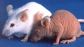
|
People with autoimmune diseases triggered by the known anti-tumor antibodies do not self-cure their tumors (though there may be modest regressions), nor do these diseases pop up in the absence of the tumors known to cause them (Lancet 341: 21, 1993).
Frankly, tumor immunology is only starting to get interesting to patient-care physicians. Although "cancer is not other, it is us", a few known antigens are distinctive (though not unique) for individual cancers, and it is against these that any effective immunotherapy will probably be directed.
* One interesting approach tags antibodies against such antigens with pseudomonas exotoxin ("immunotoxins"), selectively destroying cancer cells. Minimal side effects. So far it's worked on a few human cancers: Blood 94: 3340, 1999; Nat. Biotech. 16: 449, 1998.
* Another interesting approach will be injecting the patient with his or her own cancer cells crippled by an anti-sense gene against the tumor's autocrine growth factor (i.e., they are immunogenic but do not grow well: Ann. Surg. Onc. 8: 32, 2001, others). These cells are immunogenic but are much less tumorigenic, and there's a claim of complete remission of gliosarcomas in mice (Proc. Nat. Acad. Sci. 93: 2909, 1996).
Melanoma has a couple of antigens which are attacked by the immune system ("MART" -- "melanoma antigen recognized by T-cells"; also HMB-45/gp100 which is a popular stain to diagnose melanoma, Cancer 93: 409, 2001). Neither expressed much in health, but comes out in activated and malignant melanocytes. There are some others too (J. Imm. 168: 951, 2002). Melanoma and renal cell carcinoma are unusual in that generalized immune stimulation (i.e., by interleukin 2) is an effective therapy (Cancer 75: 905, 1995; Cancer 91: 806, 2001).
Despite the near-total failure of tumor immunology itself to help cancer patients, the era of magic bullets (targeted therapies, i.e., specific monoclonal antibodies and specific oncogene product inhibitors) in the treatment of cancer has arrived. Today's arsenal includes:
There are plenty more on the way (Cancer 94: 1593, 2002). Review Am. J. Clin. Path. 119: 472, 2003. Watch for future applications of monoclonal antibodies tagged with toxins like ricin, staph exotoxin A, others.
CANCER IN THE UNITED STATES
Despite pop claims, the overall rates of new cancers adjusted for age and smoking are about the same as always in the United States. Current talk about "a tremendous increase in cancer due to pollution and our unhealthy diet of processed foods" merely reflects the facts that (1) we are not dying in youth of violence, infection, malnutrition, or obstetrical catastrophes, but are living long enough to get cancer; (2) primitive societies do not always make the diagnosis of cancer when it strikes, and (3) people's fear of cancer makes them willing to believe obvious untruths. The myth of a "cancer epidemic": Lancet 340: 399, 1992; Br. Med. J. 308: 705, 1994. How "cancer clusters" usually result from chance: CA 54: 273, 2004.
However, rates for individual cancers are changing. The reasons must have to do with environment and/or lifestyle.
The improved prognosis is more the result of earlier detection than anything else. As physicians, you will devote much effort to screening for treatable cancers.
* Q: What's the difference between an HMO, a PPO, and the PLO?
A: You can negotiate with the PLO!
--Anonymous
CANCER AROUND THE WORLD: Science 254: 1114, 1991 is still good
Cancer is an ancient disease among humans, is ubiquitous in human societies, and is common in wild animals as well.
* In the 1960's and 1970's, the Bantu (Africa) and Hunza (Himalayan) people were portrayed by laetrile proponents as "cancer free societies, because their diets are rich in laetrile." This was just another bold lie (though it has become part of both "conservative" and "liberal-green" mythology). Both malnutrition and cancer were very common in both societies.
Around the world, different cancers are common in different countries, and in different ethnic groups. And these numbers are changing.
Breast cancer is much less common in the Third World than in the developed countries. Its incidence is highest in the U.S., and immigrants acquire the U.S. risk after a generation or so. According to some people, if a woman becomes pregnant just after menarche, she is practically immune to breast cancer for the rest of her life.
Hepatocellular carcinoma is most common where hepatitis B infection is common (the Far East, sub-saharan Africa), regardless of race. In the same region, iron overload and aflatoxin also must contribute.
Japan has a tremendously high incidence of atrophic gastritis and stomach cancer, as does Chile and many of the poor nations. The reasons are obscure. Japanese who immigrated to the U.S. have the same high rate, while their children have the same low rate as other native-born U.S. citizens.
The U.S. leads the world in incidence of colon cancer ("because of our low-fiber diet"). Immigrants have increased risk, and their children have the same high rate as other native-born U.S. citizens.
Japanese men, and men in the poor nations, seldom get prostate cancer. The incidence is somewhat increased in Japanese who move to the U.S. The incidence of prostate cancer is highest in African-Americans (MMWR 41: 401, 1992).
In parts of China and the southeastern USSR, cancer of the esophagus is the leading killer. This may have to do with nitrosamines and/or something else in the environment; it remains quite mysterious. Generally, cancer of the esophagus is many times more common in the poor nations than in the developed world. In addition to alcohol and tobacco, risk factors also include super-hot drinks and carcinogen-loaded ethic delicacies.
Epstein-Barr virus is ubiquitous around the world, but Burkitt's lymphoma is an African disease, and its distribution corresponds to regions where malaria is endemic. Immigrants to Africa are susceptible just as are the native Black peoples.
Skin cancers, notably melanomas, are commonest in light-skinned people who have heavy sun exposure at low latitudes and/or high altitudes. The author's old home area, East Tennessee (fair-skinned Scotch-Irish people, high altitudes, outdoor work), has an appallingly high rate of skin cancer.
Cancer of the uterine cervix is supposedly less common where men are circumcised (which interferes some with the growth of HPV); otherwise it has the typical epidemiology of a sexually transmitted disease.
Cervix cancer kills about 500,000 women per year worldwide; typically young mothers.
Choriocarcinoma is most common in the Far East, where molar pregnancies are so common.
African-American men almost never get cancer of the testis.
Squamous cell carcinoma is caused by bladder schistosomiasis. This is a terrible problem where the infestation is common, especially in Egypt.
Under the Romanian tyranny, transitional cell carcinoma of the renal pelvis (a component of Balkan nephropathy) was the probably the major cancer killer. Stay tuned.
It is hard to believe that diet doesn't have something to do with risks for particular cancers, and there is plenty of data, but convincing conclusions (beyond the link between stool transit time and colon cancer) have been elusive (review: Postgrad. Med. 84(5): 275, Oct. '88.
Despite the mass of conflicting results from past studies, the current work suggests that "the Western diet" just isn't the great cancer risk we've been told. For example, a huge study of from Oxford showed no real differences between carnivores and vegetarians in any major category of fatal disease (Am. J. Clin. Nutr. 78(3S): 533S, 2003).
There is some interest in carotene as an agent to prevent various squamous cell carcinomas. High levels do seem to correlate with lower risk of breast cancer (yes! Am. J. Clin. Nutr. 79: 857, 2004; no! Am. J. Clin. Nutr. 76: 883, 2002), urothelial cancer (J. Urol. 173: 1502, 2005), etc., etc.; there are also negative studies. The much touted flavenoid polyphenols fail to affect cancer risk: Am. J. Clin. Nutr. 81(s1): 317-S, 2005.
One of the "exciting new discoveries" of the 1990's that's still a subject of considerable interest is the finding that isothiocyanates like sulforaphane in broccoli and other brassica vegetables promote apoptosis and thus "help prevent cancer", especially in folks with certain enzyme variants. Update Cancer Caus. Control 15: 977, 2004.
Obesity does seem to increase the risk of esophageal, colorectal, breast, endometrial, and kidney cancers. Alcohol abuse is well-known to increase the risk for mouth, throat, laryngeal, and esophageal cancer and (to a much lesser extent) liver cancer (Lancet 360: 861, 2002).
Later on, I'll talk some about the sub-science and politics behind Uncle Sam's ever-changing directives on how to eat right. "Based on current epidemiologic knowledge, public health recommendations to decreased total fat intake for the prevention of cancer appear largely unwarranted" (Am. J. Med. 133 S9B: 63S, 2002). Any additional claims you hear about diet and cancer are much more dubious, and since people have been looking at this intensively for decades, I can't recommend getting overly concerned about "red meat", "fiber", "vitamins", and so forth.
EFFECTS OF BENIGN TUMORS ON THE PATIENT
Some benign tumors cause serious problems because of their location (one unfortunate expression is "malignant by location"). These include:
Other benign tumors cause serious problems by other means. These include:
EFFECTS OF MALIGNANT TUMORS ON THE PATIENT
{07562} neglected breast cancer
These are numerous. Remember that any cancer, unless successfully treated, will eventually kill the patient.
Many of the effects of cancer result from invasion of normal structures. For example:
Brain damage and herniation results from growing tumors (primary, metastatic) inside the brain
Pulmonary edema results from cancer blocking pulmonary lymphatics (* "pulmonary carcinomatous lymphangiosis" or "lymphatic carcinomatosis")
Bone fractures result from cancerous destruction of bone
{21118} bone fracture secondary to osteosarcoma
Thrombocytopenia, granulocytopenia, and even anemia result from bone marrow replacement by tumor. Hemorrhage at many locations results from thrombocytopenia and/or vascular invasion.
{07138} exsanguination, larynx cancer (probably a jugular-esophageal fistula in neck)
Bowel obstruction is as bad as it sounds and is a common problem with large tumors.
Pleural effusions result from cancer growing on the pleural surfaces.
Ulcers and fistulas (the latter is an abnormal communication between two epithelial surfaces) result from tumor growth.
Regardless of type, advanced malignant tumors usually cause cachexia, which is a poorly-understood syndrome of weakness, malaise, anorexia, and wasting.
Nutrient uptake by the cancer, tumor products (notably the two cachectins, of course), the acute phase reaction, and depression are all implicated.
Simply not eating (for whatever reason) is also a life-shortener (review Ca 48: 69, March-April 1998).
Paraneoplastic syndromes result from (known or presumed) elaboration of substances by the tumor, and occur remote from the tumor. The harder you look for them, the more you will find:
Fever is a common presentation for some cancers (mostly Hodgkin's and non-Hodgkin's lymphomas), and many of your "fever of unknown origin" patients -- especially the ones which seem most baffling -- will turn out to have malignant lymphoma.
Nowadays, it's commonplace to find one cytokine over-produced by cancers that cause fever (for example, interleukins 1-6 or one of the tumor necrosis factors: Case Gastroent. 107: 543, 1994).
Cushing's syndrome (too much cortisol, with all that this implies) is common in benign and malignant tumors of the adrenal cortex, and in oat cell carcinoma of the lung and occasionally other apudomas (produces ACTH).
{25669} Cushing's, before and after
{25670} Cushing's
{49426} Cushing's
Masculinization (too much male hormone) is common in certain ovarian and adrenal tumors. Obviously, this is easier to spot in a child or woman. Feminization (estrogen production) means breast development in a child or man, recurrence of uterine bleeding after menopause. It is seen most often with testicular or ovarian tumors.
Low serum sodium caused by hypersecretion of hADH is a major problem in oat cell carcinoma. In cachexia from any cause, the "osmostat may be reset" by protein wasting, and sodium lowered for this reason.
High serum calcium can be due to bony metastases from any tumor, or from parathormone-like substances produced by squamous cell carcinoma of the lungs (parathyroid-like hormone) and * HTLV-I induced malignant T-cell lymphoma (nobody knows why). Patients lapse into coma and die.
* Renal phosphate wasting is caused by the production of a hormone (usually fibroblast growth factor 23) that blocks the renal resorption of phosphate. The result is paraneoplastic osteomalacia ("oncogenic osteomalacia"), the demineralization of bone (NEJM 330: 1645, 1996; NEJM 348: 1656 & 1705, 2003; J. Clin. Endo. Metab. 85: 549, 2000).
 Low serum calcium is uncommon as a side-effect of cancer but you need to recognize it,
because it is the least-understood
component of the dreaded tumor lysis syndrome (high serum potassium, uric acid, and phosphate,
low serum calcium) seen especially in chemotherapy of lymphomas and leukemias
(Cancer 67: 2062, 1991; Am. J. Med. 94: 133, 1993; Br. J.
Cancer 77(S4): 18, 1998; Cancer 85: 1055, 1999;
Mayo Clin. Proc. 77: 722, 2002, plenty more).
I suspect the cause of the low calcium
is precipitation of calcium phosphate
in the tissues.
Low serum calcium is uncommon as a side-effect of cancer but you need to recognize it,
because it is the least-understood
component of the dreaded tumor lysis syndrome (high serum potassium, uric acid, and phosphate,
low serum calcium) seen especially in chemotherapy of lymphomas and leukemias
(Cancer 67: 2062, 1991; Am. J. Med. 94: 133, 1993; Br. J.
Cancer 77(S4): 18, 1998; Cancer 85: 1055, 1999;
Mayo Clin. Proc. 77: 722, 2002, plenty more).
I suspect the cause of the low calcium
is precipitation of calcium phosphate
in the tissues.
Hypoglycemia can result from insulinomas and rarely from other tumors. It causes obesity, feels terrible, and can kill people.
Carcinoid syndrome (paroxysms of flushing, wheezing, and diarrhea) results from production of serotonin and kinins by certain apudomas.
Erythrocytosis (excessively high red cell mass) is seen whenever a renal cell carcinoma produces excessive erythropoietin. It can cause fatal thickening of the blood.
Thrombocytosis is a poorly-understood, common finding in cancer. Usually the increase in platelets is mild.
Autoimmune hemolytic anemia affects a large percentage of patients with malignant lymphoma.
Hyperviscosity syndrome results from cancers that elaborate IgM. The very thick plasma sludges in the brain and death results.
Peripheral neuropathy is a very common paraneoplastic syndrome, and when it appears in an adult without an obvious explanation, a search for cancer might be a good idea.
Nervous system syndromes may result from metastases, electrolyte disturbances, infections (remember progressive multifocal leukoencephalopathy), or autoimmunity. Several of the latter have turned out to be mediated by antibodies which are directed again tumor antigens but which cross-react with healthy antigens (Clin. Lab. Med. 12: 61, 1992; the onconeural antigens).
* Anti-Yo disease is a paraneoplastic cerebellar degeneration seen with several different cancers (Neurology 55: 713, 2000; others can do the same thing; update Brain 126: 1409, 2003).
* Anti-Ri disease is opsoclonus secondary to breast cancer or oat-cell lung cancer (Neurology 41(S): 363, 1991; Lancet 341: 21, 1993; Brain 124: 437, 2001).
* Anti-retina antibody disease causes blindness in patients with oat-cell lung cancer (NEJM 321: 1589, 1989). One antigen is recoverin (Am. J. Ophth. 126: 230, 1998.
* Anti-Hu disease (anti-ANNA1, or anti-neuronal nuclear antibody 1 disease) is family of peripheral neuropathy and/or encephalopathy diseases usually from oat cell lung cancer (Mayo Clin. Proc. 68: 278, 1993; Mayo Clin. Proc. 66: 1209, 1991; Lancet 341: 21, 1993; still the most common cause of limbic encephalopathy Brain 123: 1481, 2000).
* Anti-Ma2 disease is a limbic and brainstem
encephalopathy of testicular cancer patients (NEJM 340: 1788, 1999).
* Anti-Ma1: Brain 122: 27, 1999.
* Anti-amphiphysin: Stiff person syndrome: Neurology 61: 195, 2003; Ann. Neuro. 55: 286, 2004.
* Anti-VGKC: limbic encephalitis Brain 127: 701, 2004.
Eaton-Lambert syndrome is a myasthenia-gravis-like NMJ problem seen in many
patients with oat-cell lung cancer.
Patients make an autoantibody against calcium channels that blocks release of acetylcholine
(Science 239: 405, 1988; NEJM 332: 1467, 1995).
 Anti-Hu
Wash U, St. Louis
Acanthosis nigricans is an accumulation of black hyperkeratotic papules in the armpits and groin. If it first appears in an adult, suspect adenocarcinoma somewhere.
* Hypertrichosis lanuginosum ("dog-faced boy syndrome"): new growth of long, dark, fine hairs on the body, is a rare phenomenon which often heralds cancer (Arch. Int. Med. 149: 273, 1989)
Dermatomyositis-polymyositis is often a marker of occult cancer.
Skin signs include the eruption of multiple seborrheic keratosis ("sign of Leser-Trelat"), sebaceous gland tumors / keratoacanthomas (* "Muir-Torre syndrome"), and the bizarre "erythema gyratum repens", snakelike red lines like Jar Jar Binks's skin.
The ability to get Muir-Torre seems to be autosomal dominant, i.e., antioncogene deletion syndrome (MSH2; Ann. Int. Med. 90: 119, 539, 1993; Am. J. Surg. 173: 37, 1997).
Clubbing of the digits ("Hippocratic change"; "hypertrophic osteoarthropathy" when it is extremely severe and involves more joints) commonly results from lung cancer, but is nonspecific (and seen in many non-cancerous diseases, notably those which cause extensive lung damage or right-to-left cardiac shunts).
{16598} clubbing
{46265} clubbing
{12337} clubbing
Venous thrombosis, not just in the legs, is a marker for pancreatic cancer ("Trousseau's other sign") and (less famously but still importantly) for many other cancers (NEJM 349: 109, 2003; JAMA 293: 715, 2005).
Disseminated intravascular coagulation is common in advanced cancer, especially when the blood vessels have been invaded
Marantic endocarditis is little fibrin vegetations on the heart valves seen in patients with any wasting disorder, but especially in adenocarcinoma patients and especially when the adenocarcinoma arises in the pancreas. They are prone to embolize.
Myasthenia gravis, immune destruction of normoblasts and suppression of plasma cells are all common in thymomas.
Plugging of the renal tubules by immunoglobulin light chains is common in cancer of the plasma cells.
Glomerular protein leakage ("the nephrotic syndrome") is a troublesome effect of various cancers.
* Paraneoplastic pemphigus: autoantibodies against the desmosomes in the epidermis cause blistering and loss: NEJM 323: 1729, 1990.
* Bowen's disease of the skin: a carcinoma-in-situ once thought to herald internal malignancy, probably doesn't (JAMA 266: 842, 1991).
What causes cancer pain?
Invasion of bone, causing microfractures is probably the most common mechanism of cancer pain.
Obstruction of a hollow organ (ureter, gut) causes colicky pain, and is usually worth treating even when the cancer is advanced.
Infiltration of nerve plexuses is less common, and the most familiar example is pancreatic cancer invading the celiac plexus. (* This isn't simply a mechanical process; perineural invasion is now known to be a complex interaction between cancer and nerve: Gastroent. 107: 219, 1994).
After surgery: Post-surgical analgesia is often still inadequate; surgeons are lawyer-shy (Lancet 341: 27, 1993).
 Psychological distress has various causes you will not want to overlook.
Even nowadays, cancer patients are still neglected by physicians, family, and friends. The reasons are complex. In my experience, most cancer patients want to be touched.
Some (most?) cancer patients and their families think the disease is contagious. (When I was caring for cancer patients, I made it a point to tell them it was not. This seemed to surprise most of them.)
Some cancer patients have other attitudes that most of us would consider superstitious. The best way to deal with all of these is frank discussion -- raise the subject if necessary.
Incredible as it may seem, in the early part of the 20th century (up through the 1950's or thereabouts), it was routine to lie to cancer patients about their diagnosis. Today, you cannot conceal the diagnosis of cancer from the patient (JAMA 266: 2550, 1991, and probably we never really could).
In the U.S. and around the world, barbaric "opiophobic" laws designed "to help control drug addiction" (they don't) make it very difficult to provide adequate morphine for the terminal cancer patient. See Lancet 341: 1061 & 1075, 1993. Thankfully, education of physicians (including those on state licensure boards) is helping the problem, and increased use of opiates is helping people die with greater comfort and dignity, without evident harm to them or to society (JAMA 283: 1710, 2000.) Nowadays pharmacies in predominantly nonwhite (their term) neighborhoods in the US usually do not stock opiates (NEJM 342: 1023, 2000).
Survivors have their own special problems, including difficulty getting or keeping jobs (Ann. Int. Med. 111: 517, 1989).
Managing cancer pain: Lancet 353: 1695, 1999. This is highly political in the U.S. and elsewhere because of Kafkaesque bureaucracies. The inadequacy of pain mangement among outpatients: NEJM 330: 592, 1994 (ECOG study, horrifying). Update on how inadequate things still are in the US: Hosp. Pract. 35: 101, April 15, 2000. Managing cancer pain in France: Br. Med. J. 310: 1034, 1995 (French physicians exhibit stoical indifference to their patients' pain.) The government of India had made opiate analgesia very difficult to prescribe or obtain obtain, again because of the "war on drugs" (Lancet 358: 139, 2001). The fact that there are still serious problems in the industrialized nations is showcased by the fact that you still have to do a prospective study to see if a shot of morphine works (Cancer 94: 3049, 2002).
Pain control is among the cheapest and easiest of medical interventions, and future generations will look on our policies as barbaric. More on this under "The War on Drugs".
What finally kills the cancer patient?
Pneumonia (bacterial lung infection) is the common pathway out of life for many (most?) cancer patients. Part of the problem is neutropenia (bone marrow replacement by tumor) and/or non-specific immunosuppression caused by cancer. Also, tumor nodules in the lung obstruct the airways, pleural effusions and just lying in bed cause atelectasis (collapse of alveoli, which get infected easily), cachectic patients are too weak to cough to keep their airways clean, bedridden patients aspirate food and vomitus, and narcotics suppress respiratory drive and cough ("the drugs we gave for pain shortened his life-span"; actually they probably don't: Cancer 86: 871, 1999).
Sepsis, especially gram-negative, leading to shock, is also common. There may or may not be an obvious source -- lung, bladder, necrotic tumor, stercoraceous ulcer from constipation (remember that last one), etc.
Hemorrhage (brain, gut, elsewhere) is common in the thrombocytopenic patient (i.e., the patient with bone marrow replacement).
Pulmonary thromboemboli kill many bedridden (and even ambulatory) cancer patients.
Renal failure (tumor infiltration, ureteral obstruction)
Certain paraneoplastic syndromes (see below) are highly lethal.
Iatrogenic disease is to be expected in patients receiving surgery, radiation, chemotherapy. Some damage is likely to persist following cure. Major review of iatrogenic disease in cancer survivors: Ann. Int. Med. 111: 411, 1989.
People started talking about a decade ago about "economically unjustifiable cancer therapy": J. Roy. Soc. Med. 83: 25, 1990; JAMA 269: 837, 1993. Managed care followed. More about this soon from the Republicans and Democrats.
US oncologists have long been criticized for continuing chemotherapy in very sick people when it is obviously not working (big meeting Br. Med. J. 322: 1267, 2001). You'll need to decide how much of this is merely the result of the common demand to "do everything possible."
Suicide and active euthanasia account for an unknown percentage of deaths in cancer patients (Lancet 335: 718, 1990).
By the time you are in practice, active euthanasia will almost certainly be lawful under some circumstances throughout most or all of the U.S. Federal appeals judge Stephen Reinhardt says it's not the government's business to interfere with it; the British Medical Journal (not a bunch of radicals) has called Jack Kevorikain a "hero" (Br. Med. J. 312: 1431, 1996), and so forth.
Before euthanizing a cancer patient, please recall that almost all cancer pain can be controlled adequately if you know how (and are allowed to do so by the do-gooders). Strangely, the subject of pain control often gets overlooked in a young physician's training, and crazy thinking is still commonplace ("I won't increase her morphine because she will die addicted", etc.)
Less common are liver failure, cardiac tamponade, etc., etc.
TUMOR MARKERS (nice review: Am. Fam. Phys. 68: 1075, 2003)
Substances produced by cancer cells, which when elevated in the serum assist in the diagnosis of cancer. Worth learning now:
Ig Light chains: Plasma cell myeloma, other B-cell tumors
Carcinoembryonic antigen: Various carcinomas (mostly adenocarcinomas and high-grade transitional cell carcinomas)
Alpha-fetoprotein: Hepatocellular carcinoma, embryonal cell carcinoma of testis / ovary
Smooth muscle actin (SMA): smooth muscle (including myoepithelium)
hCG: Placental and certain testicular carcinomas
Calcitonin: Medullary carcinoma of the thyroid
Prostatic acid phosphatase: Prostate cancer
Prostate specific antigen: Prostate cancer (review Mayo Clin. Proc. 69: 69, 1994; more about this under "Lab Testing"!)
CA-125: Ovary cancer
CA-19-9: Pancreatic cancer
* Inhibin : Granulosa cell tumors
* CA15.3: Breast cancer
* CA19.9: Stomach, colon, pancreas cancer
* CA72.4: Stomach cancer
* CAM 17.1 / WGA mucin Pancreatic cancer (Lancet 349: 389, 1997)
KNOW: Note that carcinoembryonic antigen (CEA) and alpha-fetoprotein (AFP) are "oncofetal antigens" -- they are major proteins in fetuses that the body stops making around the time of birth.
CANCERS ARISING FROM PRE-EXISTING TUMORS:
Certain well-known cancers usually seem to arise within a pre-existing, slow-growing tumor. This is entirely in keeping with what we have already learned about the nature and development of cancers.
Glioblastoma multiforme arises from low-grade astrocytomas.
Colon cancer almost always arises from a benign colonic polyp.
Poorly differentiated granulocytic leukemia is the typical fatal event in well-differentiated granulocytic leukemia ("chronic myelogenous leukemia"). This is the infamous "blast crisis".
Aggressive leukemia/lymphoma supervening on that most indolent of systemic cancers, chronic lymphocytic leukemia, is the infamous "Richter's syndrome". And aggressive lymphomas often arise from low-grade, indolent, follicular B-cell lymphomas.
Plasma cell myeloma ("multiple myeloma") often arises in the setting of longstanding "benign monoclonal gammopathy" ("monoclonal gammopathy of uncertain significance"). The latter is a quasi-benign clonal plasma-cell proliferation.
Anaplastic carcinoma of the thyroid gland arises from papillary or follicular thyroid cancer.
Yolk-sac cancer often arises in a benign teratoma (especially the familiar sacrococcygeal teratomas of newborns)
Some cancers have strong or weak hereditary tendencies:
For example, the ability to get adenocarcinoma of the colon is an autosomal dominant trait (NEJM 319: 533, 1988; NEJM 322: 904, 1990) at several loci. These include weak alleles of the syndromes below.
There are a variety of genetic cancer syndromes that are common enough for you to learn now.
Type I: pituitary, parathyroids, pancreatic islets (MEN-I "menin": update J. Clin. Endo. Metab. 88: 3845, 2003)
Type II: parathyroids, medullary thyroid CA, pheochromocytoma (RET)
Type IIb: like II, but parathyroid involvement is less common, and ganglioneuromas stud the GI tract (RET)
* These are the ones that Mr. Rifkin and Ms. Abzug campaigned against screening for in 1996, and which prompted the Clinton administration's successful effort to get the presence of tumor-suppressor gene deletions removed as "pre-exists" for insurance.
A woman with BRCA1 or BRCA2 has an 85% lifetime risk of getting a breast cancer (vs. 4% for other women). BRCA1 also gives a 40% lifetime risk for ovarian cancer (1% for other women), and increases the colon and prostate cancer risks as well. Reportedly it does not increase a man's breast cancer risk.
Despite earlier reports, BRCA2 gives a 25% or so lifetime risk for ovarian cancer; it also gives a man a 6% risk for breast cancer (Cancer 86: 821, 1999).
Removing the ovaries prophylactically seems like a good idea, and is quite acceptable nowadays: NEJM 346: 1609, 2002.
NOTE: Most of these are still tumor-suppressor gene deletion syndromes ("hereditary preneoplasia"); it's still unusual to inherit a mutated oncogene (Peutz-Jegher's, MEN-II, MET).
* Telomere shortening seems to explain the aplastic anemia: Blood 91: 3582, 1998.
Fanconi review Cancer 97: 425, 2003.
NOTE: A-T, Fanconi's, and Bloom's are fragile-chromosome syndromes (* radiation therapists take note!).
LEARN THESE TUMOR FAMILY SYNDROMES NOW:
Von Recklinghausen's neurofibromatosis is covered in "Big Robbins" under genetic disorders.
This is quite a common (1 person in 3000) autosomal dominant trait, with variable expressivity but nearly complete penetrance. It is genetically heterogeneous, caused by deletion of any one of several (known or highly probable) tumor-suppressor genes.
Type I neurofibromatosis (Mayo Clin. Proc. 73: 1071, 1998; Arch. Neurol. 56: 364, 1999) is caused by loss of an effective tumor-suppressor gene on chromosome * 17 (NF-1, codes for neurofibromin), while type II (in which acoustic neuromas are the worst problem) is caused by loss of a similar gene on chromosome * 22 (NF-2, gene cloned Nature 363: 515, 1993, homologous to proteins that link cytoskeleton and plasma membrane). There are several other types.
Patients have nerve tumors anywhere, and pigmented skin lesions ("café au lait", i.e., coffee with milk, spots; look around the armpits; supposedly six big smooth ones clinches the diagnosis).
Nerve tumors include schwannomas ("neurilemmomas"; especially common on the eighth cranial nerves) and plexiform neurofibromas (tumors which turn the body's nerves into thick, sausage-like structures and which have around a 3% chance of turning malignant, a serious problem when one has lots of them). These are the major life-shortener for the disease (Arch. Path. Lab. Med. 121: 139, 1997).
The "elephant skin", sometimes seen in these people, is caused by epidermal and dermal hyperplasia overlying neurofibromas.
 * Joseph Merrick, "the elephant man" (the name "John" is
propagated error), almost certainly had Proteus syndrome, not von Recklinghausen's
neurofibromatosis (Br. Med. J. 293: 683, 1986). Proteus syndrome
is now clearly the result of post-zygotic mutation, as pathologists have thought for decades.
It features hemihypertrophy (especially
of half the face and the corresponding hand; look at this
photo of Mr. Merrick), rib thickening (observed in his skeleton),
and various hamartomas.
Review JAMA 285: 2240, 2001. More on
its being a post-zygotic mutation: Nature 417: 10, 2002,
Arch. Derm. 140: 947, 2004; occasionally
it's PTEN, the Cowden's locus (Lancet 358: 210, 2001);
other times it's not (Am. J. Med. Genet. 130: 123, 2004).
* Joseph Merrick, "the elephant man" (the name "John" is
propagated error), almost certainly had Proteus syndrome, not von Recklinghausen's
neurofibromatosis (Br. Med. J. 293: 683, 1986). Proteus syndrome
is now clearly the result of post-zygotic mutation, as pathologists have thought for decades.
It features hemihypertrophy (especially
of half the face and the corresponding hand; look at this
photo of Mr. Merrick), rib thickening (observed in his skeleton),
and various hamartomas.
Review JAMA 285: 2240, 2001. More on
its being a post-zygotic mutation: Nature 417: 10, 2002,
Arch. Derm. 140: 947, 2004; occasionally
it's PTEN, the Cowden's locus (Lancet 358: 210, 2001);
other times it's not (Am. J. Med. Genet. 130: 123, 2004).
Pheochromocytomas are tumors of the adrenal medulla or similar masses of chromaffin, neuron-like tissue.
* Patients may also have a range of skeletal deformities, "medullary" C-cell carcinoma of the thyroid, pheochromocytoma, neurofibromas/ganglioneuromas often impinge on the renal arteries or cause GI bleeding, more myeloid leukemia in childhood (NEJM 330: 597, 1994. showcases NF-1 as a tumor-suppressor gene), etc.
* Future geneticists: look for "Lisch nodules" on the iris in type I patients. "Café au lait" spots in neurofibromatosis have smooth borders, while those in Albright's disease have irregular borders. Freckles in the armpits is also typical of neurofibromatosis.
Often the diagnosis of neurofibromatosis is not obvious at birth. Given time, however, it will declare itself.
{27883} neurofibromatosis, many neurofibromas
{27886} neurofibromatosis
{37725} neurofibromatosis
{53742} neurofibromatosis, note café au lait spots,
elephant skin on right elbow
{53743} neurofibromatosis, elephant skin over large neurofibroma
{13474} neurofibromatosis, armpit freckles
{37735} neurofibromatosis, cauda equina; all nerves seem thickened by neurofibromatous change
Von Hippel-Lindau disease is also covered in "Big Robbins" under "genetic disorders".
The deleted tumor-suppressor gene seems is at 3p25, which is regularly deleted in renal cell carcinomas. The
locus is large and complex; it's been isolated (Science 260: 1317, 1993) and named VHL.
Classically, the classic tumors are retinal hemangiomas and/or cerebellar hemangioblastomas. More
familiar tumors are renal adenomas and carcinomas, and pheochromocytomas.
These patients may have a variety of other curious tumors and cysts.
Tuberous sclerosis ("epiloia", "Bourneville's disease"; now "hereditary multisystem hamartomatosis"): Ann. N.Y. Acad. Sci. 615, 1991 (whole
issue); J. Derm 19: 914, 1992; J. Urol. 169: 1635, 2003.
A relatively common (maybe 1/6000 people
or even more if you look really close) syndrome with multiple
tumors (most are really hamartomas) and widely variable penetrance.
Notable "tumors" include:
Most patients are dull or mentally retarded, and many have seizure disorders.
Ask a pediatrician to show you a child with ash-leaf depigmented spots on the skin (ultraviolet light
makes these stand out).
Long considered "autosomal dominant with variable penetrance" and a high rate of sporadic cases
(i.e., new mutations), the genetics of tuberous sclerosis was finally worked out in the 1990's.
There are two TS loci, TSC1 and TSC2. The product of the former
is hamartin, the product
of the latter is tuberin, a GTP-ase (Am. J. Path. 150: 43, 1997;
Proc. Nat. Acad. Sci. 95: 15629, 1998); Neurology 53:
1384, 1999.
{11975} "adenoma sebaceum", mild
* {1872} candle guttering, histopathology (don't worry about it)
* "The phakomatoses" is a term for the
tumor-suppressor gene
deletion syndromes that affect the brain, i.e.,
Von Hippel-Lindau, neurofibromatosis, Sturge-Weber, tuberous sclerosis, and Cowden's.
Ataxia-telangiectasia (immunodeficiency, serpiginous blood vessels
on the eyes, tumors (outstandingly leukemia and lymphomas), extra tumor risk and radiosensitivity
even in heterozygotes)
CANCER QUACKERY (update CA: 54: 110, 2004)
A sorry monument to human greed and stupidity (mostly the latter). As a nation, we spend more
money on health frauds every year than we do on medical research.
* The major cancer frauds of the 20th century:
In my opinion, after careful review, today's claims that "cancer is caused by negative attitudes, and
meditation helps the body fight cancer" are groundless.
* New Age guru Carl Simonton, M.D., a "Silva Mind Control" buff, based his claim that "negative
emotions cause cancer" on his study of his radiation therapy patients. He told his patients they were
"responsible for having cancer", but promised each patient a good result if the patient practiced
"visualization" to enhance treatment effects. Afterwards, the patients who got good results "had
good attitudes", while the treatment failures acted bitter, hurt, and angry. Cause and effect, huh?
Read Dr. Simonton's own account in The Holistic Health Handbook, 1977.
* Simonton's cruel travesty is still the basis for
most current pop-culture claims that "a positive mental attitude helps fight cancer", etc., etc. Don't
confuse this stuff with honest studies of the mind-body link. These do exist (and generally fail to
support New Age "whole person", "you create your own reality" claims). Dr. Simonton's book,
Getting Well Again, contains a grisly account of his asking each new patient, "Why did you want to
die? Why did you need to have cancer?"
One perennial scam is Mexican clinics that offer "the same chemotherapy and immunotherapy as you would
get in the States, plus natural healing remedies that both fight the cancer and prevent chemotherapy
side effects." In fact the "methotrexate", "interferon", etc., etc., administered here are placebos --
explaining the marvelous lack of side effects!
"Live cell analysis" shows people their own cooled blood under phase
contrast microscopy on TV. As the blood cools, the plasma proteins
precipitate unpredictably, forming interesting little structures
which look and move (Brownian movement) like micro-organisms. The
operator (who may be sincere) tells the patient that the doctors missed
a serious infection that underlies his/her symptoms.
Quacks are convincing because they:
The cancer quack cannot lose. Whenever a patient
As a physician, you are betraying the public's trust
if you "keep an open mind" or "respect the views of others" when the
distinction between truth and lies is so obvious.
There are many times in which reasonable people can reach
different conclusions. This isn't one of them.
If you possess basic integrity and choose to
become involved with these people, you will be bitterly
disappointed. If you are already a scientific physician
and you try to "dialogue" or "be nice" with these people,
they will smear you. Don't play their game.
You can't win, and you don't want to be the target of a letter-writing
campaign to the licensure board or the university president's office.
Yes, this happens -- their attorneys even instruct them in how to
harrass honest physicians and scientists (NEJM 336: 1176, 1997.)
It is of no consequence whatever if some quacks are "sincere".
(Some are cynical criminals. Others are decent,
altruistic people who are misguided. Suspect a pathological grief reaction
when a scientifically-trained person becomes the
staunch advocate of a bunko campaign -- Linus Pauling, the krebiozen
testimonials).
The best response that you, as an ethical medical student, can make to quackery is to practice
honest, informed medicine while making full use the quack's main techniques:
You can find a guide to non-quack complementary cancer medicine -- such
things as diet, massage, meditation, acupuncture, aromatherapy,
and so forth -- in Curr. Opin. Onc.
9: 327, 1997; CA 49(6): 362, 1999; and Med. J. Aust. 174: 88, 2001 ("Our knowledge
regarding the potential benefit and harm of complementary and
alternative medicine is insufficient.") Less friendly: West. J. Med.
163: 463, 1995.
The similarities to, and differences from,
what's listed above are obvious. Note that unlike quacks,
decent complementary practitioners talk about helping rather than
curing, and do not offer radical new ideas about the body or cosmos.
SLICE OF LIFE REVIEW: In response to student requests, I have chosen a grab-bag of "normal"
pictures of material from the Year I curriculum content for some of the upcoming units. This will
appear at the end. For each unit, the order is by picture number. As per my "make 'em think, 'cause
they have too much to read already" policy, no long explanations will be provided. Viewing this is
strictly optional.
{03680} mediastinum, normal
 Neurofibromatosis
Patient photo
Brazilian Medical Students
Neurofibromatosis
Patient photo
Brazilian Medical Students
About 2/3 of cases are new mutations.
{37746} "adenoma sebaceum"
{11976} ash-leaf spot
{11977} ash-leaf spot, as seen under ultraviolet "Woods lamp"
{12190} ash-leaf spot
{27946} tuberous sclerosis, brain, showing tubers
(like little white potatoes)
{1828} candle gutterings, walls of ventricles
{1830} candle gutterings, walls of ventricles
This is a hot topic right now. Telomeres shorten too rapidly
in these people; in health, the gene Atm allows production of p53 and other
"don't divide until we're fixed" genes following genetic injury.
Mouse model Proc. Nat. Acad. Sci. 97:
3336, 2000).
Fraud is easily recognized by the human warmth it exudes.
-- Paul Veyne
 * Another
disturbing event in recent memory was the planting of a series of false research reports by
the Maharishi's people ("transcendental meditation") in refereed medical journals. (Suckers
included "Lancet" and "JAMA".) The pattern of deception by this particular group was massive.
(Stealing university department stationery was just the beginning....)
As one former participant put
it, "We thought we weren't doing anything wrong, because we were told it was often necessary to
deceive the unenlightened to advance our guru's plan to save the world." See JAMA 266: 1471 &
1749, 1991.
Health guru Deepak Chopra was one of the physicians who touted the quack
remedies, and submitted a blatantly false statement
that he had no financial interest in the stuff (JAMA 265: 2633, 1991 and
266: 798, 1991); when caught, he responded with the usual "persecuted humanitarian" stuff.
* Another
disturbing event in recent memory was the planting of a series of false research reports by
the Maharishi's people ("transcendental meditation") in refereed medical journals. (Suckers
included "Lancet" and "JAMA".) The pattern of deception by this particular group was massive.
(Stealing university department stationery was just the beginning....)
As one former participant put
it, "We thought we weren't doing anything wrong, because we were told it was often necessary to
deceive the unenlightened to advance our guru's plan to save the world." See JAMA 266: 1471 &
1749, 1991.
Health guru Deepak Chopra was one of the physicians who touted the quack
remedies, and submitted a blatantly false statement
that he had no financial interest in the stuff (JAMA 265: 2633, 1991 and
266: 798, 1991); when caught, he responded with the usual "persecuted humanitarian" stuff.

{06227} pulmonary arteries, normal
{09130} cilia, normal
{09131} cilia, abnormal
{13424} karyotype, normal
{13425} karyotype, normal
{13426} karyotype, normal
{14270} immunoelectrophoresis, normal
{14536} umbilical cord, normal, trichrome stain
{14541} loose connective tissue, normal
{14545} loose connective tissue, normal
{14546} loose connective tissue, normal
{14565} tendon, normal
{14687} capillary, normal
{14688} arteriole-venule, normal
{14689} arteriole-venule, normal
{14690} capillary (or venule), normal
{14691} capillary (or venule), normal
{14902} olfactory epithelium, normal
{14941} transitional epithelium, normal
{14952} microtubles, cell biology
{14954} microtubles microfilaments, cell biology
{14960} cytoskeleton membrane interactions, cell biology
{15115} adipose tissue, normal
{15116} connective tissue, loose
{15117} mast cell in loose connective tissue, #3
{15118} fibroblast, in loose connective tissue
{15402} desmosome
{15926} nerve, normal
{16497} mesothelial cells, normal
{16499} mesothelial cells, normal
{17336} neutrophilic wbc, normal em
{20234} ecg, normal
{20255} chest, normal x-ray
{20621} CT, normal CT
{20622} CT, normal CT
{20623} CT, normal CT
{20624} CT, normal CT
{20625} CT, normal CT
{20626} CT, normal CT
{20627} CT, normal CT
{20628} CT, normal CT
{20629} CT, normal CT
{20630} CT, normal CT
{20631} CT, normal CT
{20632} CT, normal CT
{20633} CT, normal CT
{20634} CT, normal CT
{20635} CT, normal CT
{20636} CT, normal CT
{20637} CT, normal CT
{20638} CT, normal CT
{20639} CT, normal CT
{20640} CT, normal CT
{20641} CT, normal CT
{20642} CT, normal CT
{20643} CT, normal CT
{20644} CT, normal CT
{20645} CT, normal CT
{20646} CT, normal CT
{20647} CT, normal CT
{20648} CT, normal CT
{20649} CT, normal CT
{20650} CT, normal CT for orientation
{20717} epithelium, stratified squamous
{20718} epithelium, stratified squamous
{20719} epithelium, transitional
{20720} epithelium, pseudostratified
{20721} epithelium, simple columnar
{20730} adipose tissue, normal
{20730} adipose tissue, normal
{20731} adipose tissue, arrow on nucleus
{20732} connective tissue, loose
{20733} mast cell, connective tissue
{20734} fibroblast, connective tissue
{20735} connective tissue, dense
{20736} connective tissue, dense
{20772} nerve, peripheral
{20773} nerve, peripheral
{20776} nerve, perineurium
{20777} nerve, longitudinal section
{20778} basophil
{20779} lymphocyte, small
{20780} red blood cell, erythrocyte
{20781} lymphocyte, large
{20782} polymorphonuclear leukocyte, normal
{20783} monocyte
{20784} platelets
{20792} epithelium, stratified squamous
{22910} capillary, normal
{24630} joint, normal
{25798} urothelium, normal
{25800} basophilic cytoplasm, urothelium - normal
{26078} urothelial cells, normal
{29914} spine, normal - cryosection
{29915} spine, normal
{29916} spine, normal - cryosection
{29917} spine, normal
{29918} spine, normal - cryosection
{29919} spine, normal
{29920} spine, normal - cryosection
{29921} spine, normal
{29922} spine, normal - cryosection
{29923} spine, normal
{29924} spine, normal - cryosection
{29925} spine, normal
{29926} spine, normal
{29927} spine, normal
{29928} spine, normal
{29929} spine, normal
{29930} spine, normal
{29931} spine, normal
{29932} spine, normal cryosection
{29933} spine, normal
{29934} spine, normal
{29935} spine, normal
{29936} spine, normal cryosection
{29937} spine, normal
{29938} spine, normal
{29939} spine, normal
{29940} spine, normal
{29941} spine, normal
{29942} spine, normal
{29943} spine, normal
{41522} glycogen stores, normal and depleted
{46429} plasma cell, normal
{46431} secretory granules, normal
{46432} nucleolus, normal
{46433} mitosis, normal
{46436} mitochondria, normal
{46437} mitochondria and rough ER, normal
{46550} karyotype, normal

| Visitors to www.pathguy.com reset Jan. 30, 2005: |
Ed says, "This world would be a sorry place if
people like me who call ourselves Christians
didn't try to act as good as
other
good people
."
Prayer Request
 Teaching Pathology
PathMax -- Shawn E. Cowper MD's
pathology education links
Ed's Autopsy Page
Notes for Good Lecturers
Small Group Teaching
Socratic
Teaching
Preventing "F"'s
Classroom Control
"I Hate Histology!"
Ed's Physiology Challenge
Pathology Identification
Keys ("Kansas City Field Guide to Pathology")
Ed's Basic Science
Trivia Quiz -- have a chuckle!
Rudolf
Virchow on Pathology Education -- humor
Curriculum Position Paper -- humor
The Pathology Blues
 Ed's Pathology Review for USMLE I
![]()
![]()
 | Pathological Chess |
 |
Taser Video 83.4 MB 7:26 min |

 Tulane Pathology Course
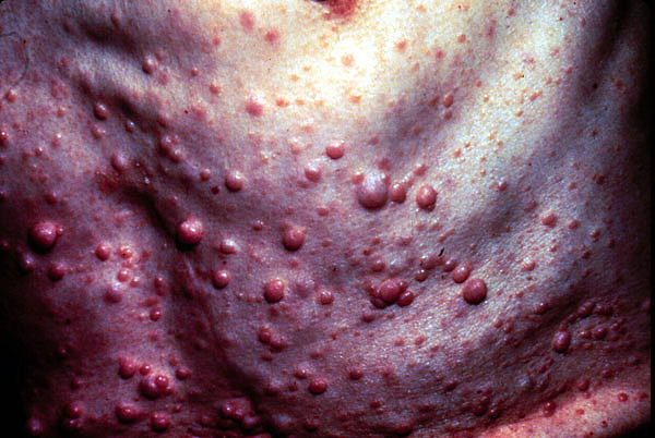 Neurofibromatosis