Ed Friedlander, M.D., Pathologist
scalpel_blade@yahoo.com


Cyberfriends: The help you're looking for is probably here.
Welcome to Ed's Pathology Notes, placed here originally for the convenience of medical students at my school. You need to check the accuracy of any information, from any source, against other credible sources. I cannot diagnose or treat over the web, I cannot comment on the health care you have already received, and these notes cannot substitute for your own doctor's care. I am good at helping people find resources and answers. If you need me, send me an E-mail at scalpel_blade@yahoo.com Your confidentiality is completely respected.
 DoctorGeorge.com is a larger, full-time service.
There is also a fee site at myphysicians.com,
and another at www.afraidtoask.com.
DoctorGeorge.com is a larger, full-time service.
There is also a fee site at myphysicians.com,
and another at www.afraidtoask.com.
Translate this page automatically
 |
With one of four large boxes of "Pathguy" replies. |
 I'm still doing my best to answer
everybody.
Sometimes I get backlogged,
sometimes my E-mail crashes, and sometimes my
literature search software crashes. If you've not heard
from me in a week, post me again. I send my most
challenging questions to the medical student pathology
interest group, minus the name, but with your E-mail
where you can receive a reply.
I'm still doing my best to answer
everybody.
Sometimes I get backlogged,
sometimes my E-mail crashes, and sometimes my
literature search software crashes. If you've not heard
from me in a week, post me again. I send my most
challenging questions to the medical student pathology
interest group, minus the name, but with your E-mail
where you can receive a reply.
Numbers in {curly braces} are from the magnificent Slice of Life videodisk. No medical student should be without access to this wonderful resource. Someday you may be able to access these pictures directly from this page.
Also:
Medmark Pathology -- massive listing of pathology sites
Freely have you received, freely give. -- Matthew 10:8. My
site receives an enormous amount of traffic, and I'm
handling about 200 requests for information weekly, all
as a public service.
Pathology's modern founder,
Rudolf
Virchow M.D., left a legacy
of realism and social conscience for the discipline. I am
a mainstream Christian, a man of science, and a proponent of
common sense and common kindness. I am an outspoken enemy
of all the make-believe and bunk that interfere with
peoples' health, reasonable freedom, and happiness. I
talk and write straight, and without apology.
Throughout these notes, I am speaking only
for myself, and not for any employer, organization,
or associate.
Special thanks to my friend and colleague,
Charles Wheeler M.D.,
pathologist and former Kansas City mayor. Thanks also
to the real Patch
Adams M.D., who wrote me encouragement when we were both
beginning our unusual medical careers.
If you're a private individual who's
enjoyed this site, and want to say, "Thank you, Ed!", then
what I'd like best is a contribution to the Episcopalian home for
abandoned, neglected, and abused kids in Nevada:
My home page
Especially if you're looking for
information on a disease with a name
that you know, here are a couple of
great places for you to go right now
and use Medline, which will
allow you to find every relevant
current scientific publication.
You owe it to yourself to learn to
use this invaluable internet resource.
Not only will you find some information
immediately, but you'll have references
to journal articles that you can obtain
by interlibrary loan, plus the names of
the world's foremost experts and their
institutions.
Alternative (complementary) medicine has made real progress since my
generally-unfavorable 1983 review linked below. If you are
interested in complementary medicine, then I would urge you
to visit my new
Alternative Medicine page.
If you are looking for something on complementary
medicine, please go first to
the American
Association of Naturopathic Physicians.
And for your enjoyment... here are some of my old pathology
exams
for medical school undergraduates.
I cannot examine every claim that my correspondents
share with me. Sometimes the independent thinkers
prove to be correct, and paradigms shift as a result.
You also know that extraordinary claims require
extraordinary evidence. When a discovery proves to
square with the observable world, scientists make
reputations by confirming it, and corporations
are soon making profits from it. When a
decades-old claim by a "persecuted genius"
finds no acceptance from mainstream science,
it probably failed some basic experimental tests designed
to eliminate self-deception. If you ask me about
something like this, I will simply invite you to
do some tests yourself, perhaps as a high-school
science project. Who knows? Perhaps
it'll be you who makes the next great discovery!
Our world is full of people who have found peace, fulfillment, and friendship
by suspending their own reasoning and
simply accepting a single authority that seems wise and good.
I've learned that they leave the movements when, and only when, they
discover they have been maliciously deceived.
In the meantime, nothing that I can say or do will
convince such people that I am a decent human being. I no longer
answer my crank mail.
This site is my hobby, and I presently have no sponsor.
This page was last updated February 6, 2006.
During the ten years my site has been online, it's proved to be
one of the most popular of all internet sites for undergraduate
physician and allied-health education. It is so well-known
that I'm not worried about borrowers.
I never refuse requests from colleagues for permission to
adapt or duplicate it for their own courses... and many do.
So, fellow-teachers,
help yourselves. Don't sell it for a profit, don't use it for a bad purpose,
and at some time in your course, mention me as author and KCUMB as my institution. Drop me a note about
your successes. And special
thanks to everyone who's helped and encouraged me, and especially the
people at KCUMB
for making it possible, and my teaching assistants over the years.
Whatever you're looking for on the web, I hope you find it,
here or elsewhere. Health and friendship!
QUIZBANK Skin (all)
LEARN FIRST
Meningococcemia?
Rocky Mountain spotted fever?
Staph toxic shock?
Strep toxic shock?
Drug rash?
Syphilis?
INTRODUCTION
Can the leopard change his spots?
-- Jeremiah 13:23
We're all of us sentenced to solitary confinement inside our own skins, for life!
-- Tennessee Williams
There was a Christian Scientist named Neil,
-- Anonymous
If it's wet, dry it.
-- Intern's joke about dermatology
{11815} skin model
The skin is our bulkiest organ, and our most vulnerable. It protects us, controls our inner temperature,
and helps the world know whether we're male or female and whether we've reached puberty.
We assume you know the layers of the epidermis (basal, spiny, granule, keratin) and dermis (papillary, reticular). A few review points about
the skin:
Melanocytes occur singly among the cells of the basal layer of the epidermis. These cells have many
branches (i.e., they are "dendritic"), and inject melanin into basal cells and other nearby cells of the
epithelium. We usually spot them by their relatively pale cytoplasm, but you can't really reliably
distinguish every melanocyte on H&E. Melanocytes crawl up to the epidermis from the nerves, and
continually are being replenished, probably from the peripheral nerve cells (really; Arch. Path. Lab. Med. 115:
115, 1991). They stain with S-100 antibody.
Langerhans cells are dendritic macrophages (i.e., antigen presenters, Birbeck granules, T1/CD6+) of the
skin. You can recognize them in the higher layers of the epidermis by their darker nuclei and relatively
clear cytoplasm. They also stain with S-100.
Merkel cells are "of neural crest origin" or at least express some oat-cell-like antigens. They are
probably there for light touch, since they're connected to axons. You can find them in the basal layer.
* Future pathologists: Distinguish them using a neuron-specific enolase stain!
* The newly-discovered "dermal dendrocyte" stains for factor XIII (!).
They're increased in inflammation. No one knows what they do, but the fact
that they make factor XIII suggests they help us consolidate scabs.
See Am. J. Path. 156: 519, 2000.
Skin biopsy site for diagnosis of a rash is best selected where the lesions are new, or at edges (why?)
How to do a punch biopsy: Am. Fam. Phys. 65(6): 1155, March 2002;
choosing your site and technique Postgrad. Med. 103: 179, 1998.
How to do a fusiform excision (skin ellipse): Am. Fam. Phys. 67: 1539, 2003.
There are more known diseases of the skin than any other organ system. This is probably because (1)
you can see the skin; (2) you can examine and biopsy the skin easily; (3) the skin interfaces more with
the environment. For some reason, most skin diseases get fancy Latin names, while most other
medical terms come from Greek.
To make matters worse, one skin disease is likely to breed another. Just scratch a rash and the
"excoriation" makes it worse. Scratch it for a long time, and you have "neurodermatitis" / "lichen
simplex chronicus", even if the other disease would have been gone.
Scratching as a "nervous habit" even starts the vicious cycle of "neurotic excoriations":
Am. Fam. Phys. 64: 1981, 2001.
Just pick a seborrheic keratosis,
and you can turn the histology into a ringer for squamous cell carcinoma. Just
introduce the wrong
staphylococci and streptococci into your poison ivy rash or mosquito bite, and you have instant
impetigo.
In this handout, we'll stick to primary diseases of the skin. There are dozens of cutaneous signs of
systemic disease. We've covered most of the important ones already.
Skin disease is seldom life-threatening. Exceptions include pemphigus vulgaris, erythema multiforme major /
toxic epidermal necrolysis / Stevens-Johnson,
aggressive bacterial infections (flesh-eating strep, pseudomonal ecthyma gangrenosum, and of course decubiti), but can be annoying and/or disfiguring. The
stigma attached to "leprosy" is notorious.
By contrast, individual owners may either like or loathe their benign nevi, birthmarks (congenital nevi
/ hemangiomas), chronic urticaria (a red-headed physician friend of mine loves his "spots"),
dermatofibromas, freckles, lentigines, male hair loss, male hirsutism, tinea versicolor, vitiligo, warts,
mild acne vulgaris, acne scars ("tough guy", Tommy Lee Jones,
Charlie Sheen, Morgan Freeman, Richard Burton, Steven Seagal, Nicholas Cage,
Sean Connery, Jet Li, others), or other scars.
Happy patients give them pet names; unhappy patients
resort to bizarre measures to remove them, etc., etc.
It's important to let patients know that non-contagious skin diseases aren't contagious, and
non-malignant skin diseases aren't malignant.
Skin words:
Macule: Spot. A flat lesion you can't feel.
Papule: A solid bump <10 mm.
Nodule: A solid bump >10 mm.
Plaque: Something that is >10 mm but either completely flat, or a plateau.
Spongiosis: Edema fluid between the cells of the epidermis (i.e., the epidermis
is inflamed). Look for separation of the epidermal cells
and tiny, fluid-filled cavities.
Vesicle: A fluid-filled cavity in or under the epidermis, <10 mm. (Often the cause is marked
spongiosis).
Blister / bulla: A fluid-filled cavity in or under the epidermis, >10 mm. (Except for friction blisters,
usually the cause is some immune business destroying cells within, or adhesion of, cells of the
epidermis.) (Blood blisters under thick keratin require no description.)
Lichen: Something has made the little lines of the skin more prominent.
Vulgaris: "Commonplace"
Acanthosis: Thickening of the spiny layer of the skin, i.e.,
the epidermis in the lesion goes deeper and/or is taller
than outside the lesion.
Papillomatosis: The dermal papillae in the lesion stick up
higher than those outside the lesion.
Hyperkeratosis: Thickening of the keratin layer of the skin. You'll see a silvery scale.
Parakeratosis: Retention of nuclei in the keratin layer of the skin.
* Retaining nuclei is OK on mucosal
surfaces, but usually not in the skin.
Parakeratotic cells might be maturing
and shedding too fast, or have their chemistry scrambled for some
other reason. Nobody really understands this. Orthokeratosis: Extra keratin without nuclei.
Acantholysis: Breaking-up of the connections between cells of the spiny layer (i.e., something's
happening to the desmosomes)
Ichthyosis: The keratin fails to shed from the skin as it should. A feature of two very
common non-diseases, several serious hereditary diseases,
and a few acquired ones.
Exocytosis: Inflammatory cells entering the epidermis.
Ectodermal dysplasia: Any of several hereditary disorders with abnormal
teeth, hair, and (often)
absent
sweat glands. Don't miss this one, especially in a sick baby.
Violaceous: Means "purple" when applied to a lesion. Usually this
means there's a lot of lymphocytes in the upper dermis.
Poikiloderma: Variably-colored skin; some hyperpigmented, some hypopigmented,
and usually with telangiectasias also.
Fissure: A crack that is very narrow and runs deep. Ulcers can hurt,
fissures usually hurt.
Erosion: Only the epidermis is gone. Erosions seldom hurt, though they
may be a bit uncomfortable.
Miliaria: The sweat duct got plugged, maybe because sweat made the keratin around its outlet swell up,
and a little drop of fluid is visible in the epidermis, or maybe the duct even popped. "Prickly heat" /
"heat rash".
{12184} miliaria
SUNLIGHT AND THE SKIN (Postgrad. Med. 89(8): 59, June 1991; Am. Fam.
Phys. 50: 32, 1994)
Ultraviolet light:
Long-wave (UVA) 320-400 nm "Black light"; tanning beds, gives the best day-glow colors; 320- 340 may burn you; some carcinogenic
effect
Medium-wave (UVB) 290-320 nm Sunburn spectrum, vitamin D activation, stronger carcinogenic effect, photoaging (* best wavelength
for burning is 297)
Short-wave (UVC) 200-290 nm Damages nucleic acid more severely than UVB; peak nucleic acid absorption and damage is 260 nm
(* "germicidal UV is 254); not much of this in sunlight.
Melanin absorbs all of these wavelengths, and a suntan should help, also.
Empirically, melanin and suntan protect from sunburn, but you must be very dark all the time
to expect much protection from skin cancer, and the worst photoaging is seen in white folks who've had
suntans most of their lives.
Nobody really knows the chemistry of sun damage (ultraviolet, "actinic" damage) to the skin, but it's
impressive. The lighter your skin, the more ultraviolet exposure you get from an afternoon at the beach.
If you biopsy your acute sunburn, you'll see individual cell apoptosis in the deeper epidermis ("sunburn
cells"; their DNA was injured and their p53 has directed apoptosis), as well as edema in the upper
dermis (the red, of course, is hyperemia).
In diseases in which there is defective DNA repair (i.e., the xeroderma pigmentosa family), patients
suffer photomutilation and many cancers of the skin.
In diseases in which sunlight falling on abnormal porphyrins
results in extra free radical generation (i.e., porphyria cutanea tarda and
its more vicious kin), the skin is heavily damaged by sunlight, though tumorigenesis isn't a prominent
feature. (This makes sense -- the porphyrins in a cell are in the mitochondria and
cytoplasm, while the DNA is in the nucleus.)
Healthy people who are exposed to lots of sunlight (tanning beds, etc., ) without good sun-screens are
also asking for skin cancers. These include the four major lesions: actinic ("solar") keratosis (carcinoma
in situ of the epidermis), squamous cell carcinoma (cancer proper of the epidermis), basal cell carcinoma
(actually cancer of hair), and malignant melanoma ("black mole" cancer of melanocytes). Sun-induced
skin changes and tumors: Prim. Care. Clin. Off. Pract. 27:
435, 2000.
Fortunately, the "healthy tan" is becoming unfashionable as the public learns more about the dangers
of sunning. This is a not-so-rare tribute to the intelligence of the U.S. public, or at least the ease with which
we become frightened (in this case, appropriately; one wonders whether the public
is more afraid of cancers or wrinkles). Teens and tanning beds: Arch. Ped. Adol. Med. 157:
854, 2003; Texans and tanning beds: South. Med. J. 96: 652, 2003.
Likewise, the Montreal and ensuing protocols
for the control of chlorofluorocarbons
that deplete the ozone layer is a rare triumph of
good science and good public policy over economic greed. Thankfully, the world
recognized the need, saving the lives of many people, especially poor
folks who work outdoors. Since skin cancer has a long latency, the benefits
won't appear for a long time. This is one that
the environmentalists got right, and because the risk is real and the cost
is reasonable, the politicians went along.
Remember that tanning doesn't protect you enough from the really bad carcinogenic rays, though it offers fair
protection from sunburn.
AGING
The epidermis thins (thin epidermis becomes shiny and wrinkles), the dermis thins (thin dermis looks and feels
thin), and the skin loses its elasticity (and this helps the wrinkling process).
A variety of minor pigmented lesions (large lentigines, cherry angiomas, and seborrheic keratosis) often
crop up.
* Chemistry of aging and photoaging: Arch. Derm. 138: 1462, 2002.
{25015} senile atrophy of the skin
ICHTHYOSIS
The X-linked recessive form is caused by lack of steroid sulfatase ("arylsulfatase
C"), which is responsible for the breakdown of the cholesteryl sulfate glue
that helps hold skin cells together.
* If for some reason you want to distinguish these two entities,
the autosomal dominant form spares the flexural creases, while the
recessive form involves them. We can tell on biopsy too.
The more severe lamellar ichthyosis is a mix of genetic diseases,
including people with defective K1 or K10 keratins,
and people with defective transglutaminases (Proc. Nat. Acad. Sci.
95: 687, 1998. The skin is red, scaly,
and blistery.
VITILIGO
A process, probably autoimmune in most cases, in which melanocytes disappear over
sharply-circumscribed patches.
The patient develops white, non-tanning, geographic spots.
Some vitiligo is familial. Occasionally, vitiligo heralds the appearance of malignant melanoma
elsewhere in the body; in this case, we assume that the immune system has become angry with all
melanocytes and chooses various battlefields.
More often, vitiligo runs with some other autoimmune disease, notably autoimmune addisonism and/or
autoimmune atrophic gastritis.
Most often, however, vitiligo occurs alone.
The process tends to spread, and it will tend to pop up at sites of injury (the "Koebner phenomenon",
more famous in psoriatic patients).
Vitiligo affects 1% of humankind. Many patients hate it (respect this), but more than a few enjoy it.
In light-skinned folks, vitiligo may only be visible when the person tans, or is examined under
ultraviolet light ("Woods lamp"; keep it handy, too, to spot ash-leaf spots and tinea versicolor). In
dark-skinned folks, the disease self-cures when the entire body is affected.
{12183} vitiligo
* Future physicians: Some patients who have residual melanocytes in the skin benefit from "PUVA"
(psoralen plus ultraviolet) therapy. (I'd refuse this for vitiligo.) There are reports of good repigmentation
using topical tacrolimus (Arch. Derm. 139: 581, 2003); this is now robust
(Br. J. Derm. 153: 498, 2005).
* There are several hereditary and acquired diseases featuring localized loss of melanocytes, from white
patches in the hair (which may mean anything from nothing to Waardenburg's, a homeobox mutation)
to the curious "piebaldism", a tyrosine kinase mutation syndrome (KIT proto-oncogene, codes for a
mast cell receptor) in humans and animals (Am. J. Hum. Genet. 51: 1058, 1992). Don't confuse any
of these with albinism, in which the melanocytes are present but never worked properly.
* Quite a few dark-completed folks get idiopathic guttate hypomelanosis
on the lower legs and/or elsewhere as they get older; the etiology is unknown.
The melanocytes in this kind of lesion lack good dendrites.
FRECKLES ("ephelides", singular is "ephelis")
Increase in pigment production by a local group of melanocytes. A freckle can be up to 1 cm. Freckles
typically occur in light-skinned (especially red-headed) kids, especially after sun exposure.
Contrary to "Big Robbins" and most other texts, a freckle (in adult or child) will indeed also contain
more melanocytes than surrounding skin. What's more, freckles are likely to show some cytologic
atypia and even stain with HMB-45 (the "melanoma antigen", actually a marker for any extra-turned-on
melanocytes). See Cancer 67: 1990, 1991. (* I hope this never makes the "Kansas City Star"; there's
enough cancer paranoia already.)
Not surprisingly, the melanocortin-1 receptor, which is the major human red-hair locus,
is also the major freckle locus (Hum. Mol. Genet. 10: 1702, 2001).
{12191} ephelis
MELASMA ("raccoon eyes", "mask of pregnancy")
Increased pigmentation of the periorbital face. Seen in patients who are pregnant, taking the oral
contraceptive pill, taking phenytoin, or "idiopathic".
* If it bothers you, there's a phenol thioester depigmenting agent (Arch. Derm. 127: 1528,
1991). This will work if, and only if, the pigment is primarily in the epidermis
rather than in the macrophages.
LENTIGINES (singular is "lentigo")
A local hyperplasia of melanocytes, with hyperpigmentation, and generally some elongation of the rete
pegs as well. Unlike freckles, they do not darken in the sunlight.
You start to acquire single lentigines as a kid (your smaller, flatter "moles" are lentigines).
As you grow older, crops of larger lentigines ("senile lentigos", "liver spots"; of course, they have
nothing to do with your liver) may erupt, especially on the backs of your hands.
* "Advertising is intended
to promote unhappiness." In the 1970's, the "Gray Panthers" (militant older folks) made a public
laughing-stock out of a skin-bleach commercial designed to cause the elderly to hate their lentigines
("Those horrible spots!....")
* Dermatosis papulosa nigra is the only name I've ever
been able to find for the multiple black facial spots
seen on many people of African ancestry (Sidney Poitier and
Bill Cosby have them), and occasionally on East Asian folks as well. They are a non-problem.
Café au lait spots feature melanocytes with
giant melanosomes. Despite their
usefulness in confirming your clinical suspicions of neurofibromatosis
or McCune-Albright's, several percent of normal people have at least one little one.
{09645} lentigo city! ("old age spots")
NEVOCELLULAR NEVI ("pigmented nevi", "melanocytic nevi", "moles";
single is "nevus")
Fascinating little lesions, somewhere in the borderland between hamartomas and tumors, in which
masses of "nevus cells" (chemically similar to melanocytes, and sometimes making melanin) pop up at
the dermal-epidermal junction and/or the upper dermis and/or elsewhere.
Melanocytic nevus: The common, garden-variety mole (i.e., there's more to it than a lentigo.)
Most people start getting theirs as young kids, and have most of them by puberty. Apparently
everybody has them; they're airbrushed out of certain photos. As we age, they tend to involute.
Nobody really knows what triggers a mole to form, but Nowell's law seems to be operating. The "West
Midlands Mole Study" found more moles in whites than in non-whites, more in boys than girls, more
in kids that burn rather than tan, more in frecklers, and an impressive correlation between number of
moles and number of holidays in hot climates, independent of sunburn history (Arch. Derm. 128: 1201,
1992). The Vancouver study found pretty much the same thing (Arch. Derm. 126: 466 & 770, 1990).
* A current mystery of medicine is why children given chemotherapy for leukemia end up with excess
melanocytic nevi, and whether these will turn to melanomas (Br. Med. J. 305: 799, 1992;
Cancer 77: 1402, 1996).
Junctional nevi feature clumps ("nests", "* theques")
of nevus cells along the dermal-epidermal junction ("junctional
activity"). Intradermal nevi stay distinctly below the basement membrane, while compound nevi feature
both patterns.
{09636} melanocytic nevus, gross
A nevus spilus is a tight cluster of benign hyperpigmented spots, often on an oval,
slightly hyperpigmented background. These are very common.
{09650} nevus spilus
Becker's nevus is a big, dark, hairy patch that pops up on a man's
upper back or shoulder (occasionally elsewhere) just after
puberty. Common, and sometimes hereditary. Keep an eye on it (small melanoma risk,
just remove 'em in a timely way before they get dangerous -- Dermatologica
182: 77, 1991), and reassure the owner that a Becker's nevus is very macho and studly.
* Sometimes the affected region also features hypoplasia
of arm and/or breast, scoliosis, lots of little skin
leiomyomas, and/or spina bifida occulta
(Am. J. Med. Genet. 68: 357, 1997). This is only a small
minority of Becker's people, but is called "Becker's nevus syndrome."
{09659} Becker's nevus
* Ota's nevus occurs around and sometimes on the eye.
Blue nevi are masses of darkly-pigmented, dendritic nevus-cells in the deep dermis.
The "blue" appearance (trust a dermatologist) results from viewing the pigment through dermis, just as
blood appears "blue" in our veins.
The vast majority of blue nevi are benign.
* Future pathologists: Big ("cellular") ones can look nasty, but
lack the anaplasia, mitoses, necrosis, and big nucleoli of a melanoma.
* The tumor recapitulates the pigmented cells of reptiles.
{24902} blue nevus, trust me, this was deep-deep in the dermis.
Halo nevi are melanocytic nevi that are under attack by the immune system (Arch. Derm. 130: 1036,
1994). Around the nevus, you'll see local vitiligo, as the bystander melanocytes get chomped, too. Halo
nevi often (not always) vanish after a while.
{09639} halo nevi, gross
Congenital nevi (1 kid in 4000) are often very large and impressive (J. Ped. 120 906, 1992).
Something in the spinal nerves causes this change, since these nevi follow dermatomes, and if you
remove them and graft new skin, the nevus recurs.
"Malignant melanomas" often arise here. These "melanomas" are usually fairly tame, though they can
kill; if you examine the cells by research techniques, many "malignant looking" melanomas prove to
be benign (Am. J. Path. 136: 817, 1990).
{12204} giant congenital pigmented nevus
Spitz nevi are bizarre, epithelioid-and-spindle-cell nevi seen in kids. They look clinically and (to the
inept pathologist) like malignant melanoma.
* Future pathologists! Confirm the diagnosis of Spitz nevus by
its symmetry, its sharp demarcation, the way the cells
look more benign deeper in the lesion, the fact that the spindle
cells tend to be oriented perpendicularly, and the fact that the
epithelioid cells tend to have a ground glass cytoplasm. There are a
host of subtypes including a malignant one.
* More than you ever wanted to know about a Spitz nevus:
Dermatology 194: 20, 1997; Cutis 58: 35, 1996.
* Help: Kids without big-time predisposing anatomic or genetic
birth defects almost never get malignant melanomas.
{24903} hairy nevus, congenital
DYSPLASTIC NEVI ("BK moles", etc.; South. Med. J. 85: 870, 1992)
"Premalignant moles." The dysplastic nevus tends to turn into a (usually superficial-spreading) melanoma,
and a person who has had a dysplastic nevus tends to develop malignant melanomas de novo as well.
Read all about it in Arch. Derm. 127: 995, 1991; Arch. Derm. 130: 993, 1994.
The typical dysplastic nevus is >4 mm across, flat or slightly raised (often areas
of both), with a variegated (multicolored)
appearance, and irregular borders. Look at the lesion when lit edgewise, and if the normal skin lines
are distorted, that's another reason to worry (Cancer 67: 3157, 1991; nice review of the whole entity).
They can occur on both sun-exposed and non-sun-exposed skin.
The typical dysplastic nevus patient suffers from a mutation in one of the melanoma genes.
The reported gene on chromosome 1 (originally called CMM1) remains unidentified.
Another gene on chromosome 16 is CDKN2A, a cycle regulator antioncogene.
See Cancer 86(S11):2464, 1999; Cancer 94: 84, 2002. Less common
is an inherited oncogene, mutant CDK4 (Cancer 94: 3192, 2002).
These patients need to have either the dermatologist or their partner keep tabs on all their nevi, and have
the nasty-looking ones off. This is excellent protection against dying of melanoma (Cancer 63: 386,
1989). Nobody's more than 50% accurate in telling which pigmented lesion is going to turn out to be
nasty (Br. Med. J. 299: 16, 1989), and some will fool everybody (JAMA 266: 3463, 1991).
Leave the histopathology of dysplastic nevi to pathologists. We look for atypical cells singly and/or in
clumps at the dermal-epidermal junction, doing
various things a normal junctional nevus won't. Another tip is elongation and fusion
of the rete pegs. Yet another is layered fibrosis in the upper dermis near the lesion.
Some "dysplastic nevi" are much nastier than others histologically.
Nowadays, you should expect your pathologist to tell you, "Should we wide-excise
this particular dysplastic nevus, or merely tell the patient that he/she has had a dysplastic
nevus?"
{12196} dysplastic nevus
MALIGNANT MELANOMA ("malignant mole", "black mole cancer"): West. J. Med. 162: 536, 1995;
Am. Fam. Phys. 63: 1359, 2001; Surg. Clin. N.A. 83: 77, 2003; melanoma atlas
Cancer 94: 3192, 2002; Lancet 365: 687, 2005
Future rural primary care physicians! Save lives at little cost by setting up a screen-you-for-skin-cancer
booth! J. Fam. Pract. 40: 471, 1995.
Cancer of melanocytes / nevus cells. The known independent risk factors are
(1) light complexion / freckler;
(2) sun exposure (that long-ago bad sunburn as a kid or teen is the greatest
risk);
(3) dysplastic nevus syndrome (all other things being equal, your risk is 7x the next person's: Arch.
Derm 127: 995, 1991);
(4) total number of benign pigmented nevi >2 mm diameter (5-10 mm, etc., rules vary; see Cancer 66:
387, 1990);
(5) xeroderma pigmentosum, etc.;
(6) having ever had a really bad (blistering) sunburn.
* (7) a few other familial syndromes, notably CDKN2 antioncogene deletion syndrome (NEJM 333: 970
& 975, 1995).
* See also Arch. Derm. 130: 1002, 1994; West. J. Med. 160: 343, 1994. Calculate your personal
melanoma risk: Lancet 2: 487, 1989.
The frequency of melanomas has increased 500% since the beginning of
the 20th century, because people suntan rather than wear cover-up clothes.
(In 1900 it was considered grossly indecent for a man to swim bare-chested
at the beach.)
* A silly flap about sunscreens causing malignant melanoma
turned out to be unfounded (Am. J. Pub. Health 92: 1173, 2002); in fact,
no relationship was found (i.e., few people use sunscreen faithfully, especially
not small children).
* Melanoma is notorious for multiple primaries, especially (but not only)
in melanoma families (JAMA 294: 1647, 2005).
The genetics of melanoma transformation is now being worked out (Nat. Rev. Cancer 3:
559, 2003). We have already mentioned the melanoma family genes.
A major player is the tyrosine kinase BRAF, especially in non-sunlight-related
cancers; it may be a pharmacologic target like bcr/abl soon (Nature 417: 949, 2002;
Lancet 363: 728, 2004).
You'll need to spot malignant melanomas. Look for irregular borders and variegated pigmentation
(Nowell's law of genetic instability; remember that any little black dot in a brown nevus can be
melanoma: Arch. Derm. 130: 1013, 1994). Of course, rapid growth and bleeding are signs, too.
Diagnosis and treatment of melanoma: Br. Med. J. 310: 492, 1995; Hosp. Pract. 29(6): 37, June 15,
1994, and 29(7): 29, July 15, 1994 (great color pictures, look at them); Am. Fam. Phys. 49: 91, 1994
(more nice pictures).
* By the time you are in your primary practice, it will probably be routine
to send digital images of worrisome pigmented lesions to your favorite
three dermatologists for their immediate recomendations about biopsy/excision
(Arch. Derm. 140: 525, 2004).
The tumor cells are usually polyhedral but may be spindle- shaped, ballooned, or dendritic, or look like
oat cells, hemangiopericytoma, signet rings (Am. J. Clin. Path. 93: 731, 1990). Many melanomas, but
by no means all, make melanin.
Telling melanomas from benign pigmented skin lesions
under the microscope is a task best left
to pathologists, unless the diagnosis is obvious. None of these are absolutely
diagnositc (J. Clin. Path. 58: 409, 2005), but we look for:
Big nucleoli are usual; future pathologists can stain with S100 and/or HMB-45
(melanoma antigen, pretty specific in skin though not elsewhere: Am. J. Ophth. 109: 696 1990) or
S-100 immunoperoxidase stains. Melanomas are usually keratin negative and vimentin positive.
* Future pathologists: Many melanomas show signs of regression,
with areas of intense inflammmation and areas of fibrosis without tumor.
These probably correspond respectively to the red and the white areas that are often seen in melanoma.
Immunostaining reveals the types of lymphocytes involved: Am. J. Clin. Path. 124:
37, 2005.
* Future pathologists: "Nuclear pseudoinclusions" are a tipoff
for melanoma, and you may perform DOPA stain for tyrosinase
on fresh-frozen sections of melanoma (turns them brown).
* The molecular probe Mitf (microphthalmia transcription factor, stains nuclei) is presently being
claimed as sensitive and specific for melanoma, in sections and smears
(Cancer 93: 337, 2001).
Most melanomas stay confined for a while to the epidermis and uppermost dermis ("in situ",
"radial growth phase"). Later (Nowell's law), they begin to invade deeper ("vertical growth phase").
Prognosis mostly depends on depth; the more lymphocytes seen "fighting" the tumor, the better the
prognosis.
Pathologists still distinguish different types of melanomas based on their appearance in the radial
growth phase:
Lentigo maligna ("Hutchinson's freckle") is a
lentigo composed of atypical melanocytes. The epidermis is
not much thickened. Stays in situ
for years.
* An alternative to removing a large portion of the
patient's face may be topical imiquimod,
the immunostimulant (Arch. Derm. 141: 510, 2005).
Superficial spreading melanoma is a junctional nevus-like lesion composed of atypical melanocytes.
Stays in situ only for months.
Acral-lentigenous melanoma is a variant that usually occurs on the palms, soles, or black-white junction
of a darkly-pigmented person's hands and feet. The epidermis is thick
and there is much atypia. Treacherous.
Nodular melanoma begins its vertical growth phase without an obvious in situ phase.
* The old "progression" of malignant melanoma, described in "Big Robbins":
1: A normal-appearing junctional nevus
2: "Lentigenous hyperplasia", i.e., extra melanocytes as singles between the junctional clumps
3: Atypical melanocytes
4: Cells entering the superficial dermis
5: Cells going down deeper into the dermis. This can now metastasize. "We have now begun the
vertical growth phase."
6: In the fat or lymphatics or has metastasized.
* The older "Clark's levels" for a melanoma (surprisingly, not listed in "Big Robbins"):
Level I: In the epidermis and junction only (radial growth phase)
100% disease-free in 5 years
Level II: In the papillary dermis, but does not fill it, and does not reach reticular dermis
90% disease-free in 5 years
Level III: Fills the papillary dermis, but does not actually invade reticular dermis
70% disease-free in 5 years
Level IV: Invades reticular dermis.
40% disease-free in 5 years
Level V: In the subcutaneous tissue.
25% disease-free in 5 years
The newer Breslow's system prognosticates a melanoma based on its thickness. Under 0.76 mm is
unlikely to have metastasized, etc. For more of this stuff, see Cancer 79: 2324, 1994;
it is superior to Clark's level for prognosticating, though both are good (Cancer 91: 983, 2001).
* An independent, additional histologic factor conferring
a bad prognosis is vascular invasion
(tumor cells just under the endothelium is bad,
tumor cells actually in lumens is worse; Arch. Derm. 137: 1169, 2001).
* A new claim is that expression of melanoma cell adhesion molecule (MCAM)
on a melanoma is a more reliable indicator of bad prognosis than any
other: Plast. Recon. Surg. 115: 367, 2005.
* Future pathologists: Little benign nevi are common on the
capsules of lymph nodes. Don't call these melanoma. Treacherous.
{09683} melanoma, gross
Clinically, malignant melanoma is notoriously capricious. The majority are now cured surgically, but
if the lesion is deep, there's the possibility of late appearance of metastases.
People under proper surveillance for melanoma are very unlikely to die of melanoma (Ann. Surg. 213:
308, 1991).
As we've seen, melanoma is the only common cancer that often expresses a "cancer-only" antigen.
Probably this is why the immune system sometimes seems to cure this cancer (Cancer 69: 1377, 1992),
and why immunotherapy is now recording a few successes (Blood 83: 56, 1994).
SEBORRHEIC KERATOSIS (* "basal cell papillomas", * "barnacles")
A badly-named local hyperplasia of the basal cells. Typically the lesions produce small masses of
keratin ("horn cysts") that resemble the pearls of squamous cell carcinoma but show no malignant
features.
The lesions usually are pigmented (remember basal cells take up melanin), usually are crusty (i.e., horn
cysts), and can be scraped off with a butter knife (only to grow back).
Microscopically, look for a flat-bottomed hyperplastic mass of benign basal cells,
with the "horn cysts" or at lesat a lot of loose keratin.
Especially if the patient picks at them ("irritated SK"), the "keratin pearls" may fool you
into thinking this is a squamous or basosquamous carcinoma.
Future primary care physicians: If many seborrheic keratoses erupt at the same time, it could be "the sign of
Leser-Trelat", herald of a visceral adenocarcinoma. Don't miss this one.
* Not standard yet, but for annoying "SK"'s, think about a trial of 1,25-dihydroxy vitaminD3,
as for psoriasis.
{12502} seborrheic keratosis
ACANTHOSIS NIGRICANS
This is seborrheic keratosis-style hyperplasia covering each armpit and/or the groin. Again, this may
herald a visceral malignancy, or be seen in some insulin-resistance syndromes,
or simply result from administration of nicotinamide.
* Some wag gave the name of "terra firma forme dermatosis" ("dirty keratin")
to the curious brown lesions that form on the backs of the necks of
some children. The pigment cannot be removed with soap, but
comes off easily with rubbing alcohol. Quite common.
{25607} acanthosis nigricans
FIBROEPITHELIAL POLYPS ("acrochordons", "skin tags", "squamous
papillomas", etc., etc.)
Mysterious little bumps of soft fibrous tissue covered with epidermis that may be slightly thick and/or
slightly pigmented.
These pop up most anywhere in older people; the favorite spot is the neck. Savvy patients may be
concerned that their acrochordons are melanomas. The most savvy patients know they aren't, and cut
them off with nail clippers. Doctors tie the base off with a thread.
{12176} acrochordon
Angiokeratomas
are hemangiomas with an overlying hyperkeratotic
epidermis. Some men gets these all over their scrotums
as they grow older; patients with Fabry's disease (Arch. Derm 140: 1440, 2004)
often have many spread all over the body.
{25116} Fabry's
EPITHELIAL CYSTS ("epidermoid inclusion cysts"; "wens"; "sebaceous
cysts")
These familiar bumps are spheres of epithelium turned inside-out. Skin products (sebum, keratin, or
both) accumulate, causing the mass to grow and grow.
Probably the epidermoid and pilar (trichilemmal)
cysts result from bits of epidermis getting pushed deep into a person
(it's a picturesque, rare, but serious complication of lumbar puncture).
* Ron Hubbard, founder of "Scientology", was obviously bothered by his epidermoid cyst toward the
end of his life. One of his prominent followers observed this, and that's how she decided that Hubbard
did not possess miraculous powers
after all. Anecdote from the biography by Hubbard's son.
When one of them ruptures (i.e., you really squeezed it good), there's often an impressive inflammatory
response, with foreign body giant cells prominent after it settles down.
{12170} epidermoid inclusion cyst, shoulder
* Future pathologists only:
Epidermoid inclusion cyst: The epithelium is garden-variety stratified squamous epithelium, and the
junk in the center is layer-upon-layer of keratin.
Pilar / tricholemmal cyst: The epithelium is more like pilosebaceous apparatus, with no granular layer,
and the junk in the center is a mix of keratin and sebum.
Dermoid cyst: Uncommon in the skin; the epithelium really is skin, and the junk in the center is a mix
of keratin, sebum, and real hair. Most often these occur at the lateral canthus of the eye. These may
result from tiny birth defects.
Milia are little epidermoid inclusion cysts that can be multiple.
Some physicians remove them simply by incising with a scalpel blade tip
and expressing the contents with a comedone extractor. (I have used
a scalpel-and-paperclip technique on myself. No anesthesia is required.)
Steatocystoma multiplex: Many sebaceous glands, either in a localized area
or all over the skin. There is an autosomal dominant form (Plast. Recon. Surg. 99:
1142, 1997), and probably the sporadoc localized lesions that you'll see on individuals
result from localized mutations. Surgery is best done by someone experienced
with plastics, and if the patient comes in often, it can be kept under good control.
Pilonidal cyst is a misnomer (should be "pilonidal sinus")
for a deeply-imbedded, chronically infected mass of hair originally trapped
in a little dimple over the sacrum. Most patients are young hairy men. It's very common and very
unpleasant.
* HIAWATHA'S MITTENS
He killed the noble Mudjokivis [beaver],
--Parody of Longfellow
KERATOACANTHOMA
A bizarre, hideous-looking, volcano-shaped, self-curing lesion of epidermis (usually the face or upper
extremities) that erupts over a week or two and produces a large (up to several cm), cone-shaped lesion with
a dense keratin plug in its "crater".
Patients will be scared that they have cancer, but it's not, and there's little or no cytologic atypia. The
good thing about this lesion is that it often goes away by itself in a few weeks or months. Puzzle that out
(virus, betcha)!
It is hard to tell the lesion from a squamous cell carcinoma
histologically. * Older studies indicate that keratoacanthomas stain for filaggrin
while squamous carciomas do not. The antibody is not in common use.
To complicate matters, squamous cell carcinoma is now known to be a frequent
complication, and nowadays dermatopathologists are starting to call these
"squamous cell carcinoma, keratoacanthoma type", indicating a high likelihood
of spontaneous regression.
Your lecturer believes that a lesion that is clinically a keratoacanthoma should
probably come off, because (1) there is a high risk that it's cancer, and (2)
there is usually a nasty scar even if it regresses. Not everybody agrees.
Consider consultation.
{12792} keratoacanthoma
SKIN ADNEXAL TUMORS ("appendage tumors"): A bewildering family of
minor skin tumors.
Cylindroma: A benign tumor of sweat glands, usually on forehead or scalp, often multiple
(* CYLD, Nat. Genet. 25: 160, 2000).
{25595} cylindromas ("turban tumor")
Syringoma: A benign tumor of sweat glands, usually appearing
in groups on and around the lower eyelids
Today, they're typically removed using lasers.
Multiple synchronous "tumors" suggests a viral infection
rather than a true neoplasm, but the familial syndrome suggests
we actually are dealing with neoplasia. Probably a circulating
growth factor awaits discovery.
{10307} syringoma
Trichoepithelioma: A benign tumor that makes little hair follicles.
* There is an anti-oncogene deletion syndrome with trichoepitheliomas and cylindromas. This can be
quite a problem for a plastic surgeon.
{11767} trichoepithelioma
Tricholemmoma: A benign tumor "arising from a different layer of the hair follicle"; a little white hair
may arise from its center
{11806} tricholemmoma
* Cystic sebaceous tumors are diagnostic of Muir-Torre (Arch. Path. Lab. Med. 127: 614, 2003),
an allele at a HNPCC Lynch locus.
Others too numerous and insignificant to merit your attention.
{24888} hidradenoma
ACTINIC KERATOSIS (review Am. Fam. Phys. 49: 817, 1994)
Localized squamous cell carcinoma in situ (or at least "atypia") of the epidermis. Grossly, you'll see
irregular atrophy and hyperkeratosis of the epidermis. Some forget how to shed old keratinocytes
altogether, and become "skin horns".
Independent risk factors include light skin / frecklers, sun exposure, and of course xeroderma
pigmentosum, as well as arsenic exposure ("arsenical keratoses" tend to be on palms and soles).
These lesion tend, if ignored, to turn into invasive squamous cell carcinomas, but only 1 in 1000 will
do this in a year (Cancer 75-S: 245, 1995). You may excise them, kill them with fluorouracil or
injected interferon (Arch. Derm. 128: 1486, 1992; it's been used for invasive squamous cancer, too),
or follow whatever is the current recommended therapy.
{12736} actinic keratosis
Bowen's disease: Hideously anaplastic carcinoma in situ of the epidermis, as a red plaque on
sun-exposed skin, oral mucosa, or genital skin. Althogh it looks terrible,
it does not penetrate the basement membrane.
Again, arsenic exposure is a risk factor.
The risk of invasion is 10% or less.
The old claim
that Bowen's of the skin is marker for a second, internal malignancy never made sense and
is discredited.
SQUAMOUS CELL CARCINOMA (Plast. Recon. Surg. 114: 82e, 2004)
The familiar histologic pattern, this time invading the skin.
There are about 100,000 of these in the United States yearly,
and abou 2500 deaths, almost all due to people ignoring them.
Independent risk factors include light skin / freckler, sun exposure,
radiation therapy,
arsenic exposure (still epidemic in some places), exposure to coal
tar (carcinogenic polycyclic compounds), xeroderma pigmentosum, the edges of chronic draining
osteomyelitis sinuses (why? Nowell's law...), longstanding pilonidal
sinuses, old burn scars (minor mystery called "Marjolin's ulcer"), and immunosuppression
for renal transplants (Lancet 346: 403, 1995.
Some of the lesions in the immunosuppressed
contain KSHV: Lancet 345: 1339, 1995).
Grossly, these are typically exophytic cancers that grow up slowly on sun-exposed skin. Squamous cell
carcinomas of the skin are common but fortunately tend to be well-differentiated and seldom metastasize
unless neglected.
{12735} squamous cell carcinoma
Children with xeroderma pigmentosum
typically develop severe disfigurement and many tumors on exposure to ultraviolet light.
These include both the usual sunlight carcinomas and some sarcomas (Arch. Path. Lab. Med. 115: 910,
1991). Death usually occurs in the teenaged years. Most versions also feature precocious death of brain
cells (Brain 114: 1335, 1991).
{25012} xeroderma pigmentosum
BASAL CELL CARCINOMA
Cancer of small, cuboidal-like cells resembling the normal basal cells of the skin. The real
"histogenesis" of this tumor is probably the hair shaft, since it doesn't occur where hair doesn't grow.
Independent risk factors include light complexion / freckler, sun exposure, xeroderma pigmentosum,
and the "patched" (PTCH)
anti-oncogene deletion syndrome "basal cell nevus" syndrome (also
mutated in sporadic cases, of course: Cancer Res. 57:
2581, 1997).
* Although sun exposure is etiologic, the most common locations are not those
that receive the most sunlight, and some other factors are involved as well: Arch. Derm. 138: 1494, 2002.
Grossly, you'll see the familiar "rodent ulcer" with a "rolled, pearly border". Or you may see a
telangiectatic papule, a small nodule, or a small pigmented area. Dermatologists are good at spotting
these when they are still tiny.
Histologically, you'll see a low-grade cancer, with its invasive masses of cells rimmed by a
picket-fence-like row of basal cells. There are subtypes. Mitotic figures and necrosis are uncommon, and
these tumors almost never metastasize (one did at K.U.: Am. J. Med. Sci. 301: 395, 1991; five neglected
huge ones did in Wisconsin Cancer 73: 328, 1994).
Rule: Most skin tumors on or above the lower eyelid are basal cell carcinomas. Most skin tumors below
the lower eyelid are squamous cell carcinomas.
* Future pathologists: The lymphocytic infiltrate can be heavy,
even simulating lymphoma.
* Future pathologists: Maybe you'll get to take part in
Mohs's microsurgery, where the surgeon takes the area apart one
sliver at a time and you tell each time whether there's cancer on it.
* Future pathologists: The banal "sebaceous nevus" does need to
come off; it's got a bit of malignant potential (Cancer Res. 59:
1834, 1999).
{12168} basal cell carcinoma, gross
MERKEL CELL CARCINOMA (neuroendocrine skin cancer; Cancer 88: 1842, 2000;
Am. J. Clin. Path. 115 S: S 68, 2001) An uncommon, bad skin cancer that (as you would expect) looks just like oat cell
carcinoma of the lung. Tumors show both epithelial and APUD differentiation (like oat cell carcinoma).
Around half of these metastasize, and 20% will kill.
BENIGN CUTANEOUS FIBROUS HISTIOCYTOMA ("dermatofibroma", "sclerosing
hemangioma")
A quasi-tumor of macrophages (?) or fibroblasts (?) in the dermis.
* Whatever the histogenesis, there's always a mix of cells that stain as fibroblasts and cells that stain as
histiocytes; nobody's found (or looked hard for) trademark genetic mutations as in a real cancer.
Dermatofibromas often follows a minor injury, bug bite, or whatever.
Patients find a palpable bump somewhere, with overlying pigment. They may want it off, or just want
reassurance. Trick: Pinch it. If it dimples, it's probably a dermatofibroma (why?). If it doesn't, it's
probably something else.
These lesions often become pigmented with hemosiderin and to become lipid-rich (many are yellow
when cut), and the overlying epidermis tends to hyper-pigment and to thicken a bit. * Future pathologists: Basal cell hyperplasia over a dermatofibroma
often mimics basal cell carcinoma microscopically.
{12203} dermatofibroma
Atypical fibroxanthoma is a dermatofibroma variant seen on sun- exposed skin (tops of the ears is a
favorite site). The histology is floridly malignant; the behavior more benign. Let the pathologists worry
about it. * Dermatofibrosarcoma protuberans is a "low-grade malignant" version of the dermatofibroma, with a
distinctive storiform (cartwheel, pinwheel, i.e., like ovarian stroma) pattern of its spindle cells.
It tends to
invade the underlying fat along the septa. Review, including histological criteria
for nastiness: Cancer 88: 2711, 2000; also Cancer 101:
2503, 2004; J. Clin. Path. 58: 751, 2005.
XANTHOMAS ("xanth-" means "yellow", "-oma" here means "bump")
Masses of triglyceride- and/or cholesterol-laden macrophages.
If you should happen to excise one, the pathologist will see masses of foamy macrophages and often
Touton giant cells (flower cells), with a ringlet of nuclei in the middle of the cell.
"Big Robbins" makes these distinctions, which I've slightly simplified:
Eruptive xanthomas: Pop up abruptly on the butt, knees, and elbows when triglyceride-rich lipoproteins
go out of control.
Tuberous xanthomas: Pop up on the backs of the heels and fingers when cholesterol-rich lipoproteins
go out of control.
* Plane xanthomas: Pop in the lines of the palms in primary biliary cirrhosis.
Xanthelasma: Pop up around the eyes when LDL is up, or for no known reason.
{09741} xanthomas
Pseudoxanthoma elasticum is a recessive disorder with little yellow plaques in skin folds (check armpits
and groin). They are made of elastin, not lipid; under the microscope, they are
often calcified. The biggest problem is angioid streaks in the retina,
which tend to bleed; elastic arteries may also dilate. The gene
is also a coronary risk locus: Circulation 106: 773, 2002.
{05937} pseudoxanthoma elasticum
VASCULAR LESIONS
{12510} cherry angiomas
* Future pathologists: The key to recognizing early Kaposi's is that it's little clusters of little blood vessels
sprouting directly off the big vessels. Tough call; otherwise, it looks like granulation tissue. Remember
African Kaposi's is common in Africa, doesn't mean AIDS, and tends to spread to the lymph nodes.
Virus, you know.
MYCOSIS FUNGOIDES
We've already examined this entity under "White Cells".
* Why are some cases of T-cell lymphoma epidermotrophic? The molecular differences in integrins:
Am. J. Path. 141: 855, 1992.
* HTLV-I and HTLV-II as possible etiologic agents in some cases of mycosis fungoides: Blood 80:
1537, 1992.
* Leave the other skin lymphomas (and pseudolymphomas, among which Lyme
disease is notorious) to pathologists.
{09041} mycosis fungoides
MASTOCYTOSIS
Localized or generalized hyperplasias of mast cells. There are at least five subtypes.
The familiar forms are "urticaria pigmentosa" (multiple pigmented lesions
that swell when rubbed;
Am. J. Clin. Path. 109: 279, 1998)
and the solitary mastocytoma.
("Tickle the baby's spot and he goes into shock!").
Generalized systemic mastocytosis is more ominous, and poorly understood. Symptoms can be due
to excess histamine (what would that do?), GI bleeding or epistaxis (remember mast cells make
heparin), and bone lesions.
* A serum marker for mastocytosis is serum tryptase.
URTICARIA ("hives")
You're already familiar with this lesion when caused by type I immune injury. Vessels dilate and
leak in the dermis, typically because histamine has been released here. Eosinophils are usually numerous.
Cold urticaria patients have mast cells that degranulate too easily in the cold. ("I'm allergic to the
cold!" "I'm allergic to popsicles.") Some people get non-IgE dependent urticaria from sun, exercise,
opiates, prostaglandin inhibitors (why?), or type III immune injury.
Many redheads have dermatographism (i.e., urticaria develops on slight pressure; for some reason
"Big Robbins" discusses this very common normal variant under "mastocytosis") or chronic
urticaria (i.e., all the time, for no reason). In dermatographism, the "hives"
are likely to be linear.
Some non-redheads develop chronic urticaria. Usually you'll
never find the cause, but you can try an elimination diet for starters.
Autoantibodies against IgE receptors account for maybe half of cases.
We await easy tests for autoantibodies against mast cells and their surface
components.
{09716} urticaria
Angioedema is urticaria that extends down into the subcutaneous fat. You remember that if you
lack C1 esterase inhibitor, you'll suffer episodes of angioedema.
THE ECZEMA FAMILY (Postgrad. Med. 89(8): 75, June 1991)
Acute-type inflammation of the epidermis. The many members of this family all present as red
(hyperemia in the dermis), oozy (i.e., the epidermis is gone in places), crusty (i.e., some protein from
spongiosis fluid has dried on the surface) papules (i.e., there's no extra tissue) and vesicles (i.e., there
is spongiosis/edema separating some of the epidermal cells).
With time, the lesions stop oozing and instead become elevated and scaly (i.e., protective
hyperplasia, with acanthosis and hyperkeratosis). At this stage, the more you scratch, the worse
your lesion gets ("lichen simplex chronicus", etc.)
{12298} lichen simplex chronicus
Contact dermatitis
Most often this results from type IV immune injury, the injurious agent is a hapten, and of course the
presenting cell is the Langerhans cell. It doesn't help if there's also exposure to some irritant.
Contact dermatitis itches bad and looks bad. Worldly-wise dermatologists can tell the
offending agent by the location on the patient. Ask one about "patch tests" to pinpoint the bad
chemical.
Atopic dermatitis (Br. Med. J. 310: 843, 1995)
Eczema, typically in the flexural creases of elbows and knees, typically in kids, typically caused by
is widely believed to be
food allergy (nobody knows how; ask about personal or family history of hay fever, food allergy, or
asthma).
The best treatment is supposed to be an elimination diet, but very
few people are going to comply with this. Topical
glucocorticoids keep most cases under control, but if the disease is extensive, they can be absorbed and make the
child sick.
* Staph superantigen toxins seem to make this worse, but only about half
of children are infected. Stay tuned: J. Allerg. Clin. Imm. 105: 814, 2000.
Watch for topical therapy with macrolactams (tacrolimus family) in the near future
(JAMA 285: 724, 2001).
Phytodermatitis ("fruit mask", "Club Med" dermatitis)
results from the photosensitizing
effects of psoralens (furocoumarins) from plants (limes, lemons and celery are
notorious) on the skin.
Drug rashes
Biopsy a common drug rash and it may turn out to be:
{08161} "eczema"
ERYTHEMA MULTIFORME ("EM"; Am. Fam. Phys. 46: 1171, 1992; Postgrad. Med. 107: 87, 2000)
A very important, generally self-limited problem (especially if the cause can be eliminated). Not
everyone will agree with "Big Robbins'" statement that this is "uncommon" (around 1 admission in
300; see Arch. Derm. 126: 43, 1990).
The common denominator of "EM" is that the T8+ cytolytic (T-CTL) lymphocytes become angry
with the epidermis and the dermal blood vessels.
A person may develop "EM" as a result of
1. Herpes simplex (it's in the lesions themselves: Pediatrics 89: 32, 1992)
2. Any serious infection that activates the chronic inflammatory cells (leprosy, deep fungi,
mycoplasma)
3. Drug rash (penicillins, sulfa drugs including "Septra/Bactrim", phenytoin, barbiturates, the old
anti-inflammatory phenylbutazone, many others)
4. Paraneoplastic (especially lymphomas, but any cancer can do it)
5. Lupus / polyarteritis / dermatomyositis. 6. Or the darned thing may be idiopathic, and keep
coming and going throughout your life.
As the name implies, the lesions can have many different gross appearances. Most famous is the
"target lesion" ("iris lesion"; the pale center is necrotic, the red surrounding region is inflamed).
Most erythema multiforme is "minor". The bad form ("major") is the "Stevens-Johnson" syndrome,
a serious febrile systemic illness with purpura and involvement
of the mucosal surfaces. (* A few dermatologists distinguish
"severe erythema multiforme" and SJS based on how the patient
looks clinically, but the pathology is about the same: Arch. Derm. 138: 1019, 2002).
Microscopically, the lesions exhibit lymphocytes attacking epidermis and vessels. {09698} erythema multiforme
Toxic epidermal necrolysis is a dermatological emergency
with full-thickness necrosis of 30% or more of the epidermis.
Early lesions show vacuolization and necrosis of the basal layer.
Soon the entire epidermis has undergone coagulation necrosis. There is little
inflammation microscopically.
As you would expect, the epidermis blisters and sloughs; separation is at the dermal-epidermal junction.
Leukocytoclastic vasculitis invites contrast with
erythema mutiforme. This is a vasculitis (usually type III immune injury)
in which dead polys surround
hurt-or-dying vessels (i.e., you see fibrinoid) in the dermis. The usual cause is
taking medication; less often, cryoglobulinemia or vasculitis.
Remember that if you see purpura and can also feel it, it's very likely
that there's a vasculitis. Don't ignore it... you don't want gangrene next.
{13313} leukocytoclastic vasculitis
UNDER THE DERMIS
Fat suffers few diseases. "Panniculitis" is inflammation of the fat under the dermis. These tend to be
self-limited and self-healing, but are uncomfortable and unsightly lesions.
Erythema nodosum presents as tender red bumps on the shins. If you biopsy it, you'll see fibrosis of
the septa of the fatty panniculus, with a mixed inflammatory infiltration.
The pathophysiology is unknown. Notable causes include any disease the chronic inflammatory cell
systems (deep infections, TB, leprosy, rheumatic fever, ulcerative colitis, Crohn's, cancer), or oral
contractive pills or sulfa drugs. Most cases
follow a strep infection or are
"idiopathic". Most cases resolve spontaneously after a
few weeks.
{12484} erythema nodosum
* Erythema induratum resembles erythema nodosum, but is less intense, more granulomatous, more
vasculitic, and more prone to undergo necrosis and to ulcerate. It is usually idiopathic.
* Weber-Christian disease is a nasty pediatric panniculitis, granuloma annulare features collagen
with dead fibroblasts surrounded by a ring ("annulare") of granulomas, and there are a variety of
other uncommon inflammatory diseases of subcutaneous fat.
* Chilblains is a poorly-understood process that must involve excessive reactivity of the vessels to
cold and/or a cryoglobulin. Feet and ankles exposed to the cold develop painful purple areas
(ischemic damage). These resolve spontaneously. Read about it: J. Am. Acad. Derm. 23: 257,
1990; Med. J. Aust. 154: 406, 1991.
Pyoderma gangrenosum is full-thickness necrosis of a patch of skin for some reason. Look for it in
ulcerative colitis, diabetes, or by itself.
PSORIASIS (Madison Avenue's "heartbreak", etc.; Lancet 361: 1197, 2003)
A common, usually banal, exacerbating-and-remitting, mysterious illness generally arising in young
adult life, affecting maybe one person in 50.
Nobody knows the cause (* ask whether the patient is taking lithium or quinidine), but one
distinctive feature is rapid cell turnover in the epidermis. An epidermal cell ordinarily lives about a
month before it's shed; in psoriasis, its life is shortened to three days or so.
The pathogenesis remains obscure.
T-cells in the upper dermis produce cytokines that cause the excessive
proliferation of epithelial cells.
Clones of activated T-cells from psoriatic lesions induce CDw60 on the
keratinocytes; stay tuned: Am. J. Path. 150: 675, 1997.
* Beyond this, you'll go crazy if you try to keep track of the various intercellular adhesion molecules,
iommune disturbances,
and so forth, in various stages of psoriasis (Arch. Path. Lab. Med. 127:
178, 2003; J. Clin. Inv. 113: 1664, 2004, others)
others).
Efalizumab for psoriasis: Arch. Derm. 141: 31, 2005.
Infliximab for psoriasis: Arch. Derm. 138: 644, 2002.
However, in some people treated for other causes,
we are now seeing spectacularly severe cases of psoriasis following initiation
of anti-tumor necrosis factor therapies (Arth. Rheum. 52: 2513, 2005.)
Most people are familiar with the sharply-demarcated salmon-pink, silver-scaled psoriasis plaques.
Look especially on the elbows.
If you pick off the scales, you'll see multiple punctate bleeding points ("Auspitz's sign"). These are,
of course, the tips of the dermal papillae.
The histology is distinctive. Look for:
There are a variety of other common findings in, and variations on, psoriasis.
Nail changes range from pitting and/or discoloration to crumbling.
Koebner's phenomenon is common in psoriasis, with lesions popping up wherever the skin is
scratched or otherwise damaged. (* Idea: Activation of genes, production of cytokines that keep one
another going, and all that. Note that both psoriasis and morphea are exaggerations of processes that
normally occur in healing.)
Psoriatic arthritis is usually mild, but can produce serious deformity. HLA-B27 positive patients are
prone to get ankylosing spondylitis, etc., etc.
Total-body confluent plaques of psoriasis is one cause of erythroderma
(the other common identifiable cause is Szary's / mycosis
fungoides).
People consider and even commit suicide over this sort of thing
(Br. J. Derm. 139: 846, 1998).
Scaly patches on the scalp can be psoriasis, seborrhea, or something in-between ("seborrhiasis").
* AIDS patients are prone to vicious psoriasis. Like almost everything else about psoriasis, this is
mysterious.
{12264} psoriasis
* Everybody knows about the various older remedies for psoriasis. The newest entry is a new
vitamin D analogue calcipotriene (Mayo Clin. Proc. 68: 835, 1993). Omega-3's flop: NEJM 328:
1812, 1993.
LICHEN PLANUS (Am. Fam. Phys. 61: 3319, 2000)
Another mysterious illness. This time, the immune system seems to attack
the basal layer.
In contrast to psoriasis, the cell turnover rate is slower
than normal. Physical diagnosticians describe "patches of purple, pruritic, polygonal papules". Good places to
look are the glans penis, the flexor surfaces of the wrist and the inner surface of the mouth. In the
last location, it will present as a distinctive white filigree of lines.
Microscopically, you'll see hyperkeratosis (probably not parakeratosis),
a thick granular layer, a band-like infiltrate of
lymphocytes in the upper dermis (hence the purple), conspicuously attacking the basal cells, which usually show
hydropic change ("vacuolized"). This also transforms the rete pegs to a sawtooth pattern.
Apoptotic basal cells (* "Civatte bodies" / * "colloid bodies") tend to drop into the dermis.
{12280} lichen planus
LICHEN SCLEROSUS ET ATROPHICUS
A mild, chronic, idiopathic lesion featuring a
sharply-circumscribed, hypopigmented plaque on the
bottom, typically extending around the genitals (especially on a man's glans) and anus and perhaps
up the intergluteal cleft.
Under the microscope, you'll see edema and loss of structure
of the normal collagen fibers in the upper dermis.
The lesion can appear at any age. As you would expect, it gets misdiagnosed as "proof of sexual
abuse" in children (Am. J. Dis. Child. 145: 1058, 1991).
Management is difficult. Most recently, topical glucocorticoids are reported
to work better than topical sex steroids.
{12188} lichen sclerosus et atrophicus
* There once was a man from Antarctica
-- Author fortunately unknown! You'll hear this limerick frequently in the clinic.
DISCOID LUPUS: Very common
You remember that patients with systemic lupus may get a butterfly rash and/or a discoid rash. Or
someone may have the discoid rash, and not have systemic lupus. (The situations are about equally
common.)
The discoid rash consists of sharply-circumscribed, scaly (hyperkeratotic), shiny (thin epidermis),
depigmented (maybe hyperpigmented at the edges), at least semi-bald,
red patches that are exacerbated by sunlight.
Histologically, discoid rash is distinctive. You will see:
{12273} discoid lupus
ACNE VULGARIS
If you never had acne, you probably weren't a teen. Many men keep some until old age. Terms:
Comedone: A solid mass of sebum and keratin from a problem follicle
Open comedone: The familiar blackhead. The black on the surface is oxidized lipid from exposure
to the atmosphere.
Closed comedone: The familiar plug buried under the epidermis, out-of-contact with air. Squeeze it
before the follicle ruptures, and you'll get the familiar "filament". When the follicle ruptures,
inflammation is likely, producing the familiar ripe whitehead; you'll find the plug when you squeeze
the pus.
Propionibacterium acnes: A diphtheroid bacillus that enjoys cleaving the triglycerides in sebum into
irritating fatty acids.
When you hit puberty, your sebaceous glands undergo hyperplasia in response to androgens, and the
dead keratin layer on your epidermis thickens in response to androgens. Young men get both effects
more than do young women. Keratin plugs follicles, more keratin accumulates in plugged follicles.
Eventually the bacterium becomes involved, and the fatty acids produced by its metabolism generate
much of the inflammatory response. The neutrophils enter; in severe cases ("conglobate acne", etc.)
they leave the familiar craters (formerly much more common).
Ways to make acne flare include taking extra androgens (i.e., at the gym), taking glucocorticoids,
iodine (for your sporotrichosis, perhaps), or bromide (ancient sedative), or getting in agent orange
("chloracne", more and bigger blackheads than you thought possible). Acne mechanica mostly results from
sports (less often, physical work) where the clothes rub and the follicles end up occluded.
"Controlled studies showing no link between eating chocolate and getting acne" (two from
the late sixties, uh, sure)
surprised this former teen.
Acne isn't the result of not washing.
As long as there's an adult level of androgen on board,
acne-types can't expect a cure.
Temporary relief comes from killing off the propionibacterium (tetracycline or various other
antibiotics work wonders), making the sebaceous glands go away (various vitamin A compounds,
systemic or topical), and removing the dead keratin layer from the skin (ineffective enough to be
available over-the-counter).
A quacky laser-therapy for acne fails utterly: JAMA 292: 2834, 2004.
{09722} acne
* Acne fulminans: Type III immune injury involving propionobacteria. Nasty. See Br. Med. J. 308:
833, 1994.
Acne rosacea is a poorly-understood acne variant over the malar areas that simulates the butterfly of
lupus and dermatomyositis. Topical metronidazole helps for rosacea.
{12149} acne rosacea
PEMPHIGUS VULGARIS (Lancet 354: 667, 1999) and the other blistering
disorders
Pemphigus vulgaris is autoimmune destruction of the desmosomes of stratified squamous
epithelium. A serious, potentially-lethal blistering disease. We don't know all the steps, but
eventually the epidermal cells cease to stick to one another.
The autoantigen is desmoglein 3, a cadherin ("calcium-dependent adhesion molecule"), and the epitope has been
found at the sticky spot (J. Clin. Invest. 102: 775, 1998).
Patients are tormented by blistering on the skin and mucosal surfaces. Nikolsky's sign is the
appearance of a blister on minor rubbing of the skin. Eventually much of the skin may become lost
and/or infected, and death can result.
Histologically, you'll see impressive acantholysis, worst immediately above the basal layer. The top
of the blister is where the dead cells are stuck together. Look for the familiar "tombstones", basal
cells remaining attached to the basement membrane as all other epidermal cells separate from one
another; i.e., the blister is caused by "suprabasal acantholysis".
Immunofluorescence stain, of course, shows IgG between the cells of the epidermis, though not along
the basement membrane.
{12123} pemphigus vulgaris
* Pemphigus foliaceous,
has a different plakoglobin antigen (desmoglein 1). In the US, it is mostly a
sporadic disease of older folks, and may transition to and from pemphigus vulgaris.
Paraneoplastic pemphigus involves yet a different autoantibody (anti-desmoplakin).
triggered by "tumor immunity" (small comfort). Review of autoimmunity against
desmosomes and hemidesmosomes: Clin. Exp. Immunol. 107 S-1: 9, 1997.
It can also involve the lungs, with lethal effect: Arch. Derm. 137: 193, 2001.
* Hailey-Hailey benign familial pemphigus is an autosomal dominant disease.
It is a groin-and-armpit problem. The histopathology
is a "dilapidated brick wall".
The gene is ATC2C1, an epidermis calcium pump: Nat. Genet. 24: 61, 2000.
(BULLOUS) PEMPHIGOID
Another autoimmune, blistering disease, milder than pemphigus, with negative Nikolsky's sign. It
may be localized or generalized. In bullous pemphigoid, the attachment of basal cells to the
basement membrane is selectively damaged. As you would expect, blisters are "subepidermal" and
"non-acantholytic". Immunofluorescence shows a linear (not granular, as in lupus) deposit of
immunoglobulin and complement along the basement membrane.
The autoantigen is hemidesmosome collagen (J. Clin. Invest. 87: 734, 1991; J. Imm. 145: 3728,
1990). * The good news is that the disease responds to nicotinamide plus tetracycline, an innocuous
regimen (Arch. Derm. 130: 753, 1994).
* Cicatricial (benign mucosal) pemphigoid involves either of at least
two different antigens, and affects mucosal surfaces, most troublesome being
eyes and larynx.
* Herpes gestationis is a similar, very itchy, thankfully
rare disease seen in pregnancy.
The placenta triggers production of an antibody that cross-reacts
with the basal layer. Of course it has nothing to do with herpes virus.
{12125} pemphigoid
* Misdiagnosis of bullous pemphigoid as "sexual abuse of a little girl": Arch. Derm. 128: 804, 1992.
COMMENT: It's terrible to miss true sexual abuse, and it's also terrible when a physician's or nurse's
arrogant ignorance traumatizes a family emotionally and economically. For much, much more on
this, see Md. Ec. March 8, 1993, p. 79 (chickenpox called cigaret burns, normal anatomic variants
illustrated in a child-abuse manual as "proof of sexual abuse", etc., etc.); also Pediatrics 91: 423,
1993. All-too-much of my own pro-bono work results from this kind of stupidity.
DERMATITIS HERPETIFORMIS
An autoimmune disease that still presents intriguing riddles. The essential problem seems to be
autoantibodies against reticulin (!), which anchors the basement membrane to the dermis proper.
Patients come in with symmetric (right-left) patches of little vesicle clusters ("as in real herpes")
and/or hives that itch bad.
If you don't recognize the lesion on clinical grounds
and decide to biopsy it, you'll see (on H&E) masses of fibrin and
groups of polys in the tips of the dermal papillae, and (on immunofluorescence) clumps of IgA at the tips of
the dermal papillae. Blistering (if present) occurs underneath the epidermis and its basement
membrane.
Almost all of these patients have at least subclinical gluten (gliadin) enteropathy, and if you want the
dermatitis herpetiformis to go away, you need to prescribe a gluten-free diet.
And now you sit in your bathtub, testing the validity
of the proposition, "The more you scratch, the
more you itch."
{12122} dermatitis herpetiformis
EPIDERMOLYSIS BULLOSA
Another subepidermal blistering disease, this one caused by autoantibodies against type VII collagen
of the basement membrane (Arch. Derm. 128: 58, 1992), and/or inheritance of a defective form of
type VII collagen, or a defective basal epidermal keratin (Proc* (Nat. Acad. Derm. 90: 7414, 1993) or
integrin (Nat. Genet. 13: 366 & 370, 1996; Am. J. Path. 152: 935, 1998), or overproduction of collagenase,
or any of several other defects. There are several variants.
The worst hereditary form makes it impossible to safely handle the child without causing it severe
pain.
WARTS ("verrucae")
Contagious, auto-inoculable, and sometimes serious. A few terms:
Flat wart ("plana"): Most anyplace. Prone to regress spontaneously; some people claim "suggestion"
sometimes works.
Plantar wart: On the sole of the foot, and ground in deep
Condyloma acuminatum ("venereal wart"): At best, a cosmetic problem. At worst, a breeding-ground
for cancer.
Bowenoid papulosis: carcinoma in-situ with a tendency to spontaneously regress, caused by HPV
16.
The gross appearance of the wart requires no description. You
can appreciate the papillomatosis, especially if you peel
off the surface keratin.
You remember that a DNA virus family
(HPV) is responsible; common warts are HPV-2. Condyloma acuminatum
is often HPV-6.
The immunosuppressed may be covered with many, many warts.
Histologically, look for acanthosis,
papillomatosis, and hyperkeratosis. Vacuolization (koilocytosis) of some cells
produces perinuclear halos. Electron microscopy shows human papillomavirus in the nucleus.
{08203} warts
Any man who knows his trade, does not fear ghosts, has read fifty good books, and practices the
common decencies stands out as brilliantly as a wart on a bald head.
-- H.L. Mencken, 1922
MOLLUSCUM CONTAGIOSUM ("dimple warts")
A minor, self-curing, annoying disease caused by a pox-virus spread by touching.
Grossly, you'll see itchy little papules up to 4 mm (bigger in those who are perhaps extra
vulnerable). The nodule has a central dimple, and when squeezed or scratched (and the virus knows
you will squeeze or scratch it!), a chunk of cheese-like material comes out. This is pox-viruses
eager to be spread on your hands to somewhere or someone else.
Microscopically, look for marked acanthosis forming a raised nodule. Infected cells produce an
intracytoplasmic molluscum body (pox virus inclusion) as they mature, and as they die, they deposit
the virus in the center.
There are a variety of Rx's that seem to work okay. Sodium nitrite
with salicylic acid sounds best so far (Br. J. Derm. 141:
1051, 1999).
{08205} molluscum contagiosum
A relatively common disease, mostly of young adults. The etiology remains obscure; probably it is
some virus or other.
The initial lesion is the "herald patch", a pink plaque with its long axis along a dermatome. After a
while, this fades, only to be replaced by a widespread eruption of smaller plaques, with their long
axes all oriented along the dermatome, too.
Nobody dies of this, but it itches like crazy for a few weeks. A mild sunburn is therapeutic.
{12279} pityriasis rosea
IMPETIGO
Bacterial infection of the epidermis, almost always staphylococci with or without help from
β-hemolytic streptococci (the latter can give you glomerulonephritis just like a strep cellulitis can).
Grossly, dermatologists admire the honey-colored crusts (i.e., the usual dried-spongiosis fluid crust
seen when epidermis and its basement membrane are damaged, plus honey-color from greenish-yellow dead neutrophils tinged
with red cells). Eczema and most other diseases involving breaks in
the epidermis are prone to impetigenize, i.e., become secondarily infected with bacteria.
Impetigo is a little bit contagious. If you are foolish enough to biopsy impetigo, you'll see
neutrophils just under the stratum corneum.
{24970} impetigo
* Factitious dermatitis ("dermatitis artefacta") is self-inflicted and usually
obvious enough to a skilled physician. English beggars actually had
a technology ("cleymes") to cause horrid-appearing sores; they would use
a mix of calcium salts, soap, iron rust, and sometimes arsenic
under an occlusive dressing.
* Pitted keratolysis is a corynebacterial infection
of macerated super-thick keratin on the soles of the feet,
especially in people who go barefoot or wear wet shoes or
have super-tight boots.
It looks warty and smells like decomposition.
THE EXFOLIATINS
* There is a disagreement in the literature as to whether these staph toxins are
also superantigens. Yes: J. Imm. 164: 2207, 2000; No: Infect. Immun.
68: 3048, 2000.
* SWEET'S SYNDROME
This is usually a paraneoplastic syndrome. Nobody
understands why it happens.
HIDRADENITIS SUPPURATIVA
A deep, longstanding, troublesome,
hard-to-treat inflammation of skin bearing apocrine sweat glands (i.e.,
armpits, crotch).
The pathology always centers around obstruction of the glands, with
cyst-like change of the adnexal structures, inflammation and scarring.
Bacteria are present, at least in most cases.
The most common aerobe is staph epidermidis (Br. J. Derm. 140: 90, 1999),
but a host of bacteria, especially anaerobes (J. Med. Micro. 48: 103, 1999)
have been demonstrated. Often there is a mixed flora.
Whether the bacteria are etiologic, help the process keep going, or are simply
commensals is still unclear, but once the process is underway,
the response to antibiotics is very poor.
Hidradenitis suppurative often ends up requiring surgery, or more recently laser work.
The results are not always satisfactory.
* There are anecdotal reports of good responses to infliximab, the
immunomodulating antibody that has been so effective in Crohn's disease,
which of course is another mysterious inflammatory illness.
J. Derm. Treat. 16: 58, 2005.
{12230} hidradenitis suppurativa, vulva
SUPERFICIAL FUNGI ("dermatophytes")
Lots of fungi thrive in the cool, nutrient-rich environment of the epidermis. To find them, take a bit
of scale and treat it with KOH, then look under your microscope.
Seborrheic dermatitis (Arch. Derm. 126: 1497, 1990; Am. Fam. Phys. 52: 149, 1995) is a flaky,
oily dermatitis that tends to occur in the nasolabial folds, the center of the chest, and the scalp
("dandruff" is part of the seborrheic dermatitis picture).
Long considered "idiopathic" or "caused by excess sebum" (plain wrong: Br. J. Derm 122: 71,
1990), and even "psychogenic", we now know that a principal
culprit is Pityrosporum yeast ("P. ovale", "P. orbicularis",
"Malassezia furfur"), which thrives on sebum, and that people with seborrhea are those who
have difficulty doing cell-mediated immunity against this yeast (J. Am. Acad. Derm. 23: 82, 739, &
1106, 1990).
"Atopic eczema most prominent on the face" is likely to be childhood seborrhea.
Babies have more sebum production than adults.
{12499} seborrheic dermatitis
Candida yeast infection takes a variety of forms. Especially when a skinfold in involved,
satellite lesions are typical just past the edges of the main rash.
{12348} candida paronychia
Ringworm ("athlete's foot", others, depending on location) is caused by a variety of fungi in the dead
layer of the epidermis.
The immune response to the fungus can look like eczema, but of course glucocorticoids can make it worse
rather than better.
Fungal infections on hairy areas are especially hard to treat topically,
as the preparations don't reach the hair follicles.
The curious "id reaction" is a mysterious vesicular process involving the fingers in patients with
superficial fungus infection somewhere else. (Your lecturer had this as a would-be athlete with the appropriate foot
infection.)
{14234} tinea capitis ("ringworm of the scalp")
Erythrasma is a curious jock-itch caused by a corynebacterium; it fluoresces
red. Onychomycosis ("onychogryposis")
Crusty, thick nails. You've seen these in the elderly. The result of lowering the temperature and
resistance of the nail by poor blood supply. (One example of A. T. Still's thinking being "right on
the nail".)
Tinea versicolor ("pityriasis versicolor") My choice as "the world's mildest disease", this is a common, faint skin eruption of the upper torso
and perhaps elsewhere, caused by * Malasezia furfur / * Pityrosporum.
You might never notice it. As the name implies, it "reverses color". Skin with tinea versicolor does
not tan. It is darker than untanned skin, lighter than tanned skin. The pattern resembles raindrops
("guttate pattern"). A variant involves the hair follicles,
with reddening and sometimes pus formation.
There are a variety of cheap topical fungicides that
work remarkably well for temporary control. Or give a single ketoconazole
tablet and have the patient work up a sweat one hour later.
* Throughout his teens, your instructor had
pityrosporum folliculitis
everyplace where his hair was starting to thicken. He was tricolored in summer --
red, tan, white (postinflammatory depigmentation) in about an even mix, like a checked tablecloth.
Baffled the town's "best
dermatologist", and was never appropriately treated, which was unfortunate.
BUGS
Scabies is an epidemic, transmissible infection by the sarcoptes mite, which likes to tunnel through
keratin. Favorite sites are the fingers and penis. It itches like crazy.
{09753} scabies
Pediculosis (head, body, and/or pubic crab lice) requires no description. Look for eggs on the hairs.
Mosquito bites you know. Spider bites look like urticaria but hurt and usually occur in pairs; brown
recluse spider bites can undergo necrosis.
{12222} bug bites
"Delusional parasitosis" has long been diagnosed in people who insist
they have bugs but the doctor cannot see them. I was always suspicious,
and now it turns out that 18 of 20 (in one series) have collembola springtails
on board (controls were negative). Click here
for the article.
HAIR LOSS
All normally-masculinized men remodel their temporal
hairlines at about age 20, losing it on their temples and accentuating the
forward spike at the mid-forehead level. All but the smoothest
men have progression sooner or later. Male pattern baldness
has required no description since ancient times (* Lev. 13: 40
and the difficult 2 Kings 2: 23-24.)
Generally, the hairier the guy's upper body is during his 20's, the more rapidly
he gets male-pattern baldness. The correlation is very close -- just look
around you. In my opinion, the stuff about "diet", "how fast your hair grows", "what to
put on it", and so forth is just seeing faces in the clouds. Hairy-balding
guys are supposedly at increased coronary risk but it's slight (Arch.
Int. Med. 160: 165, 2000); this may be because they're insulin
resistant (Lancet 356: 1165, 2000;
uh, is this a tie-in with Stein Leventhal? -- Ed).
The major gene for hirsutism-and-baldness seems to be
the androgen receptor gene itself (J. Inv. Derm. 116: 452, 2001).
It is ironic that hundreds of thousand of U.S. men are paying $1000/year each for topical minoxidil
or propecia, while many super-macho bodybuilders and
fighter-types shave themselves from crown to toe. But the stuff works:
Aus. Fam. Phys. 28: 248, 1999.
If you or someone you love is worried about masculine hair loss, the most effective way to halt the
process was cited 2500 years ago by Hippocrates, who observed
that eunuchs almost never go bald.
Alopecia areata is localized or generalized loss of hair for no obvious reason. In the most extreme
cases, even the eyebrows are lost. Widespread cases are likely to remain permanent. Inconsistent
evidence of autoimmunity and anecdotal accounts of the problem beginning after an episode of
extreme fright are interesting, but the illness remain mysterious. Every large city has a club for these
folks.
{12327} alopecia areata
To be distinguished from alopecia areata is trichotillomania, habitual tugging on hair. Some
psychiatrists can treat this; don't expect much success in general medical office practice.
{12328} trichotillomania
A SAMPLER OF OTHER SKIN LESIONS
We'll talk about TB elsewhere, but if you hear the term "lupus vulgaris",
remember it's a strange name for cutaneous tuberculosis.
{08157} fifth disease
* Worth reading: Wallace "Levels" Clark's delightful introduction to his
skin chapter in "Rubin & Farber". BEWARE: The chapter deals almost
entirely with current research topics. It does not appear to be
directed toward medical undergraduates, but toward practicing
pathologists. Nevertheless, it's thoroughly enjoyable.
* Pseudoscience: Over the past several years, an elaborate mythology
about melanin (as the essence of human intelligence, a superconductor, the source of psychic
powers, etc., etc.) has been promoted. There are "melanin conferences", etc. It's obvious to any real
scientist or thinking physician that this is simply a crooked
way to make money.
Ironically, until recently, U.S. minorities were traditional victims of the same caliber
of racist pseudoscience. Stay tuned, this business isn't funny.
STILL MORE SKIN STUFF:
{12189} balanitis xerotica
* SLICE OF LIFE REVIEW
11760
skin, normal
Ed's Autopsy Page
 I am presently adding clickable links to
images in these notes. Let me know about good online
sources in addition to these:
I am presently adding clickable links to
images in these notes. Let me know about good online
sources in addition to these:
Pathology Education Instructional Resource -- U. of Alabama; includes a digital library
Houston Pathology -- loads of great pictures for student doctors
Pathopic -- Swiss site; great resource for the truly hard-core
Syracuse -- pathology cases
Walter Reed -- surgical cases
Alabama's Interactive Pathology Lab
"Companion to Big Robbins" -- very little here yet
Alberta
Pathology Images --hard-core!
Cornell
Image Collection -- great site
Bristol Biomedical
Image Archive
EMBBS Clinical
Photo Library
Chilean Image Bank -- General Pathology -- en Español
Chilean Image Bank -- Systemic Pathology -- en Español
Connecticut
Virtual Pathology Museum
Australian
Interactive Pathology Museum
Semmelweis U.,
Budapest -- enormous pathology photo collection
Iowa Skin
Pathology
Loyola
Dermatology
History of Medicine -- National Library of Medicine
KU
Pathology Home
Page -- friends of mine
The Medical Algorithms Project -- not so much pathology, but worth a visit
National Museum of Health & Medicine -- Armed Forces Institute of Pathology
Telmeds -- brilliant site by the medical students of Panama (Spanish language)
U of
Iowa Dermatology Images
U Wash
Cytogenetics Image Gallery
Urbana
Atlas of Pathology -- great site
Visible
Human Project at NLM
WebPath:
Internet Pathology
Laboratory -- great site My team:
Ed Lulo's Pathology Gallery
Bryan Lee's Pathology Museum
Dino Laporte: Pathology Museum
Tom Demark: Pathology Museum
Dan Hammoudi's Site
Claude Roofian's Site
Pathology Handout -- Korean student-generated site; I am pleased to permit their use of my cartoons
Estimating the Time of Death -- computer program right on a webpage
Pathology Field Guide -- recognizing anatomic lesions, no pictures

St.
Jude's Ranch for Children
I've spent time there and they are good. Write "Thanks
Ed" on your check.
PO Box 60100
Boulder City, NV 89006--0100
More of my notes
My medical students
Clinical
Queries -- PubMed from the National Institutes of Health.
Take your questions here first.
HealthWorld
Yahoo! Medline lists other sites that may work well for you

We comply with the
HONcode standard for health trust worthy
information:
verify
here. 

![]()
 Tulane Pathology Course
Great for this unit
Exact links are always changing
Dermatlas
From Johns Hopkins
For more advanced learners
Skin Exhibit
Virtual Pathology Museum
University of Connecticut
Pathology of Biowarfare
Great review and photos,
including Saddam's old facility
Piette on Dermatology
Virtual Hospital
Iowa
Disorders of the Skin
Mark W. Braun, M.D.
Photomicrographs
Skin I
Introductory Pathology Course
University of Texas, Houston
Skin II
Introductory Pathology Course
University of Texas, Houston
Dermatology has few emergencies, but there are several rashes that you DON'T
want to "just watch". As you approach this unit, think for each illness,
"Could I confuse it with one of these?"
Who said, "Although pain isn't real,
When I sit in a pin
And it punctures my skin,
I don't like what I imagine that I feel!"
If it's dry, wet it.
If it's ugly, cover it up.
If you don't know what it is, put glucocorticoids on it.
Those "natural holistic Chinese herbal skin creams" are really
the glucocorticoid
dexamethasone: Br. Med. J. 318: 563, 1999. Follow-up: Arch. Dis. Child 88: 1056, 2003
(almost every one out of dozens was some glucocorticoid). So
what did you expect?
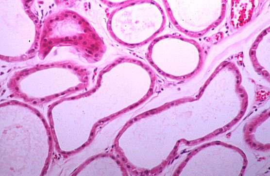 Apocrine glands
From somebody's armpit
Photo by Ed
{00690} epidermis, histology
{11760} skin, histology
* Future physicians: The Latin has the nuance of "all too familiar".
The English word suggests, instead, crude and disgusting. Be sure this
does not generate misunderstandings.
You do not beg the sun for mercy.
-- Stilgar in "Dune Messiah"
Some people don't tan;
in non-redheads, it's
genetic spectrum
at the melanocortin-1-receptor MC1R gene Lancet 355:
1072, 2000.
* You remember that the trademark mutation for sun damage is CC->TT double-base substitution.
 In skin exposed to heavy sunlight for long periods of time ("weather-beaten", "photo-aged"), the upper
layer of the dermis exhibits a poorly-understood change in its fibers and ground- substance. Pale
basophilic material that stains like elastin is deposited ("solar elastosis"), causing the skin to become
deeply furrowed. It's now clear that much of the problem is greatly decreased type I collagen synthesis,
and that tretinoin (retinoic acid) works by restimulating this synthesis (NEJM 329: 530, 1994;
* metalloproteinase induced by ultraviolet and blocked by retinoic acid
NEJM 337: 1419, 1997).
In skin exposed to heavy sunlight for long periods of time ("weather-beaten", "photo-aged"), the upper
layer of the dermis exhibits a poorly-understood change in its fibers and ground- substance. Pale
basophilic material that stains like elastin is deposited ("solar elastosis"), causing the skin to become
deeply furrowed. It's now clear that much of the problem is greatly decreased type I collagen synthesis,
and that tretinoin (retinoic acid) works by restimulating this synthesis (NEJM 329: 530, 1994;
* metalloproteinase induced by ultraviolet and blocked by retinoic acid
NEJM 337: 1419, 1997).
Common ichthyosis ("ichthyosis vulgaris") produces dry skin
and (if you look closely), alligator-like pattern of scales, mostly
on the extremities.
The autosomal dominant form of common ichthyosis
is caused by defective synthesis of
filaggrin, the histidine-rich protein of keratohyalin granules.
For some reason, the desmosomes stay intact longer than they should,
delaying the shedding of the dead epidermal cells.
This semi-disease is extremely common, with 1 person
in 250 affected.
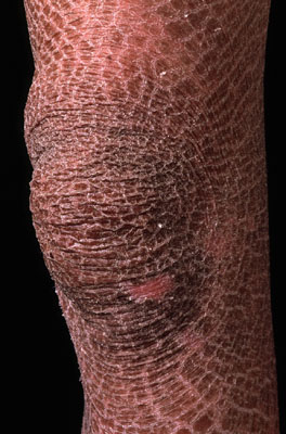 Common ichthyosis
Prize photograph
Institute of Medical Illustrators
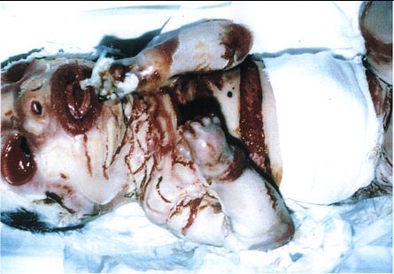 Ichthyosis "Collodion baby"
From a Saddam-era Iraqi
propaganda website (!)
* Refsum's disease (inability to break down phytanic acid) produces
ichthyosis, retinitis pigmentosa, and movement problems. And so forth.
{12474} vitiligo
{12489} vitiligo
 Vitiligo
WebPath Case of the Week
A patch of non-pigmented hair is "poliosis"; usually it is idiopathic though
you may see it with various syndromes

{09647} lentigo
{09648} lentigo
{12192} lentigines
{12525} lentigo city
{09638} melanocytic nevus, gross
{12177} melanocytic nevus, gross; this was intradermal
{12194} melanocytic nevus, gross; this was compound
{12195} melanocytic nevus, gross; this was intradermal
{08150} intradermal nevus, histology
{08153} intradermal nevus, histology
{25680} junctional nevus with melanophages
{25681} junctional nevus with melanophages
{08156} nevus cells
{09651} nevus spilus
{09653} nevus spilus
{09656} nevus spilus
* Richard Gere has one, which he does not cover with makeup during his
love scenes. Celebrities
Patrick Muldoon and
Heath Ledger also display
theirs without applying makeup.
 Becker's Nevus
Richard Gere in "Breathless"
 Becker's Nevus
Richard Gere in
"An Officer and a Gentleman"
{09641} halo nevus, gross
{09642} halo nevus, gross
{24625} giant congenital pigmented nevus
{24904} hairy nevus, congenital
{25108} compound nevus, histology
Nevus sebaceus ("nevus sebaceous") is a common, often large congenital hamartoma
(skin, hair, sebaceous glands, apocrine glands),
usually on the hairline on the temple.
Thre are syndromes with very large lesions.
There is much controversy about the rate of malignancy, but the tendency
nowadays is to excise them.
* Amelanotic dysplastic nevi are rare but do occur: Lancet 359: 1999, 2002.
{12197} dysplastic nevus
{09663} dysplastic nevus
* Affected celebrities include Sen. John McCain, football star
Troy Aikman, and Ronald Reagan's daughter Maureen.
Satellite lesions, i.e., little black patches just beyond the borders of the
main lesion, are extremely suggestive of melanoma. If you see them
microscopically, it means the melanoma is more likely to be aggressive
(Arch. Derm. 141: 739, 2005).
* Today patients with level 4 or ulcerated melanomas
are likely to have a sentinel lymph node biopsied for
staging, and to receive interferon alfa-2b therapy if the node is positive
or the tumor is thicker than 4 mm. To find the sentinel node, inject
carbon dye first: Cancer 92: 535, 2001. Consensus conference Arch. Derm. 137: 1217, 2001.
Don't just watch it: Arch. Derm. 128: 657, 1992.
How to do the surgery: Cancer 73: 2964, 1994.
 Melanomas
Photos from a laser group
Not for young or sensitive visitors.
Malignant Melanoma
Australian Pathology Museum
High-tech gross photos
{09684} melanoma, gross
{09687} melanoma, gross
{09689} melanoma, gross
{09690} melanoma, gross
{09692} melanoma, gross
{09695} melanoma, gross
{09098} melanoma, gross, amelanotic (i.e., this one makes no melanin)
{09099} melanoma, gross, amelanotic
{24916} melanoma, gross
{24741} superficial spreading melanoma, vertical growth phase
{10940} superficial spreading melanoma, gross
{12198} superficial spreading melanoma, gross
{24914} superficial spreading melanoma, vertical
{24915} superficial spreading melanoma, vertical growth phase
{25601} superficial spreading melanoma
{12199} nodular melanoma
{25516} nodular melanoma
{25600} nodular melanoma
{24911} spindle cell melanoma
{12200} lentigo maligna
{24912} lentigo maligna
{24913} lentigo maligna
{13121} lentigo maligna
{13122} melanoma
{13123} melanoma
{13306} melanoma
{13307} melanoma
{13308} melanoma
{15400} melanoma, electron micrograph showing melanosomes
{13309} superficial spreading melanoma
{13310} superficial spreading melanoma
{12201} acral lentigenous melanoma
{08967} invasive malignant melanoma
{08968} invasive malignant melanoma
{08969} invasive malignant melanoma
{10546} melanoma, metastatic in liver
{08035} melanoma, metastatic in heart
 Melanoma metastases
Only one side of the body is involved
New England Journal of Medicine
{12765} seborrheic keratosis
{25496} seborrheic keratosis
{24895} seborrheic keratosis, histology
{24896} seborrheic keratosis, histology
{12771} seborrheic keratoses, think of Leser-Trelat
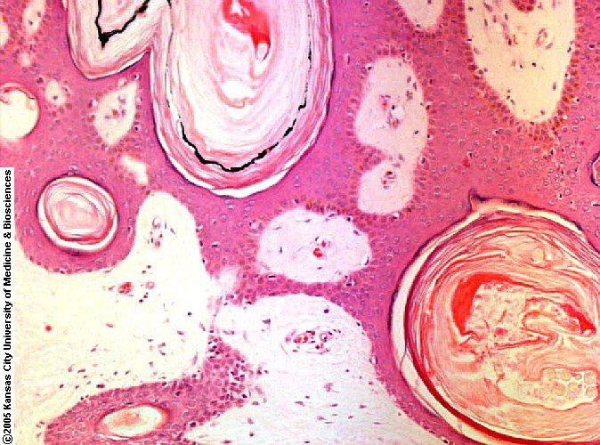 Seborrheic keratosis
Horn cysts
ERF/KCUMB
{25608} acanthosis nigricans
{12780} acrochordons
{12781} acrochordons
{12206} brown skin tag
{10310} epidermoid inclusion cyst
{12504} epidermoid inclusion cyst
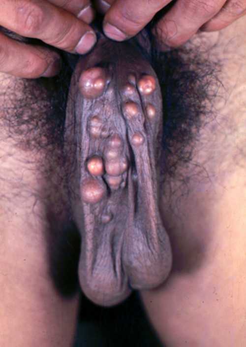 Steatocystoma multiplex
Scrotum
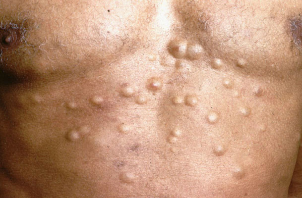 Steatocystoma multiplex
Prize photograph
Institute of Medical Illustrators
Of the skin he made him mittens,
Made them with the fur side inside,
Made them with the skin side outside,
He, to get the warm side inside,
Put the inside skin side outside;
He, to get the cold side outside,
Put the warm side fur side inside.
That's why he put the fur side inside,
Why he put the skin side outside,
Then he turned them inside outside.
The presence of an epithelial lip, and sharp demarcation of the stroma
favor a keratoacanthoma. Ulcerated lesions, many mitotic figures, and
obvious anaplasia usually means squamous cell carcinoma instead (Dermatology 199:
208, 1999).
{12795} keratoacanthoma
{12799} keratoacanthoma
{24892} keratoacanthoma, histology
{24893} keratoacanthoma, histology
 Keratoacanthoma
Prize photograph
Institute of Medical Illustrators
 Keratoacanthoma
Eye pathology site
{25596} cylindromas
These are extremely familiar. There's an autosomal dominant syndrome,
and a variant in which they erupt in large numbers all over the trunk.
{11788} trichofolliculoma
{24889} hidradenoma
* {24920} calcifying epithelioma of Malherbe ("pilomatrixoma")
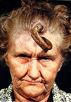 Skin horn
Source unknown
Not for young or sensitive visitors.
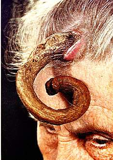 Skin horn
Source unknown
Not for young or sensitive visitors.
{12738} actinic keratosis
{25506} actinic keratosis

On the penis, it's erythroplasia of Queyrat, and has about a 30%
chance of invading.
{12733} neglected squamous cell carcinoma
{21858} squamous cell carcinoma
{21859} squamous cell carcinoma of eye
{12169} squamous cell CA, lip
Squamous cell carcinoma
Eye pathology site
Squamous Cell Carcinomas
Photos from a laser group
Not for young or sensitive visitors.
You remember xeroderma pigmentosum, a heterogeneous (Am. J. Hum. Genet. 54: 191,
1994) family of hereditary diseases in which the body has difficulty repairing actinic (and probably
other) damage to DNA. Most common gene: Arch. Derm. 128: 971, 1992; the family Arch. Derm.
128: 1233, 1992.

It is extremely common, and by no means rare even in adults under age 40
(JAMA 294: 681, 2005).
Basal Cell Carcinomas
Photos from a laser group
Not for young or sensitive visitors.
Basal Cell Carcinomas
Operative procedures
Not for young or sensitive visitors.
 Basal cell carcinoma
Photo and mini-review
Brown U.
{12706} basal cell carcinoma
{12711} basal cell carcinoma
{12714} basal cell carcinoma
{12723} basal cell carcinoma
{21847} basal cell carcinoma
{21850} basal cell carcinoma, histology
{21856} basal cell carcinoma, histology, sclerosing variant with desmoplasia
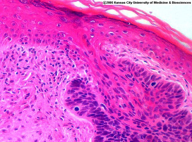 Basal cell carcinoma
Arising off a hair shaft
ERF/KCUMB
 Nevoid basal cell carcinoma syndrome
Pittsburgh Pathology Cases
Merkel Cell Carcioma
Electron micrographs
VCU Pathology
* Ask a pathologist
to show you the famous "dirty fingers" elongation and pigmentation of rete pegs at the margins of the
tumor.
{12772} dermatofibroma
{12774} dermatofibroma
{12775} dermatofibroma
{12777} dermatofibroma
{25593} dermatofibroma
* The trademark CC->TT mutation in p53 in these tumors is the first proof that sunshine can
cause a sarcoma, as well as a carcinoma: Am. J. Path. 145: 11, 1994.
{09744} xanthomas
{12219} xanthoma
{24886} xanthoma
{05926} spider angioma
{05928} spider angiomas
{12235} hemangioma
{12237} Kasabach-Merritt hemangioma
{12754} mycosis fungoides
{12751} mycosis fungoides, tumor stage
{12757} mycosis fungoides (this was a Sezary case)
{12759} mycosis fungoides, Pautrier micro-abscess
{09042} mycosis fungoides, convoluted T-cell as seen on electron microscopy
Mastocytosis
Virginia Pathology Cases
{09717} urticaria
{09720} urticaria
{12241} dermographism
{13114} lichen simplex chronicus
Botanical dermatology
Some fascinating pictures
of plants and people
{12498} "eczema"
{24990} "eczema"
{12523} eczema secondary to photodermatitis
{12293} atopic dermatitis
{12294} atopic dermatitis
{12531} atopic dermatitis
{12534} atopic dermatitis, lichenified
{08160} contact dermatitis, hat band
{25581} she was allergic to her tincture of benzoin
{19349} contact dermatitis, earring
{12127} contact dermatitis
{12538} contact dermatitis, weepy
{12542} contact dermatitis, metal hapten
{12544} contact dermatitis, shoe
{12546} contact dermatitis, spray-on deodorant
{19358} contact dermatitis, balloon cells
{08154} poison ivy
{12562} nummular dermatitis

In both Stevens-Johnson and common erythema multiforme, the lymphocytes
appear to be causing apoptosis of epidermal cells.
In SJS (but not in the milder variant), the lymphocytes stain richly
for perforin
(Arch. Derm. 133: 845, 1997).
{09699} erythema multiforme
{09701} erythema multiforme
{09702} erythema multiforme
{09704} erythema multiforme
{09705} erythema multiforme
{12261} erythema multiforme
{12516} erythema multiforme, vesicular
{12126}erythema multiforme bullosum
{12529} erythema multiforme, purpuric
{09710} bad erythema multiforme
{05943} Stevens-Johnson syndrome, skin
{05944} Stevens-Johnson syndrome, oral ("cheilitis")
{10304} toxic epidermal necrolysis
{09711} toxic epidermal necrolysis
{09713} toxic epidermal necrolysis
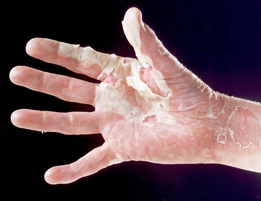 Pustular dermatitis
Prize photograph
Institute of Medical Illustrators
The usual cause is taking medicines (notoriously sulfa, allopurinol,
or phenytoin).
{12259} leukocytoclastic vasculitis, palpable purpura
* Infliximab for pyoderma gangrenosum: Am. J. Gastro. 98: 1821, 2003.
(clusters of neutrophils in the lower
epidermis) and/or "Munro's microabscesses"
(clusters of four or more neutrophils in the scale itself)
{12574} psoriasis
{12576} psoriasis
{12579} psoriasis
{12580} psoriasis
{12582} psoriasis
{12585} psoriasis
{19367} psoriasis
{19370} psoriasis
{19385} psoriasis
{24875} psoriasis
{12475} psoriasis, Koebner phenomenon
{12513} exfoliative erythroderma, maybe psoriasis, maybe mycosis fungoides, maybe who-knows?
Usually it is "idiopathic", but certain medications can
trigger it, and it is especially common in patients with hepatitis C virus
on board.
{12476} lichen planus
{12591} lichen planus
{12592} lichen planus
{12597} lichen planus
{12600} lichen planus
{12601} lichen planus
{12591} lichen planus
{12604} lichen planus, filigree lesions in mouth
{24988} lichen sclerosus et atrophicus
Who had balanitis xerotica;
His wife had kraurosis
And lichen sclerosus
So neither of them got any erotica!
* Lichen nitidus features innumerable tiny, same-size flesh-colored papules
on the genitals; they'll also appear anywhere that you scratch ("Koebner phenomenon").
The etiology is unknown; I've seen it a couple of times.
The old story is that
if you stain for IgG or complement, you'll see granules along the dermal-epidermal junction ("a
positive lupus band test"). If the patient has only cutaneous lupus, you'll see this only on
sun-exposed skin; if you see this on all skin, it's systemic lupus. It is probably not true:
Clin. Exp. Rheum. 17: 427, 1999.
{25674} discoid lupus
{25676} discoid lupus
{12324} alopecia secondary to discoid lupus
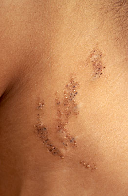 Comedone nevus
Prize photograph
Institute of Medical Illustrators
{09725} acne
{12144} acne
{12145} acne
{24919} acne, face; your lecturer age 13
{24981} acne, blackhead city
{12495} acne, closed and open comedones
{12148} acne, open comedone, world's largest blackhead
{12221} acne, open comedone, world's second largest blackhead
{25672} histology of the comedone!
{25673} histology of the comedone!
{09728} acne rosacea
 Rhinophyma ("W.C. Fields' nose", Shakespeare's Bardolph), supposedly a variant of acne rosacea, involves hyperplasia of
sebaceous glands, notably on the nose. There can be a conjunctivitis too. Again, the process is mysterious.
Rhinophyma ("W.C. Fields' nose", Shakespeare's Bardolph), supposedly a variant of acne rosacea, involves hyperplasia of
sebaceous glands, notably on the nose. There can be a conjunctivitis too. Again, the process is mysterious.
{12124} pemphigus vulgaris
{12651} pemphigus vulgaris
{12657} pemphigus vulgaris
{12658} pemphigus vulgaris
Pemphigus
Great photos
Bill Weems MD / "Dermpath India"
"Endemic pemphigus foliaceous" or "fogo selvagem", a endemic disease of young
people in rural Latin America, is probably a different disease, though the antibody
is the same. Big review: NEJM 343: 23, 2000.
{12633} pemphigoid
{12634} pemphigoid
{12636} pemphigoid
{12637} pemphigoid
{12640} pemphigoid
{12643} pemphigoid, histology
{12644} pemphigoid, immunofluorescence (yellow)
{12694} cicatricial pemphigoid
Pompholyx ("dyshydrotic eczema") is a common, banal, but annoying disease in which little blisters
cover the palms and soles. A minor mystery of medicine, there's a link to nickel allergy (J. Derm.
19: 964, 1994; Cutis 47: 157, 1991), or other metal allergy, in people with sweaty palms.
In the clinic, you may order antibodies against endomysium and/or
tissue transglutaminase, as for sprue.
* Jean-Paul Marat, amateur scientist and
quack doctor turned far-left-wing ideologue,
was the French Revolution's first mass-murderer.
His skin disease was intensely pruritic, blistering,
began in the perianal region,
and was associated with weight loss leading to emaciation.
He was sick with it for the three years prior to his assassination,
and spent most of this time in his bathtub.
My choice is dermatitis herpetiformis.
Another pathologist agrees: Am. J. Dermpath. 1: 251, 1979.
-- Marat-Sade

{12663} dermatitis herpetiformis
{12524} dermatitis herpetiformis, eczematized
{12672} dermatitis herpetiformis, immunofluorescence for IgA
This disease is a frequent focus for "everybody can have
a good quality of life" ethics discussions:
J. Med. Ethics 24: 200, 1998.
* Update on the molecular biology of the subepidermal blistering diseases,
both autoimmune and hereditary: Virchows Arch. 443: 184, 2003.
Epidermolysis Bullosa
{12138} wart
{24894} wart
{08983} wart, histology
{08986} wart, histology
{12164} condyloma acuminata
{12174} molluscum contagiosum
{12175} molluscum contagiosum
{24730} molluscum contagiosum
{24731} molluscum contagiosum
Molluscum contagiosum
WebPath Case of the Week
Molluscum Contagiosum
Electron micrographs
VCU Pathology
 PITYRIASIS ROSEA
* Erythromycin is an empirical remedy (J Am. Acad. Derm 42: 241, 2000).
An attempt to blame Herpes 7 for pityriasis rosea in the late 1990's was a flop.
Pityriasis rosea
Patient photo
Thanks Brian!
{12487} pityriasis rosea
{12610} pityriasis rosea
{12616} pityriasis rosea
{24971} impetigo
{24972} impetigo
{24973} impetigo
{12128} impetigo, bullous
Gym types: If you've got ringworm or impetigo, it's basic courtesy to keep it covered with clothing
while you are throwing the weights around. Active herpes gets passed from wrestler to wrestler
("herpes gladiatorum") and might survive on mats, gym machines, and so forth, so keep your herpes
blisters covered or off the equipment, too. Nobody knows how pityriasis rosea is spread, but it's also
worth covering. Molluscum, scabies, and pediculosis
are probably also transmissble this way.
If you have pseudomas ecthyma, I'm going to assume you are not in the gym.
Since the agents of candida, warts, and tinea versicolor are
ubiquitous, I would not worry about gym infections.
Beyond this, gym transmission of disease seems highly improbable.
You've studied
them in microbiology. Usually these poisons are produced by staph.
Staphylococcal epidermolytic toxin A and B produce staph
scalded-skin syndrome. It's a pediatric disease, since most adults
have antibodies. Review Arch. Dis. Child. 78: 85, 1998.
Clinically, there is spectacular blistering.
Under the microscope, expect to see mostly acantholysis, especially in a
cleavage plane between the spiny and granular layers, i.e., the toxin
attacks filaggrin granules, which is why mucosal surfaces are spared.
Don't confuse this with toxic shock syndrome, which during
the acute phase merely looks like a sunburn despite the patient being
severely and systemically ill. Late in toxic shock syndrome, there's
some epidermal inflammation and necrosis and it
often desquamates over the palms and soles.
Neutrophils invade and damage the epidermis, without any
underlying vasculitis.
Antifungals are now the mainstay of therapy. See Lancet 358: 170, 2001.
{12565} seborrheic dermatitis
{12571} seborrheic dermatitis
{14238} candida, penis
{12507} candida exacerbating intertrigo
{08163} moniliasis of skin ("diaper rash")
{08148} tinea pedis ("athlete's foot")
{12268} tinea pedis
{14227} tinea pedis
{12270} tinea cruris ("crotch-rot")
{12477} tinea corporis ("ringworm of the body")
{14221} tinea corporis
{14223} tinea corporis, leg
{08149} ringworm, id reaction
{08152} more ringworm
{12625} pityriasis rubra pilaris (mysterious disease that
simulates ringworm)
{12182} tinea versicolor
{12505} tinea versicolor
{12519} tinea versicolor
{14247} tinea versicolor
{14250} tinea versicolor
 Pityrosporum, KOH prep
from a doctor buddy with
folliculitis of his beard area
{43077} scabies
{43079} scabies mite
{24872} scabies
 Yes, there is a little mite named
"Demodex" that lives head-down in your follicles.
Its ability to harm us remains uncertain.
It is one of the "usual suspects" in rosacea and in the itchy-eyelids
problems of older folks.
Yes, there is a little mite named
"Demodex" that lives head-down in your follicles.
Its ability to harm us remains uncertain.
It is one of the "usual suspects" in rosacea and in the itchy-eyelids
problems of older folks.
We don't have room for many of the skin diseases, including some common
entities like Darier's (an ATPase mutation that produces many warty lesions;
review Br. J. Derm. 152: 920, 2005).
Some skin diseases have no anatomic changes; these include the bizarre
"aquagenic pruritus" ( see also aquagenic urticaria), in which contact with water at any temperature is followed
by intense itching; some neuropeptide in the epidermis must be involved.
{08158} fifth disease
{08167} erythema chronicum migrans
{08170} heroin tracks
{08199} yaws, skin disease
{08200} yaws, skin disease
{08389} cutaneous larva migrans
{08393} leishmaniasis
{09734} pyoderma gangrenosum
{09737} pyoderma gangrenosum
{09738} pyoderma gangrenosum
{10067} abscess
{10081} squamous cell carcinoma, keratin pearl
{12215} amyloid of the skin
{12229} erysipelas, strep infection
{12486} squamous cell carcinoma
{12513} exfoliative erythroderma
{12523} photodermatitis
{12805} keloid
{13120} dermatomyositis, Gottron's sign
{13338} herpes, skin
{13343} leprosy
{14145} herpes zoster
{14146} herpes zoster
{14148} herpes zoster
{14200} folliculitis. ERF had this ages 11-17, probably
tinea versicolor folliculitis.
{14202} folliculitis, ditto
{24917} scleroderma, skin
{12226} granuloma annulare
{24918} granuloma annulare
{24923} blastomycosis, skin
{24933} histology of chicken pox
{53760} linear nevus sebaceus (nevus sebaceous)
{12190} tuberous sclerosis ash-leaf spots
{12169} squamous cell CA, lip
{12214} necrobiosis lipoidica diabeticorum
{12226} granuloma annulare
{12227} furuncle
{12230} hidradenitis suppurativa, vulva
{5926} spider angioma
{5928} spider angioma
{12235} hemangioma
{12237} Kasabach-Merritt hemangioma
{12274} lupus, butterfly rash
{12292} measles (rubeola)
{12337} clubbing
{12305} abetalipoproteinemia, RBC's ("ET-finger cells")
{12668} nutmeg liver
{12762} Kaposi's
{12801} scar
{12807} keloid
{13053} granuloma annulare, gross
{13120} dermatomyositis, Gottron's sign
{13338} herpes, skin
{13343} leprosy
{12786} pyogenic granuloma
{12789} pyogenic granuloma
{12805} keloid
{13613} amyloid vessel, H&E
{13392} cystic hygroma
{13370} flea-bitten kidneys
{14137} varicella
{18250} phenylketonuria
{18253} phenylketonuria
{19377} melanoma of the eye
{21859} squamous cell carcinoma of eye
{24626} scleroderma, hands
{24854} hyperplastic arteriolar sclerosis in scleroderma
{24917} scleroderma, skin
{24918} granuloma annulare
{24923} blastomycosis, skin
{24933} histology of chicken pox
{25116} Fabry's
{25024} fatty streaks
{25675} lupus, butterfly rash
11761
skin, normal
14788
skin, thick
14789
skin, thick
14790
melanin in the skin, normal
14791
melanin in the skin, normal
14792
skin, thick
14793
skin, thick
14794
skin, thick
14795
skin, thick
14796
sebaceous gland, normal
14797
sebaceous gland, normal
14798
dermal papilla, normal
14799
dermal papilla, norma
14800
meissner's corpuscle
14801
meissner's corpuscle
15092
skin, normal
15093
skin, normal
15305
skin, thin
15306
skin, thin
15307
skin, thin
15308
skin, thin
15309
skin, thin
15310
skin, thin
15311
skin, thin
15313
skin, thick
15315
skin, stratum corneum and lucida
20914
skin
20916
skin, eccrine gland
20917
sebaceous gland, skin
20918
skin, thick
20919
skin, thick
20920
skin, thick
20921
skin, thick
20922
pacinian corpuscle, skin
20923
skin, thick
24860
skin, normal
25083
skin, normal
25084
skin, normal epidermis
The Pathology Blues
 Ed's Pathology Review for USMLE I


| Visitors to www.pathguy.com reset Jan. 30, 2005: |
 Drop by
and meet Ed Drop by
and meet Ed
|

|
 Teaching Pathology
![]()
![]()
 | Pathological Chess |
 |
Taser Video 83.4 MB 7:26 min |

)window.location='http://www.indigo.com/software/gphpcd/pat2.jpg') Basal Cell Carcinomas
Basal Cell Carcinomas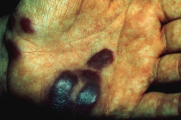 Kaposi's sarcoma
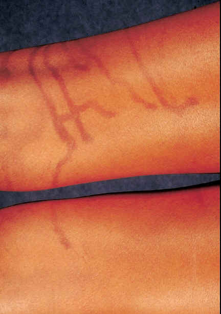 Phytodermatitis from a lime on a cocktail
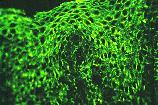 Pemphigus