Ed Friedlander, M.D., Pathologist
scalpel_blade@yahoo.com


Cyberfriends: The help you're looking for is probably here.
Welcome to Ed's Pathology Notes, placed here originally for the convenience of medical students at my school. You need to check the accuracy of any information, from any source, against other credible sources. I cannot diagnose or treat over the web, I cannot comment on the health care you have already received, and these notes cannot substitute for your own doctor's care. I am good at helping people find resources and answers. If you need me, send me an E-mail at scalpel_blade@yahoo.com Your confidentiality is completely respected.
 DoctorGeorge.com is a larger, full-time service.
There is also a fee site at myphysicians.com,
and another at www.afraidtoask.com.
DoctorGeorge.com is a larger, full-time service.
There is also a fee site at myphysicians.com,
and another at www.afraidtoask.com.
Translate this page automatically
 |
With one of four large boxes of "Pathguy" replies. |
 I'm still doing my best to answer
everybody.
Sometimes I get backlogged,
sometimes my E-mail crashes, and sometimes my
literature search software crashes. If you've not heard
from me in a week, post me again. I send my most
challenging questions to the medical student pathology
interest group, minus the name, but with your E-mail
where you can receive a reply.
I'm still doing my best to answer
everybody.
Sometimes I get backlogged,
sometimes my E-mail crashes, and sometimes my
literature search software crashes. If you've not heard
from me in a week, post me again. I send my most
challenging questions to the medical student pathology
interest group, minus the name, but with your E-mail
where you can receive a reply.
Numbers in {curly braces} are from the magnificent Slice of Life videodisk. No medical student should be without access to this wonderful resource. Someday you may be able to access these pictures directly from this page.
Also:
Medmark Pathology -- massive listing of pathology sites
Freely have you received, freely give. -- Matthew 10:8. My
site receives an enormous amount of traffic, and I'm
handling about 200 requests for information weekly, all
as a public service.
Pathology's modern founder,
Rudolf
Virchow M.D., left a legacy
of realism and social conscience for the discipline. I am
a mainstream Christian, a man of science, and a proponent of
common sense and common kindness. I am an outspoken enemy
of all the make-believe and bunk which interfere with
peoples' health, reasonable freedom, and happiness. I
talk and write straight, and without apology.
Throughout these notes, I am speaking only
for myself, and not for any employer, organization,
or associate.
Special thanks to my friend and colleague,
Charles Wheeler M.D.,
pathologist and former Kansas City mayor. Thanks also
to the real Patch
Adams M.D., who wrote me encouragement when we were both
beginning our unusual medical careers.
If you're a private individual who's
enjoyed this site, and want to say, "Thank you, Ed!", then
what I'd like best is a contribution to the Episcopalian home for
abandoned, neglected, and abused kids in Nevada:
My home page
Especially if you're looking for
information on a disease with a name
that you know, here are a couple of
great places for you to go right now
and use Medline, which will
allow you to find every relevant
current scientific publication.
You owe it to yourself to learn to
use this invaluable internet resource.
Not only will you find some information
immediately, but you'll have references
to journal articles which you can obtain
by interlibrary loan, plus the names of
the world's foremost experts and their
institutions.
Alternative (complementary) medicine has made real progress since my
generally-unfavorable 1983 review linked below. If you are
interested in complementary medicine, then I would urge you
to visit my new
Alternative Medicine page.
If you are looking for something on complementary
medicine, please go first to
the American
Association of Naturopathic Physicians.
And for your enjoyment... here are some of my old pathology
exams
for medical school undergraduates.
I cannot examine every claim which my correspondents
share with me. Sometimes the independent thinkers
prove to be correct, and paradigms shift as a result.
You also know that extraordinary claims require
extraordinary evidence. When a discovery proves to
square with the observable world, scientists make
reputations by confirming it, and corporations
are soon making profits from it. When a
decades-old claim by a "persecuted genius"
finds no acceptance from mainstream science,
it probably failed some basic experimental tests designed
to eliminate self-deception. If you ask me about
something like this, I will simply invite you to
do some tests yourself, perhaps as a high-school
science project. Who knows? Perhaps
it'll be you who makes the next great discovery!
Our world is full of people who have found peace, fulfillment, and friendship
by suspending their own reasoning and
simply accepting a single authority which seems wise and good.
I've learned that they leave the movements when, and only when, they
discover they have been maliciously deceived.
In the meantime, nothing that I can say or do will
convince such people that I am a decent human being. I no longer
answer my crank mail.
This site is my hobby, and I presently have no sponsor.
This page was last updated February 6, 2006.
During the ten years my site has been online, it's proved to be
one of the most popular of all internet sites for undergraduate
physician and allied-health education. It is so well-known
that I'm not worried about borrowers.
I never refuse requests from colleagues for permission to
adapt or duplicate it for their own courses... and many do.
So, fellow-teachers,
help yourselves. Don't sell it for a profit, don't use it for a bad purpose,
and at some time in your course, mention me as author and KCUMB as my institution. Drop me a note about
your successes. And special
thanks to everyone who's helped and encouraged me, and especially the
people at KCUMB
for making it possible, and my teaching assistants over the years.
Whatever you're looking for on the web, I hope you find it,
here or elsewhere. Health and friendship!
QUIZBANK: Respiratory
Breakdown of the "Respiratory" quizbank items:
 I am presently adding clickable links to
images in these notes. Let me know about good online
sources in addition to these:
I am presently adding clickable links to
images in these notes. Let me know about good online
sources in addition to these:
Pathology Education Instructional Resource -- U. of Alabama; includes a digital library
Houston Pathology -- loads of great pictures for student doctors
Pathopic -- Swiss site; great resource for the truly hard-core
Syracuse -- pathology cases
Walter Reed -- surgical cases
Alabama's Interactive Pathology Lab
"Companion to Big Robbins" -- very little here yet
Alberta
Pathology Images --hard-core!
Cornell
Image Collection -- great site
Bristol Biomedical
Image Archive
Chilean Image Bank -- General Pathology -- en Español
Chilean Image Bank -- Systemic Pathology -- en Español
Connecticut
Virtual Pathology Museum
Australian
Interactive Pathology Museum
Semmelweis U.,
Budapest -- enormous pathology photo collection
Iowa Skin
Pathology
Loyola
Dermatology
History of Medicine -- National Library of Medicine
KU
Pathology Home
Page -- friends of mine
The Medical Algorithms Project -- not so much pathology, but worth a visit
National Museum of Health & Medicine -- Armed Forces Institute of Pathology
Telmeds -- brilliant site by the medical students of Panama (Spanish language)
U of
Iowa Dermatology Images
U Wash
Cytogenetics Image Gallery
Urbana
Atlas of Pathology -- great site
Visible
Human Project at NLM
WebPath:
Internet Pathology
Laboratory -- great site My team:
Ed Lulo's Pathology Gallery
Bryan Lee's Pathology Museum
Dino Laporte: Pathology Museum
Tom Demark: Pathology Museum
Dan Hammoudi's Site
Claude Roofian's Site
Pathology Handout -- Korean student-generated site; I am pleased to permit their use of my cartoons
Estimating the Time of Death -- computer program right on a webpage
Pathology Field Guide -- recognizing anatomic lesions, no pictures

St.
Jude's Ranch for Children
I've spent time there and they are good. Write "Thanks
Ed" on your check.
PO Box 60100
Boulder City, NV 89006--0100
More of my notes
My medical students
Clinical
Queries -- PubMed from the National Institutes of Health.
Take your questions here first.
HealthWorld
Yahoo! Medline lists other sites which may work well for you

We comply with the
HONcode standard for health trust worthy
information:
verify
here. 

![]()
 Lung Exhibit
Virtual Pathology Museum
University of Connecticut
Includes some gunshot wounds
Lung Transplant Pictures
Great site
Transplant Pathology Internet Services
Tulane Pathology Course
Great for this unit
Exact links are always changing
Respiratory Pathology
Virginia Commonwealth U.
Great pictures
Introduction to Lung Pathology 1-42, 170-198
Interstitial Disease 235-249
Lung Cancer 199-234
Tobacco 43-65
Occupational Disease 150-169
Obstructive Disease 132-149
Infectious Disease 66-131

Chest wall problems
structural... THE CHEST DEFORMITIES
neuromuscular...THE PARALYSIS & WEAKNESS SYNDROMES
Obstructed upper airway
structural... QUINSY ("PERITONSILLAR ABSCESS"); CROUP ("LTB"")
functional...THE SLEEP APNEAS
Obstructed large bronchi
all, subtotal... CHRONIC BRONCHITIS
one, total... OBSTRUCTIVE ATELECTASIS; ENDOGENOUS LIPID PNEUMONIA
Constricted small bronchi
mast-cell / inflammation mediated... THE ASTHMAS
platelet-mediated... PULMONARY EMBOLUS
apudoma products... CARCINOID SYNDROME
dense collagen... SOME CHRONIC BRONCHITIS VARIANTS
Fibrotic respiratory bronchioles... SILICOSIS
Collapsed respiratory bronchioles... EMPHYSEMA/"CHRONIC BRONCHITIS"
Fluid-filled alveolar spaces
transudate... ALVEOLAR PULMONARY EDEMA
exudate & pus... THE PNEUMONIAS
exudate, fibrin, debris... THE RESPIRATORY DISTRESS SYNDROMES
surfactant... ALVEOLAR LIPOPROTEINOSIS; ENDOGENOUS LIPID PNEUMONIAS
other lipid... EXOGENOUS LIPID PNEUMONIAS
blood... GOODPASTURE'S DISEASE; ANTI-NEUTROPHIL CYTOPLASMIC ANTIBODY DISEASES, ACUTE MOUNTAIN SICKNESS, OTHER PULMONARY BLEED SYNDROMES
organisms alone... PNEUMOCYSTOSIS, CRYPTOCOCCOSIS
Fluid-filled alveolar septa
transudate... INTERSTITIAL PULMONARY EDEMA
exudate...THE PNEUMONITIS FAMILY; VIRUSES; MYCOPLASMA
Fibrosis around ulcerated bronchi...BRONCHIECTASIS
Fibrosis of alveolar septa
slow... THE INTERSTITIAL RESTRICTIVE LUNG DISEASES; (Hamman-Rich, rheumatoid lung, sarcoid, asbestosis, many others)
fast... THE RESPIRATORY DISTRESS SYNDROMES
Collapsed alveoli
large-airway disease... OBSTRUCTIVE ATELECTASIS
alveolar disease... THE RESPIRATORY DISTRESS SYNDROMES
ischemia... PULMONARY EMBOLUS, SEVERE SHOCK
Necrotic lung ("cavities", etc.)
infarction... PULMONARY EMBOLUS (COMPLICATED)
suppurative... NECROTIZING PNEUMONIAS; LUNG ABSCESS
caseous... TUBERCULOSIS, HISTOPLASMOSIS, BLASTOMYCOSIS, COCCIDIOIDOMYCOSIS
weird immune... WEGENER'S GRANULOMATOSIS
malignant... LUNG CANCER
Pulmonary Hypertension
secondary to low alveolar oxygen... see above; also MOUNTAIN DWELLERS
secondary to alveolar fibrosis... see above
Left-to-right shunts / Eisenmenger's
primary... PULMONARY EMBOLUS; VASCULITIS; IDIOPATHIC
High PaCO2... all whole-lung ventilation problems
Low PO2...
all whole-lung ventilation problems
perfusing non-ventilated lung
fluid/fibrosis in alveolar septa
 MOUNTAIN DWELLERS
STUDY OBJECTIVES
Describe the essential gross and microscopic anatomy of the airways, from trachea to alveolar sacs. Distinguish the two principal types of pneumocytes (I and II). Describe the anatomic and functional barrier to gas exchange at the alveolar-capillary level.
Describe the factors that influence PaCO2 and PaO2. Describe how PaO2 correlates with the actual oxygen content of the blood. Give the conditions when cyanosis will appear.
List the principal causes of edema in the lung, and compare these to things that cause edema anywhere else in the body. Distinguish interstitial and alveolar edema. Explain why edema of the lung is bad for one's health.
Describe pulmonary congestion, and mention its pathologic sequelae.
Review the pathology and pathophysiology of pulmonary thromboemboli. Describe their frequency and clinical correlations.
List the common causes of increased resistance in the pulmonary arteries (the usual cause of "pulmonary hypertension"), and other causes of pulmonary hypertension. Explain why these things are so harmful. Explain the hypoxic vascular response, why it is useful in health, and why it is such a problem in disease.
Define adult respiratory distress syndrome and list a few of the many synonyms. Tell about the etiologies, gross and microscopic pathology, the pathophysiology, and the common clinical picture.
Explain the pathophysiology and clinical correlations of neonatal respiratory distress syndrome.
Define atelectasis. Tell how lung collapses due to obstruction, compression, and lack of surfactant, and give clinical examples of each situation.
Define "sudden infant death syndrome". Briefly describe what we think causes genuine "SIDS", and give a "differential diagnosis".
 * Review how to order "arterial blood gases", what you get,
and what they tell you. (Be able to do
this at 3 AM as the only doctor on the ward.) This might be a
good time to look at the
Blood
Gases" handout.
* Review how to order "arterial blood gases", what you get,
and what they tell you. (Be able to do
this at 3 AM as the only doctor on the ward.) This might be a
good time to look at the
Blood
Gases" handout.
Describe the abnormal anatomy and functional problems that every cigaret smoker should expect.
Explain the importance of elastic recoil in keeping respiratory bronchioles patent during exhalation. Explain how this relates to the classic definition of emphysema as "an abnormal, permanent dilatation of part of all of the acinus, with destruction of alveolar walls."
Distinguish the two "classic" types of emphysema, and mention their alleged causes. Tell what we think causes emphysema in cigaret smokers and alpha-1 antitrypsin deficient patients. Tell what a "pink puffer" looks like clinically, and how emphysematous lungs look at autopsy. Describe the complication of "bullous emphysema".
Define "chronic bronchitis" and mention its common causes. Describe the gross and microscopic pathology and the pathophysiology. Be able to define the "Reid Index". Tell what a "blue bloater" looks like clinically, and mention the common organisms that superinfect these patients' lungs.
Define bronchial asthma. Describe its important causes, and distinguish "allergic" and "idiosyncratic" kinds. Describe the common pathophysiology. Tell what you will see at the autopsy of an asthmatic. Mention other causes of wheezing.
Define bronchiectasis. Describe the important causes, the abnormal anatomy, and the typical clinical picture.
Describe the various breathing problems that occur during sleep. Recognize sleep apnea as a common cause of several illnesses.
Describe the normal flora of the lungs in non-smokers and smokers, and recognize the range of micro-organisms that have caused lung infections. Recognize the tremendous clinical importance of lung infections.
Distinguish bronchopneumonia, lobar pneumonia, and pneumonitis. Describe the typical histopathology of lung infections caused by various agents.
List the etiologic agents of lobar pneumonia, the classic stages in its progression, the major complications, and those at risk for each form.
Describe the causes, underlying problems, pathophysiology, and morbid anatomy of bronchopneumonia, aspiration pneumonia, legionellosis, pneumocystosis, lung abscess, and viral and mycoplasmal pneumonias. Describe the distinctive features of hantavirus infection and SARS (the 2003 epidemic).
Describe the anatomic pathology, pathophysiology, and clinical picture of the "idiopathic pulmonary fibrosis" family of diseases. Identify "Hamman-Rich" syndrome, and mention its likely cause.
Define sarcoidosis, and describe a typical sarcoidosis patient. Explain the usual effects of sarcoidosis on the lungs, skin, and eyes. Mention the serious consequences of untreated sarcoidosis. Describe the histology, and give a differential diagnosis for a granuloma found on biopsy. Explain how sarcoidosis causes abnormalities of calcium metabolism. Recognize the Kveim test as of limited usefulness. Tell how to make the diagnosis of sarcoidosis, and how to treat sarcoid patients.
Explain the essential lesion of Goodpasture's disease involving the lung, and mention the clinical picture and diagnostic lab test, and essential treatment. Mention some "related" (?) causes of bleeding from the pulmonary alveoli.
Briefly describe the eosinophilic pneumonias, and the various lipid pneumonias and lipoproteinosis, focusing on their histopathology.
Give the numbers of new cases of lung cancer in U.S. men and women expected this year. Explain how rates are changing, and why. Describe the risk factors for lung cancer, mentioning the importance of cigaret smoking, industrial exposure, radon in the home, and indoor air pollution.
 Explain how and why pathologists subclassify lung cancers.
Recognize each of the nine members of the WHO-1999 classification:
Explain how and why pathologists subclassify lung cancers.
Recognize each of the nine members of the WHO-1999 classification:
Describe the important distinctions among the various types. Identify the common lung cancers under the microscope. Explain how pathologists use each of these to distinguish primary lung tumors:
Tell how bronchogenic carcinomas present. Describe the various paraneoplastic syndromes seen with lung cancer, especially the hypercalcemia syndromes and the small cell undifferentiated carcinoma syndromes.
Identify bronchial carcinoid, tell how it looks grossly and microscopically, how to recognize it, and describe its origin and its variants.
List the common problems that affect the larynx or trachea. List the different kinds of pleural effusions, and tell the significance of each. Describe the various kinds of pneumothorax and why they are important. Tell how pleural plaques look and what causes them.
Identify the cell of origin, risk factor, gross and microscopic appearance, and prognosis for malignant mesothelioma.
Mention the basic biology of ciliary dyskinesia syndromes, tell when you would suspect one, and how you would verify it.
As usual, given a gross lung or larynx, or a biopsy of any level of the respiratory tract, recognize any of the lesions exhibited in this section with at least 70% accuracy.
|
|

Life is not measured by the number of breaths you take, but by the number of moments that take your breath away.NORMAL ANATOMY AND PHYSIOLOGY
-- Attributed to George Carlin
* Click here for Dr. Karius's scheme for normal respiration. You must know this perfectly if you are going to be able to make sense out of lung pathology. Thanks Diane!
All about the uvula. A human's is much bigger than any other mammal's, and the uvula's job is probably to keep us from getting hoarse while talking (amazing, Yearbook of Path 1994, p. 97 describes its anatomy).
Review the gross anatomy of the respiratory system, and the general histology of a bronchus.
Bronchi are usually defined to be the airways with cartilage and/or complex glands (precise usage varies). The orders of bronchi end in membranous bronchioles.
The bronchioles divide further, leading to the terminal bronchioles, the last division with a continuous pseudostratified ciliated epithelium.
Remember the importance of the mucociliary elevator provided by this epithelium. The cilia are supposed to be extremely easy to damage and hard to recover, but they almost always are present in my autopsies, even on people who have been very sick for a very long time.
The portion of lung parenchyma supplied by one terminal bronchiole is called an acinus ("respiratory unit", diameter about 7 mm).
From the terminal bronchiole arise several orders of respiratory bronchioles, which have part of their walls alveolated and part with pseudostratified respiratory epithelium. These in turn give rise to alveolar ducts and then atria. The alveoli themselves are wide-mouthed sacs that open into all three of these divisions.
Remember that the elastic recoil of the surrounding lung tissue is what keeps the respiratory bronchioles from collapsing when a person starts to exhale.
Schematic diagram of the alveolar wall:
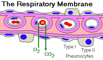
Type I pneumocytes: simple squamous, stretchy, permeable to O2 and CO2, easily injured, do not divide
Type II pneumocytes: reserve cells; cuboidal, granular cytoplasm, produce and process surfactant ("lamellar bodies" in the cytoplasm), divide and flatten to become new type I pneumocytes.
The interstitium contains a few fibroblasts, smooth muscle cells, collagen, elastin.
There are probably no lymphatics in the respiratory membranes themselves, but there are many lymphatics in the fibrous septa between groups of alveoli.
Contrast the alveolar epithelium with that of the bronchi and bronchioles, which includes ciliated, goblet, reserve (basal), Kulchitsky ("K-"), Clara, and other types of cells.
Macrophages in the alveoli spaces ("alveolar macrophages") and in the septa ("interstitial macrophages") eat surfactant and most anything else that comes along, in addition to modulating the immune response locally.
Remember that the lung receives a dual blood supply, from the pulmonary and bronchial arteries. The bronchial arterial supply is one of the first to stop in low-output states (left heart failure, shock; why?)
Bronchial lymphoid tissue (BALT) shouldn't be present until a baby is a few months old. Lymphocyte clusters in a newborn's large airways means infection. Smokers have much more "BALT" than do non-smokers.
Remember blood PaCO2 is almost entirely a function of overall alveolar ventilation.
Remember blood PaO2 is a function of the quality of ventilation-perfusion matching, alveolar septal thickening (if excessive), and overall ventilation. And remember that lung structures are generally much less permeable to O2 than to CO2 (a fact which becomes important only in disease.)
Don't forget either that blood with PaO2 of 65 torr is carrying almost as much oxygen as blood with PaO2 of 100 torr or higher, because most oxygen is bound to hemoglobin (recall the hemoglobin-oxygen dissociation curve....) It's when PaO2 drops to around 60 that you start getting into trouble, because the hemoglobin stops binding oxygen as well as it should.
Cyanosis appears when the arterial blood contains 5 gm % or more of unoxygenated hemoglobin.
All of the first group of diseases on the handout (except pulmonary thromboemboli) are patterns of lung injury, each of which has many different causes.
PULMONARY CONGESTION AND EDEMA
{37956} pulmonary edema gestalt
Pulmonary edema classically results from the same factors that cause edema in the rest of the body. These are:
1. Increased venous hydrostatic pressure (left-sided heart failure)
2. Fluid overload (renal failure, iatrogenic, etc.)
3. Decreased albumin in the blood (liver disease, nephrotic syndrome, poor nutrition, etc.)
4. Lymphatic obstruction (cancer, etc.)
5. Endothelial damage (i.e., the pneumonias; most striking is the hantavirus that first struck in the US southwest)
6. Getting strangled / physically asphyxiated (negative pressure)
You have to learn these causes of pulmonary edema for which the mechanisms are not well understood:
7. Acute CNS injury
8. Opiate overdose
 9. Exposure to high altitudes (unacclimatized people)
However, this can't be the full story, because of the pharmacology (Br. Med. J. 321: 267, 2000). Dexamethasone (the familiar glucocorticoid) apparently prevents acute hypoxia from making endothelial cells more permeable, while how acetazolamide (a carbonic anhydrase inhibitor) works is anybody's guess. The finding that a beta-agonist helps is attributed to increased active transport of sodium from the alveolar fluid into the bloodstream (NEJM 346: 1631, 2002).
* The roles of various mediators are being worked out (endothelin 1: Circulation 99: 2665, 1999), and the whole business remains rather puzzling (see for example Lancet 357: 1342, 2001.)
* Populations that inhabit high mountains seem to survive only by making huge amounts of nitric oxide, which you can measure on their breath. See Nature 414: 411, 2001. Natural selection or physiologic adaptation? We don't yet know. It's finally documented that mountain dwellers have greatly expanded chest cavities: Resp. Phys. Neuro. 132: 223, 2002.
In each case, when the capacity of the lymphatics to drain the interstitial fluid is exceeded, interstitial edema develops, with loss of lung compliance and a barrier to oxygen exchange (alveolar-capillary block.)
As the interstitial pressure rises still further, the tight junctions between the alveolar epithelial cells open and fluid pours into the alveolar spaces, causing alveolar edema and stopping ventilation.
Alveolar edema fluid is a good culture medium for bacteria. Secondary pneumonia is common.
When you hear crackles (rales) through your stethoscope, you're hearing the little air bubbles in the alveoli. Your patient has alveolar pulmonary edema, and you must figure out why.
 {10145} pulmonary edema (just enough protein
content to stain...)
{10145} pulmonary edema (just enough protein
content to stain...)
{11666} pulmonary edema
|
|
Pulmonary congestion of course results from increased pulmonary venous hydrostatic pressure, typically from left-sided heart failure especially mitral valve disease.
If marked or longstanding, microhemorrhages, fibrosis, and iron pigmentation ("brown induration") occur in the lungs. (Hemosiderin-laden macrophages are called "heart-failure cells.")
|
|
PULMONARY EMBOLIZATION ("embolism") AND INFARCTION (NEJM 339: 93, 1998; Lancet 363: 1295, 2004)
Pulmonary thromboemboli are very common (and still under-diagnosed -- see Arch. Int. Med. 148: 1345, 1988, also still good). Thrombi in unoperated pulmonary arteries are almost always emboli.
Most originate in the deep veins of the legs; they may also come from the pelvic veins.
Thrombosis in a leg vein can be uncomfortable ("thrombophlebitis"), but is most often asymptomatic. As a junior clerk, you will compare calf circumferences, check Homan's sign (be careful you don't break the thrombus off), etc.
The majority of pulmonary thromboemboli do no harm and eventually organize or lyse; many are fatal.
Pathologists report pulmonary emboli in 8-25% of autopsies on hospital patients. But in 3-5% of autopsies (figures vary), the embolus is the fatal event, and estimates of the number of U.S. deaths from pulmonary emboli are in the 50,000-150,000 range. (Without an autopsy, the clinicians will often tell the family, "heart attack".)
Virchow's triad. Typical settings for deep vein thrombosis and pulmonary thromboemboli include:
2. hypercoagulable states
3. damaged endothelium
For some reason, babies very seldom get or die of pulmonary thromboemboli: Arch. Path. Lab. Med. 114: 142, 1990.
Pulmonary thromboemboli cause several types of problems.
Pulmonary infarcts are peripheral and hemorrhagic (and can even cause hemolytic jaundice if the patient survives. Rising bilirubin and rising LDH-3 -- think of a pulmonary infarct.) Listen for a friction rub; look for fibrin on the pleural surface at autopsy (why?). They can be distinguished best from regular intrapulmonary hemorrhages by the necrosis. Most will have the classic "wedge" shape.
Infected thromboemboli can cause "septic infarcts" which may become lung abscesses.
Any patient with sudden anxiety, chest pain, dyspnea, cough, hemoptysis or sudden death should make you think of pulmonary emboli. You'll learn how to make the diagnosis while on rotations -- but don't get over-confident; pulmonary embolization is the most-often-missed diagnosis in the hospital (Arch. Path. Lab. Med. 129: 201, 2005.
Fibrin D-dimer is sensitive, but not specific: Arch. Path. Lab. Med. 123: 235, 1999. Some folks consider this the best screening test: Lancet 363: 1295, 2004.
As we have already mentioned, pulmonary emboli organize into fibrous bands which remain in the lung for the rest of the person's life (Arch. Path. Lab. Med. 123: 170, 1999).
{29052} pulmonary embolus, ancient, organized and turned into fibrous bands
 Pulmonary Thromboembolus
Australian Pathology Museum High-tech gross photos
|
|
|
Other causes of lung infarction (immune or infection vasculitis, other things embolizing) are rare but not unheard-of (Chest 127: 1178, 2005).
Fat embolization remains mysterious. You're familiar with this dread complication of a bony fracture. Today's pathologists make the diagnosis during life by finding fat in the cells obtained by pulmonary lavage.
The deadly chest syndrome in sickle cell anemia may also result (at least sometimes) from fat embolization after a bony infarct (NEJM 342: 1904, 2000). The same picture can appear in sicklers with bacterial or viral infection or thromboembolization; often no cause is found. I suspect that cells sickling in the presence of reduced oxygen tension is part of the cause.
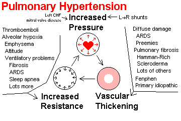
PULMONARY HYPERTENSION
|
|
Generally analogous to systemic arterial hypertension, pulmonary hypertension also results from a variety of causes.
1. Left-sided heart failure (especially mitral stenosis)
2. Increased blood flow into the pulmonary arteries (i.e., congenital cardiac malformations with left-to-right shunts, or after resection of a lung)
3. Increased pulmonary vascular resistance from any cause.
NOTE: This is important. The "hypoxic vascular response" causes constriction of arterioles supplying underoxygenated alveoli. If all the alveoli are underoxygenated, all the arterioles constrict, and the pulmonary arterial blood pressure has to rise. To this day, nobody knows which mediator produces the response.
Persistent pulmonary hypertension of the newborn results from hyperplasia of the smooth muscle in the arteries, present at birth (usually term). It is likely to prevent closure of the foramen ovale and/or ductus, and produce a cyanotic baby. It is quite common, often causes serious brain damage, and has been overlooked for too long. One cause seems to be SSRI antidepressants taken late in pregnancy (NEJM 354: 579, 2006).
Treating it with calcium channel blockers (helps, no panacea): NEJM 327: 76, 1992; nitric oxide and continuous intravenous prostacyclin are new remedies (Chest 119: 970, 2001; Am. J. Card. 75: 51A, 1995; big review); also Lancet 352: 719, 1998; NEJM 338: 273, 1998; aerosolized iloprost (prostacyclin analogue: NEJM 342: 1866, 2000; reversal in a mouse model using a serine elastase inhibitor: Nat. Med. 6: 698, 2000.
* Bosentan, the endothelin receptor antagonist, seems to help "idiopathic" primary pulmonary hypertension: Lancet 358: 1119, 2001; NEJM 346: 896, 2002.
NOTE: You'll frequently hear, "This patient is disabled because of pulmonary hypertension" or "This patient has right sided heart failure due to pulmonary hypertension". Of course, the real problem is increased pulmonary vascular resistance from thickened and narrowed arterioles.
Sustained pulmonary hypertension results in changes in the anatomy of the pulmonary arteries and arterioles, some of which are irreversible.
The current, principal molecular suspect is endothelin 1, derived from damaged endothelium. It is a potent vasoconstrictor, and makes smooth muscle proliferate. See Ann. Int. Med. 114: 213, 1991; NEJM 328: 1732, 1993.
* By contrast, the ability of the lung endothelium to produce nitric oxide ("endothelium derived relaxation factor", EDRF), a potent vasodilator, is much diminished in emphysema. There's also maybe a lack of prostacyclin. Nobody really understands this.
Unlike in "benign" systemic hypertension, some of these changes clearly contribute to the ongoing process.
Lesions include:
Main arteries: atherosclerosis (never severe)
Small arteries: hyalinization ("hyaline arteriolar sclerosis"), intimal onion-skinning ("hyperplastic arteriolar sclerosis"), extra elastic, extra muscle, fibrinoid.
Arterioles: muscle appears (should be none), plexiform lesions (bad sign)
Pulmonary hypertension (especially, pulmonary hypertension due
to increased pulmonary vascular
resistance) is common and under-appreciated.
A very common mechanism of sudden death in generalized lung
disease is an arrhythmia arising in
the strained right ventricle.
And no matter what the underlying disease, the pulmonary vascular
resistance is likely to determine the
patient's exercise tolerance ("quality of life"). This commonly
takes precedence over the "respiratory
function tests" of spirometrists (Chest 92: 387, 1987).
Using nitric oxide to control pulmonary hypertension in ARDS
seems natural and physiologic:
NEJM 328: 399, 1993. More: Chest 115: 1407, 1999.
 Plexiform lesions
Lung pathology series
Dr. Warnock's Collection
Of course, the right ventricle undergoes hypertrophy; *
morphologists
see Am. J. Card. 78: 584, 1996.
|
|
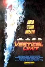
ACUTE ADULT RESPIRATORY DISTRESS SYNDROME (ARDS -- review NEJM 332: 27, 1995).
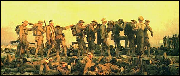 |
Gassed WWI painting Mustard gas victims John Singer Sargent |
This very common problem, deadly, and expensive problem results from anything that severely injures the Type I pneumocytes and capillary endothelial cells throughout the lungs.
There are at least 150,000 ARDS cases in the U.S. every year (as an autopsy pathologist, I'd say this about right).
 ARDS first came to be recognized during the Vietnam War.
It was called "Da Nang lung". Originally there was concern about biological warfare. But it soon became clear that the pulmonary changes were remote effects of injuries which previous soldiers did not survive.
ARDS has many causes. These include...
The "multiple organ injury syndrome" seen in survivors of severe trauma commonly produces ARDS in a week or so. Nobody knows why this is; probably widespread activation of complement throughout the body is part of the problem.
Mechanisms of injury are complex, with free radicals, complement, enzymes from marginated polys (can't be the whole story, since the neutropenic can and do get ARDS), microthrombi, aggregation of polys, "shock toxins", and many other ideas.
The major experimental model involves reperfusion of an animal's hindlimb rendered ischemic for a considerable time (J. Traum. 31: 760, 1991).
Interleukin-8, a great neutrophil attractant, abounds in fluid from ARDS lungs. We don't know why, but this probably has a lot to do with the problem.
Biopsy is often helpful to establish an underlying diagnosis even though these people are ver sick (Chest 125: 197, 2004. Here are some tips for guessing the cause of ARDS
Whatever causes the injury, the result pretty much the same:
When the alveolar cells are injured fluid leaks into the
interstitial spaces and alveolar air spaces --
this is pulmonary edema.
Later, with cell necrosis, fibrin is released into the
alveoli, producing hyaline membranes. Of course
there is loss of surfactant, so many alveoli
collapse.
During this early stage, the patient is very tachypneic and
dyspneic, but the chest x-ray looks
normal. (Why?)
NOTE: In "respiratory distress syndrome" or "hyaline membrane
disease" of low-birth-weight
infants, the lack of surfactant is one primary problem, though
not the only one. In ARDS, surfactant
is decreased secondary to diffuse alveolar damage.
* Surfactant aerosol-replacement flops for ARDS: NEJM
334: 1448, 1996.
As Type I pneumocytes are destroyed, Type II pneumocytes
divide to replace them ("regenerative
epithelial hyperplasia" or "cuboidalization" of alveolar
epithelium.) Of course, they are not so permeable to oxygen as the healthy
type I cells.
Fibrosis ensues as the intra-alveolar hyaline membranes
and the interstitial exudate organize (Am. J.
Path. 126: 171, 1987).
Of course, fibrotic lung (from any cause) is prone to develop bacterial
infections (bronchopneumonia), since it is harder to mobilize the exudate and
perhaps the neutrophils must travel farther in pursuit of the microbes.
Even where the alveoli remain ventilated, these factors
combine to cause poor pulmonary
compliance plus poor response to oxygen therapy
("alveolar-capillary block" in damaged alveolar
walls.) Further, the pulmonary vascular bed is
progressively obliterated.
Hantavirus Pneumonia
Immunoperoxidase
Dr. Hjelle
Hantavirus
Electron micrograph showing
damaged basement membrane
{06359} ARDS
The outcome depends on whether the patient can be supported and the underlying problem successfully treated before fibrosis becomes extensive.
High-dose steroids were tried in the '80's and failed to affect the outcome: NEJM 317: 1565, 1987.
Nobody knows exactly why (maybe relieving the weight pressing on the pulmonary veins), but putting the patient prone often helps oxygenation (thought not long-term survival: NEJM 345: 568, 2001).
About 50% of patients with ARDS die from it. Long, agonizing periods on the ventilator are fairly common.
| * With more patients surviving ARDS nowadays, there is considerable interest in the quality of these survivals. There is often brain damage sufficient to impair the quality of life (AJRCCM 171: 340, 2005), post-traumatic stress disorder (Am. J. Psych. 161: 45, 2004), and of course impaired lung function (Chest 123: 845, 2003; NEJM 348: 683, 2003). |  |
NEONATAL RESPIRATORY DISTRESS SYNDROME ("hyaline membrane disease", HMD, RDS, etc.)
This is the common cause of respiratory distress in premature infants (usually 1500 gm or under), beginning a few hours after birth.
In addition to prematurity, risk factors include maternal diabetes, caesarean sections, premature rupture of the membranes. Heroin addiction or glucocorticoid administration in the mother protects from RDS.
The pathophysiology is lack of surfactant plus high permeability of the immature pulmonary epithelium. A few air spaces are hyperinflated, the rest of the lung is collapsed.
The principal lesion is probably necrosis of the respiratory epithelial cells; why this happens to premature infants is unclear but probably has something to do with their being unable to tolerate even normal amounts of inspired oxygen.
Surfactant is also deficient in these patients and this accounts for part of the problem. Surfactant -- dipalmitoyl lecithin -- is the stuff that keeps the alveoli uniform in size. Recall the "two balloons on joining pipes" model in physiology.
In RDS, hyaline membranes line the open alveoli (mostly along the respiratory bronchioles). As in ARDS, they result from plasma proteins exuding through the alveolar walls. (* These kids usually have patent ductus arteriosus with marked left-to-right shunting, which greatly exacerbates this problem.)
As in ARDS, the presence of hyaline membranes is both a marker for alveolar injury, and a further barrier to gas exchange.
{11427} hyaline membrane disease, newborn
{20014} hyaline membrane disease, newborn
{20015} hyaline membrane disease, newborn
|
|
To test for maturity of an unborn child's lungs, check the lecithin-to-sphingomyelin (L/S) ratio in amniotic fluid obtained by amniocentesis -- should be 1.5 or more.
Until recently, treatment was limited to ventilatory support and oxygen, plus * drugs to dilate the pulmonary arterioles.
During the last decade, administering surfactant into the lungs, before the first breath, has become standard. This is not a panacea.
A significant number of low-birth-weight kids do suffer brain damage from hyaline membrane disease hypoxemia: NEJM 325: 276, 1991.
* The extracorporeal membrane oxygenator (ECMO) is a modified heart-lung machine for preemies. The economic-ethical nightmares still happen.
Big doses of oxygen cause further alveolar damage, resulting in fibrosis, which is given the unfortunate name of bronchopulmonary dysplasia (it is neither incipient cancer, nor a birth defect). Today it is better to call it chronic lung disease of infants.
Some of these kids get better in a few months, but the outcome
is usually bad for both lung and brain
(Pediatrics 77: 345, 1986). Many of these babies spend
months or years on ventilators before
finally dying.
Bronchopulmonary "dysplasia"
WebPath Photo
* Other problems of preemies: patent ductus arteriosus, meconium aspiration, necrotizing enterocolitis, cerebral hemorrhages from the germinal zone of the subependymal plate, preemie retinopathy (oxygen, bright lights).
* In genetic absence of surfactant, babies born at term are unable to inflate their lungs. The disease is fatal: NEJM 350: 1296, 2004.
ATELECTASIS
Collapse (or incomplete expansion) of pulmonary acini from any cause.
Obstructive atelectasis (Baby Robbins and others call it absorption atelectasis) results from non-ventilation of alveoli that are still perfused; the alveolar gas is carried away by the bloodstream.
Seen distal to tumors, foreign bodies, mucus blobs, post-surgical discomfort preventing cough, enlarged hilar nodes (cancer, TB -- producing "the right middle lobe syndrome") etc. Before antibiotics, this was a setup for bronchiectasis.
If an airway is obstructed, expect that over time the alveoli will fill with surfactant, which will mostly be engulved by macrophages. This is the "obstructive pneumonia" or "golden pneumonia", and often is the first x-ray sign of lung cancer.
Compressive atelectasis results from something in the pleural cavity (blood, exudate, tumor, air.)
If extensive and unilateral, obstructive and compressive atelectasis can be distinguished by the direction in which the mediastinum is shifted on x-ray (think about it).
A deficiency of surfactant produces "patchy atelectasis" in both hyaline membrane disease ("fetal atelectasis" -- * a term also used for lungs of stillborns who never breathed) and ARDS (see below.)
{10228} atelectasis (left lung
of a baby whose left bronchus failed to form)
* Round atelectasis ("folded lung", "shrinking
pleuritis"): a vanishing coin lesion, resulting when
normal lung parenchyma is crunched into a little ball beneath a
shrinking pleural scar. Familiar to
radiologists, sometimes operated for
diagnosis.
SUDDEN INFANT DEATH SYNDROME ("SIDS", "crib death", "cot
death", "today, the most
common cause of death during infancy" -- Br. Med. J. 298:
689, 1989; "the seventh leading cause of
years of potential life lost before age 65, commensurate with
AIDS" -- MMWR 37: 644, 1988, etc.,
etc.). The following will upset you.
Sudden death in an apparently healthy baby, less
than one year old, with no explanation even after autopsy.
Sources in the 1970's and 1980's claimed that this affected
2-3 per 1000 live births, "the single most common cause
of infant death" (Baby Robbins,
etc., etc.) -- slightly more common than baby death due to birth
defects.
There is no question that genuine SIDS cases exist, i.e.,
some babies do die from disturbed pathophysiology
without any environmental or anatomic explanation.
A few causes are known (i.e., channelopathy) or suspected
(actual problems with respiratory drive; these must be rare).
But to understand
"sudden infant death",
we first need to sort out deaths that aren't "SIDS" at all.
Thankfully, this is now happening.
During the 1960's, mostly
because of the militancy of a single pediatrician-activist,
certain anti-common-sense ideas about the sudden deaths of infants were
suddenly and uncritically accepted, both by the medical community
and by the public. Looking back, this had a lot to do with
the "feel good / no blame / flower power" mentality of the times.
It was dogma that:
That this was wishful thinking should have been obvious, even at the time.
The classical autopsy finding in SIDS is petechiae over the
thymus, lungs, and heart, without other
abnormalities... or with nasal hemorrhages too. And
this is exactly
what you'd expect to find in a baby who has been suffocated -- accidentally,
intentionally, on the bedclothes, or against the mattress (JAMA
263: 2865, 1990; Arch. Dis. Child. 85: 116, 2001).
And it was already well-known that
"higher rates are encountered in many
developing countries" (Baby Robbins), and
especially among the underclass in industrial countries
(Practitioner 232: 577, 1988; Arch. Dis.
Child 65: 830, 1990; risk for First Americans correlates
with underclass risk factors rather than
genes J. Ped. 121: 242, 1992; also & JAMA 288: 2717, 2002).
Some classic reported SIDS
death rates:
0.04%... Rich New York suburbanites
0.4%... Poor New York slum dwellers (both from NEJM
315: 100 & 126, 1986)
0.35%... North Plains Indian reservations (JAMA, above)
2% (!!) Children of British criminal offenders
(Arch. Dis. Child. 62: 146, 1987.
near zero... Hong Kong -- attributed to zero privacy and many
helpers for mothers, no junk in the cribs, "nobody
lets her baby sleep prone", "no unwanted babies": Lancet
2: 1346, 1985; Br. Med. J. 298: 721,
1989.
It is now obvious that many (if not most)
deaths signed out as "SIDS"
during the decades of ignorance were the results of negligence
or abuse by family members. This was substantiated
in the 1980's by careful death-scene
investigators (filicide Arch. Dis. Child. 60: 505, 1985;
NEJM, above; Lancet 1: 313, 1986; Lancet
1: 199, 1989; J. Ped. 117: 351, 1990.)
There was never any basis for believing that overlying
cannot kill a baby. (King Solomon's judgement; Yeats's Moll Magee;
most cultures historically have simply accepted that overlying
kills babies.)
See also Am. J.
Forensic Med. & Path. (8): 256,
1987;
when we
stop kidding ourselves, overlying turns out to be common: Am. J.
Dis. Child. 146: 968, 1992.)
Sharing a bed with a parent as a risk for SIDS: Br. Med. J. 319: 1457, 1999;
Arch. Path. Lab. Med. 126 343, 2002; J. Ped. 147: 32, 2005;
Arch. Dis. Child. 88: 1058, 2003;
Lancet 363: 185, 2004 (painfully obvious),
sofas are worse and so is sleeping with Mom when she is drunk or on drugs
(go figure; Arch. Dis. Child. 88: 112, 2003).
Drug use by Mom ("during pregnancy", and of course most
likely continuing after) is a very
powerful risk factor for SIDS, especially when the drug is heroin
or methadone, and when other risk
factors (poverty, smoking, and so forth) are controlled-for (J.
Ped. 123: 120, 1993). A classic
observation is a higher incidence (+ 60% or so) of "SIDS" on
weekends (Aust. N.Z. J. Med. 18:
861, 1988; still true Arch. Dis. Child. 89: 670, 2004), when Mom and Dad/current boyfriend are more likely to be drunk or stoned.
This is every bit as true today as before the "back to sleep" campaign
(Arch. Dis. Child. 89: 670, 2004 -- no surprise).
And children of schizophrenic mothers have around five times the risk of "SIDS"
(Arch. Gen. Psych. 58: 674, 2001).
The fact that "SIDS is more than ten times more frequent if a
sibling has already died of SIDS"
(Arch. Dis. Child. 64: 179, 1989) hardly proves that "bad
genes" are responsible. (No adoption
studies are available yet....) The fact that SIDS rates are more than double for very young
parents (J. Ped. 116: 520, 1990)
hardly proves that some mysterious intra-uterine factor is
involved. Nor does the fact that SIDS
rates are double if the parents smoke (Am. J. Pub. Health
80: 29, 1990) prove that the cause is
subtle irritation of the airways.
And the fact that twins used to die of
"SIDS" at exactly the same time
invites an obvious
conclusion (Am. J. Forens. Med. Path. 10: 200, 1989;
AJFMP 19: 195, 1998). The fact that this no longer happens
(Arch. Ped. Adol. Med. 153: 736, 1999) just says to me that
pathologists are no longer stupid enough to call these "SIDS".
The traditional wisdom that "SIDS" typically follows a minor
illness now seems to be unfounded
(Lancet 300: 1237, 1990).
The claims about immunization being a risk factor are clearly untrue;
statistically, children actually seem protected (Br. Med. J. 322:
822, 2001; the role of coincidence Pediatrics 115: e643, 2005).
And the familiar junk-science claim by
breast-feeding militants that SIDS is due to
bottle feeding simply isn't true: Br. Med. J. 310: 88,
1995. At best, the relationship is statistical, and it's weak.
However, the SIDS organizations still tell people that
breast-feeding protects children from SIDS, which must put a terrible
burden of guilt on people whose babies died of the real thing.
Possibly a few infants do die from failure of respiratory
drive during sleep. "Near-miss" apnea and
related respiratory rhythm disturbances are demonstrable in some
siblings, though the findings are
notoriously un-reproducible.
* I was impressed only by Hum. Genet. 73: 39, 1986 -- a
family with very little myelin in the
respiratory centers of their medullas). Since this hasn't been
replicated, I wonder about the
neuropathologist's myelin stain. Finding hypoplasia of the
arcuate (CO2 sensor) nucleus of the
medulla in a subset of SIDS is interesting: J. Neuropath.
51: 394, 1992; I'd bet these brains are those
that don't respond to having the mouth and nose up against the
mattress.
An Italian group claims to have found "frequent alterations, mainly
cogenital, of the autonomic nervous system" (Am. J. Clin. Path. 124:
259, 2005; also J. Clin. Path. 58: 77, 2005). Stay tuned.
Otherwise, "near-miss apnea seen in siblings" is peculiarly
non-lethal: Science 264: 197, 1994. I
suspect that, in most cases, "near-miss SIDS" is Cheyne-Stokes
respirations (as a junior med student
sharing a call room, I was told I do this while asleep) or
transient obstructive sleep apnea. It is now obvious that some cases of SIDS result from laying
babies prone (Pediatrics 93: 814, 1994;
Pediatrics 105: 650 2000;
JAMA 273: 783, 1995, lots more).
Most plausibly,
sleeping babies might simply fail to move
when their faces lie flat against mattress, and smother in this
way.... (gee whiz) ... or rebreathe carbon
dioxide, which is pretty much the same thing (Am. J. Dis. Child.
147: 642, 1993; J. Ped. 122: 874,
1993). This is consistent with some studies which strongly
suggest prolonged hypoxia has occurred
prior to death in many SIDS cases (Pediatrics 87: 306,
1991, others).
There has been about a 50%
reduction in "SIDS" in countries where there's been a campaign to
get parents not to place their
babies prone. The impact is extremely
obvious (Lancet 363: 185, 2004).
In the U.S., where "SIDS activists"
insisted for two decades that "SIDS is not your fault
and a child cannot smother against the mattress", the campaign was
delayed.
Figure out yourself how many children died as a result.
"Suffocated prone: the iatrogenic
tragedy of SIDS": Am. J. Pub. Health 90: 527, 2000.
Strangely, in the US, breast-feeding militants are still pressuring
women to sleep with their infants.
There's some interesting work on facial morphology in these
kids -- their upper and lower jaws seem to be, on average,
farther back, and perhaps this makes it easier for them
to smother on their mattresses (Lancet 317:
293, 1998). The relationship with obstructive sleep apnea --
which this group claims is more common in SIDS families --
is going to take more study to work out.
When you are working in the neonatal intensive care unit, you
will frequently see preemies that have
trouble breathing or even stop breathing ("apnea of prematurity")
or suffer cardiac rhythm
disturbances. These have nothing to do with SIDS (Pediatrics
77: 811, 1986; Pediatrics 78: 787,
1986).
Public awareness of "SIDS" resulted in a huge "apnea
monitor" industry (racket, or "expensive
cover-your-butt stuff", as almost everybody will tell you nowadays).
Today, the people who
really know about SIDS laugh at the
idea that monitors save lives (see, for example, Chest 91:
898, 1987; Can. Med. Assoc. J. 140:
1072, 1989), but they did keep parents from staying up all night
watching their babies breathe.
"Consensus document" on home monitoring: Pediatrics 79:
292, 1987; noncompliance or worse:
Am. J. Dis. Child. 142: 1037, 1988. By 1994, the idea
that apnea is an important cause of SIDS is
dismissed by almost the entire scientific community, though still
believed by physicians (Science
264: 197, 1994); however it's still routinely prescribed
(Arch. Dis. Child. 84: 270, 2001).
To clinch the matter, Waneta E. Hoyte,
the mother whose "tragic story" led to the
paper (Ped. 50, 646, 1972) that spawned the apnea monitor
business confessed in 1994 to having
smothered her five children. ("Their screaming made her feel
useless": Ped. 93: 944, 1994). Some "clever"
parents learn to stop their kid from crying by smothering it
into unconsciousness; this will load the
lungs with hemosiderin-laden macrophages (why?).
Eventually, these kids are likely to die.
Pathologists
are now routinely signing "SIDS" cases with lots of these macrophages
as "manner
of death undetermined" (J. Clin. Path. 52: 581, 1999)
and staining all infant lungs to see hemosiderin
(Am. J. For. Med. Path. 23: 360, 2002.
* Believe it or not, some "ethicists" got very upset over the invasion of the
British parents' privacy
and "breach of trust" between the medical community and the abusers
(Med. J. Aust. 160: 352, 1994).
The British estimate that 1 in every 10 SIDS cases is a covert homicide
(Arch. Dis. Child. 89: 443, 2004); whatever you think of the estimate,
common-sense tells you when to suspect murder.
SIDS "research" and political agendas: Arch. Dis. Child 88: 1085, 2003.
Before you write "SIDS" on a death certificate, try to rule
out:
* Kids with febrile seizures are not at increased risk for SIDS,
so it is probably unacceptable to invoke a seizure as causal: Arch. Dis. Child. 86:
125, 2002.
But after you've done all this, can your autopsy rule out
smothering or all other forms of lethal
trauma? In a word, "No" (Am. J. Dis. Child. 144: 137,
1990). The SIDS autopsy: J. Clin. Path.
45(S-11): 11, 1992.
* The pathology lab has nothing to offer for the differential
diagnosis of "near-miss SIDS" (Am. J.
Dis. Child. 140: 484, 1986).
Protocol for the SIDS autopsy: J. Clin.
Path. 40: 481, 1987.
When a second SIDS death takes place in a home, the pathologist's
task is especially difficult. One big study concluded
that most of these are natural, and that many are homicides,
and that sorting them out definitively is impossible (Lancet 365:
29, 2005). Probably you have to call the child protection folks
(Arch. Dis. Child. 88: 699, 2003).
CHRONIC OBSTRUCTIVE PULMONARY DISEASE (COPD, chronic air flow
obstruction,
* COAD, * CAWO, * chronic airway obstruction):
The most
common cause of activity-restricting disability in the U.S.
Morphology and pathophysiology
review: Lancet 364: 709, 2004.
Clinical review (no surprises): Lancet 362: 1053, 2003.
"Obstructive lung disease" is a traditional and irrational
grouping of four illnesses:
1. chronic bronchitis
The term generates tremendous confusion, and I urge you not to
use it.
Chronic bronchitis causes obstruction to air flow
because of edema, necrosis, fibrosis, and recurring
infections in the bronchial tree.
The walls do get thicker and this narrows
the bronchial lumens.
This has been confirmed by some good pathology studies
(Am. Rev. Resp. Dis. 143: 1152,
1991; Chest 97(S2): 6S, 1990), and now we see it on high-resolution
imaging studies. Although narrowing
of the bronchial lumens ("thickening of the bronchial walls")
interferes some with breathing,
it's usually the severity of the
co-existing emphysema that really disables these
people.
Increased mucous secretions and hyperplasia of
mucous-secreting glands by themselves, though typical of chronic
bronchitis, are now thought not to contribute
significantly to obstruction (i.e., it's not prolonged suffocating
on hockers). Am. Rev. Resp. Dis. 128:
491, 1983; Am. Rev. Resp. Dis. 133: 942, 1986.
* Constrictive bronchiolitis" is a striking lesion seen in a minority
of smokers, and in some other lung diseases. It's dense fibrosis
around bronchioles with serious compromise of the lumens.
Emphysema causes obstruction to air flow because loss
of the lung's elastic recoil allows small
airways to collapse at the beginning of expiration. We all lose
some elasticity as we age, but
smokers and people who are alpha-1 antitrypsin deficient
lose it much more, and also have
some destruction of their septa, which is not part of normal
aging.
NOTE: Some people include all asthma cases as COPD, but
regular asthma has a much better
prognosis. Today, even chronic "asthmatic bronchitis" is
distinguished from other forms of chronic
bronchitis, which are much more lethal: NEJM 317: 1309,
1987.
Both emphysema and chronic bronchitis are most commonly caused
by cigaret smoking. Most
smokers with one have the other, too.
Smokers should be prepared for:
Baby Robbins's estimate of "10,000 to 20,000 deaths in the
U.S." yearly due to COPD is much too
low. COPD probably contributes in a major way to around 100,000.
* Supposedly the neuropathies seen in these patients
are due to hypoxemia itself.
* Not a trivial problem -- advising the COPD patient about air
travel: Ann. Int. Med. 111: 362,
1989.
EMPHYSEMA
{11506} emphysema
The old-fashioned anatomists define "emphysema" as an
abnormal, permanent dilatation of part of
all of the acinus, with eventual destruction of many of the
alveolar walls.
I predict that the definition will eventually be changed to
reflect the true sine qua non -- loss of
elasticity of the lung. The new definition may also include the
requirement that there be some
destruction of the alveoli.
You'll diagnose emphysema on lung function tests by noting
prolongation of the time
required for a full forced expiration,
in the absence of asthma.
Two common forms are distinguished:
Centrilobular (centriacinar) emphysema shows
more dilatation of the respiratory bronchioles and
their alveoli.
Traditionally, this is seen
in early smoker's emphysema "because cigaret
smoke exposure is heaviest in the centers
of the acini". And "it is worse in the upper lobes, because they
get more smoke exposure" (why?).
Panlobular (panacinar) emphysema involves the
acinus uniformly.
Traditionally, this is caused by alpha1-protease
inhibitor ("antitrypsin") deficiency "because the
blood brings neutrophils to all of the lung uniformly". And "it is
worse in the lower lobes, because
they get more blood" (why?)
By the time emphysema patients come to autopsy,
the damage is so severe that you won't be able to make the
distinction, and it's not really something
that matters to the
clinician, radiologist, or patient.
There are about
2 million diagnosed emphysema patients in the U.S. at any time, and the
real prevalence of this disease is
much higher.
The pathogenesis is pretty clear.
Emphysema is not caused by air trapping behind inflamed
bronchioles (asthmatics don't develop
emphysema from it) or playing wind instruments (Chest 88:
201, 1985).
The problem is damage to the elastic fibers
of the lung by elastases from polys,
monos, possibly pancreas. Of course, smoking cigarets brings
lots of polys to the lung.
Deficiency of serum alpha-1-antitrypsin
(our major * "serpin" -- serine protease
inhibitor) is an inborn error of metabolism in which severe
panacinar emphysema develops in
non-smokers.
This results from inability to efficiently release an abnormal
anti-protease from the hepatocytes.
Inclusions develop in the cells. The defects is the result of
alleles at the Pi ("protease-inhibitor")
locus -- "M" is the common allele, while "S", "Z", and others
result in less product getting into the
bloodstream. Homozygous patients may also get cirrhosis. Whether
heterozygotes are more at risk
for emphysema if they smoke is the subject of much discussion, as
they have intermediate levels of
anti-protease. Replacement therapy with the enzyme ("Prolastin",
watch for "Respitin") is now in use
for these patients, at a cost of
around $36,000 per year (Chest 119: 745, 2001; third-party payers
are of course balking and estimates of the cost of a year of life saved
vary widely: Chest 117: 875, 2000).
* Social factors in disease: The Swedes screened all newborns
for SZ and ZZ and warned all parents
of these children to stop smoking. They discovered that none of
the parents complied (Thorax 43:
505, 1988).
Because of loss of elastic recoil, small airways collapse
during forced expiration.
The classic "emphysema" patient is a "pink puffer", with normal PaCO2, barrel chest, pursed lips,
dyspneic, tachypneic, thin (he's working hard all the time),
miserable. The only consistent finding
on physical exam is slowing of forced expiration ("Can you blow
out a match at six inches with your
mouth wide open?").
Pink puffers learn to keep their lungs hyperinflated to keep the
respiratory bronchioles from collapsing,
and this eventually changes the shape of the chest itself
("barrel chest", "increased AP diameter",
"increased total lung volume").
These people eventually start getting bacterial lung
infections, and die of cor pulmonale, pneumothorax, or pneumonia.
At autopsy, the lungs are hyperinflated and relatively
bloodless. Eventually, broken alveolar septa
dangle in the breeze, and many of the capillaries in the septa
are gone (we don't really know why the
latter occurs).
In 1996, the NEJM (334: 1095, 1996) published an
article showing that the newly-popular "lung
reduction surgery" (lung-lift, like face lift or certain other
cosmetic procedures to reverse sagging, $75,000)
produces improved exercise ability for a
while. Just as you'd expect from what you've learned
about emphysema, people who have little inspiratory
resistance (i.e., wide open larger airways) do nicely,
while those with really badly-narrowed bronchi are not benefited
(NEJM 338: 1181, 1998). Update NEJM 343: 239, 2000
(better exercise tolerance, no proof of longer survival).
It is now very common and the benefits are obvious (Chest 123: 1838,
2003).
Changing times: Medicaid decided every
smoker on welfare did not have a
"right" to this surgery, and surprisingly few people were
outraged.
Bullous emphysema produces air-filled blebs (if >2
cm, you can call them "bullae") containing little
or no lung tissue, usually at the apices, sometimes (but by no
means always) at the sites of old TB
scars.
Most cases probably result from common emphysema, with the
inelastic lung "collapsing under its
own weight" (Thorax 44: 533, 1989); the upper lobes have
more contact more tobacco smoke
because they are better ventilated.
The blebs may be removed surgically, with improvement in the
"pink puffer"'s puffing.
Blebs are also prone to rupture, causing pneumothorax and
sudden death. Iatrogenic disease: "IPPB
breathing treatments" are irrational therapy for uncomplicated
emphysema, and kill patients by
blowing out blebs.
{10778} blebs in emphysema
Other forms of "emphysema":
* Paraseptal (distal acinar): rare, blamed for spontaneous
pneumothorax in young people. I suspect
this is a mythical process.
Compensatory (i.e., following removal of a lobe of a lung, the
other lobes expand; this is a
misnomer, as there is no destruction of alveoli, and no loss of
elasticity.)
"Irregular" or "tractional" (i.e., after scarring, etc.;
another misnomer.)
"Senile" (loss of elasticity without loss of lung substance,
from "old age")
"Interstitial emphysema" doesn't even refer to lung. It means
air has been forced into the fibrous
tissues of the body, often as the result of tearing of the lung
itself (by real emphysema, by severe
coughing, by a respirator, by a broken rib, by barotrauma). Listen for the
"milkman's crunch" sign as the heart
beats, palpate the little bubbles under the skin, and reassure
the patient that it will reabsorb.
CHRONIC BRONCHITIS ("smoker's cough" -- never trivial) Defined clinically, as persistent cough with sputum
production for at least three months in at least
two consecutive years. (Worth committing to memory.)
It's not the cough
Again, the usual cause is cigarets.
Marijuana isn't exactly good for the
lungs either (gee whiz, JAMA 259: 966, 1988;
NEJM 318: 547, 1988) but so far hasn't produced wards full
of respiratory cripples.
The "classic chronic bronchitis patient" is a "blue bloater",
with increased PaCO2, obese, edematous
(cor pulmonale), cyanotic, producing much sputum, happy
(CO2 narcosis.)
The distinguishing feature of this kind of patient is an
acquired tolerance for the hypercarbia that
poor ventilation (i.e., from emphysema) ultimately causes.
Unlike "pink puffers" (who retain their
hypercarbic drive), these patients no longer really struggle to
breathe, so long as they have adequate
oxygen.
Because of the poorer alveolar ventilation, (* and perhaps
with a contribution from scarring around
airways and vessels, and the fact that they present
later in the course of their disease),
pulmonary hypertension supervenes earlier
in "chronic bronchitis" patients
than in emphysema patients.
Exacerbations and death often follow infection with S.
pneumoniae or H. influenzae.
Death may also result from cor pulmonale or from apnea brought
about by breathing oxygen
(remember, hypercarbia no longer stimulates respiration in these
patients.)
Many clinicians today are treating chronic bronchitis with
glucocorticoids "to control the
component of bronchospasm, as in asthma". I suspect they're
actually making the walls of the inflamed large bronchi thinner,
with even a little bit of widening the lumens helping a lot.
As an autopsy
pathologist in the 1980's, I was very impressed with the ability
of systemic glucocorticoid side effects to kill these people.
Today's pulmonologists are switching to inhaled glucocorticoids
(Lancet 351: 773, 1998).
Genetic factors seem to determine whether a smoker becomes a
"pink puffer" or a "blue bloater."
See Am. Rev. Resp. Dis. 129: 207, 1984. The onset of
"chronic bronchitis" is supposed to be earlier
than emphysema, which is understandable since it will be
diagnosed by hearing the patient rattle all those
hockers, while emphysema is diagnosed only when shortness of
breath becomes profound.
At autopsy we find copious secretions in the airways, even in
the absence of pneumonia. The
trachea itself may be almost filled with yellow slime.
Microscopically, we see thickening of the bronchial basement
membrane (seen also in asthma),
proliferation of goblet cells, hypertrophy and hyperplasia of
mucous glands, more of the various inflammatory
cells, more lymphoid aggregates and follicles than usual,
and often a considerable amount
of scarring (histopathology update on "smoker's small airways disease":
NEJM 350: 2645, 2004).
"Reid index" is ratio of thickness of submucosal mucous glands
to entire submucosa. Normal is up
to .4; increased in chronic bronchitis.
* In the worst cases, we see widespread obliteration of the
lumens of the terminal bronchioles (i.e.,
bronchiolitis obliterans or denser scarring).
{08761} chronic bronchitis (chronic
inflammation, missing epithelium)
* "Coarse breath sounds" / "coarse rhonchi"
are hockers moving around in the big
airways. Make the patient cough,
and they'll perhaps go away or at least change.
Despite elaborate systems of testing pulmonary function, the
ultimate diagnosis of "COPD" is made
on history and physical exam. This applies to most other
disease, too (Am. J. Med. 94: 188, 1993).
BRONCHIAL ASTHMA (some lump it with
"small airways disease"; BMJ 314: 45,
1997; NEJM 325: 425, 1991)
Common syndrome (10% of kids, 5% of adults) in which the small
bronchi are abnormally
responsive to various stimuli which cause constriction and/or are
considerably inflamed (usually both). This
produces episodes of dyspnea, wheezing, cough.
You'll hear plenty of wheeze sounds through your stethoscope;
the sound is air rushing through
narrow airways, making noises like the wind section of the
orchestra playing out of tune.
Asthma kills around 3000 people in the U.S. each year.
Very few
of these people took good care of themselves (no-nonsense
article: Thorax 44: 97, 1989); and
asthma death is primarily an underclass phenomenon (NEJM
331: 1542, 1994). Likewise, the asthmatic children
who die are for the most part the underclass ones, as a result
of smoky homes and "disease mismanagement":
Ped. Clin. N.A. 50: 65, 2003.
Lately there's been much interest in chlamydia TWAR, as a
chronic infection, as cause of asthma -- and six months
on a macrolide to effect a cure.
Update AJRCCM 171: 1083, 2005 (many childern harbor it);
AJRCCM 167: 406, 2004 (but few adults); Chest 121: 1782, 2002
(macrolides help if and only if chlamydia are present).
Mast cell factors appear to mediate the bronchoconstriction
regardless of what triggers the attack.
These factors include histamine, bradykinin, leukotrienes
("SRS-A", etc.), prostaglandins (must be present: Science 287:
2013, 2000), probably
others.
We're still learning what's in those mast cells.
* If you actually look inside (rather than below) the
epithelium, asthmatics
actually average ten times as many intraepithelial mast cells as
their
non-asthmatic counterparts (Am. Rev. Resp. Dis. 148: 80,
1993).
All about arachidonic acid metabolites in lung disease: Am.
Rev. Resp. Dis. 143: 188, 1991.
Leukotrienes are of course notorious for being chemotactic for
inflammatory cells, and for
tightening smooth muscle.
Asthmatic attacks are often triggered by:
* "I wasn't surprised": Allergy shots offer no benefits for
asthmatic kids getting appropriate medical
treatment: NEJM 336: 324, 1997
Allergic asthma is said to be present when the
patient's attacks are typically triggered by
IgE-mediated hypersensitivity.
This often is severely disabling in childhood, though it
generally gets better in adult life. An old
misnomer is "extrinsic asthma".
Remember both the familiar inhalants and food allergy (J. Allerg. Clin. Imm. 112: 168, 2003).
Remember histamine, leukotrienes, prostaglandin D2,
and platelet activating factor as the major
contributors to this kind of wheezing, with leukotrienes perhaps
most important.
Industrial asthma is a serious problem.
The worst offenders are cedar wood
platinum salts, anhydrides (epoxy hardeners) and
isocyanates,
followed by
proteolytic enzymes, epoxy resins themselves,
lab animals, vinyl chloride used in meat packing, flour, crab
processing, oil mists,
and penicillin. Formaldehyde asthma is a problem for a few unlucky
pathologists.
* Platelet-activating factor antagonists are on the way: Chest
108: 529, 1995. Montelukast / zafirlukast
for leukotriene receptor blockade
(JAMA 279: 455 & 1181, 1998) -- works wonders for
exercise-induced bronchoconstruction
(chilling and drying?)
and mild allergic asthma (NEJM 339: 1998).
We believe that some of the longstanding epithelial havoc is
wrought by major basic protein of
eosinophils recruited to the sites of the reaction. Be this as
it may, another eosinophil protein
crystallizes as "Charcot-Leyden crystals" in the sputum of
allergic asthmatics.
If a chronic aspergillus infection gets established in an
asthmatic's lungs,
allergy to this fungus is likely to make the
asthma much worse).
As many as 15% of severe
cases get this. In "allergic bronchopulmonary aspergillosis",
the fungi actually find safe haven inside the plugs, creating a vicious
cycle.
Idiosyncratic asthma is said
to be present when the patient's attacks are
typically triggered by exposure to aspirin (Chest 88: 387,
1985), another cyclo-oxygenase inhibitor,
and/or tartrazine yellow.
These people's small airways are teeming with eosinophils and
mast cells
(Am. J. Resp. CCM. 153: 90, 1996, NEJM 346: 1699, 2002), the obvious source for
the
extra leukotrienes.
This is more likely to begin in adult life. An old misnomer
is "intrinsic asthma". (Look for nasal
polyps in the aspirin-sensitive patient.)
Reactive airways disease is a current concept (some might
say a fad) in which asthma follows a single noxious exposure of the
bronchial mucosa to something hurtful (i.e. poison or hot gas, poisonous
fumes). Other folks use the term as a synonym for triggerable asthma.
Regardless of cause, the pathology in asthma is inflammation
of the bronchial mucosa, with eosinophils, and
(probably also, maybe as a result) increased fragility of the
epithelium. Review Chest 124: 32, 2003.
The smooth muscle is
also likely to be hyperplastic. This (and some increase in
collagenization) is the principal histologic feature
correlating with severity (AJRCCM 167: 1360, 2003).
Part of the trouble, we may reasonably think, is irritation of
the C-fibers that travel up the vagus
nerve and mediate bronchospasm by an autonomic reflex.
The worse the asthma, the worse the inflammation (Am. J. Resp.
CCM. 154:
24, 1996). No surprise. Anatomy of asthma, with a focus on the
long term
"repair" changes which actually make things worse: J. Allerg.
Clin. Imm. 98:
S278, 1996.
Only a small percent of asthmatics lack eosinophils, and it's not
clear whether this is the same disease: Am. J. Med. 115(S-A3): 49S, 2003.
In the animal model, the airways do not undergo much fibrous thickening
if the animal has no eosinophils (Science 305: 1776, 2004).
Fatal cases ("status asthmaticus") show mucosal edema, many
eos, polys, mucous secretions,
desquamation, and obvious basement membrane thickening (see
Thorax 87: 152-S, 1985).
A study of non-fatal asthma cases showed that all had
thick bronchiolar epithelial basement
membranes. Non-asthmatic controls had no mast cells or
eosinophils, while these were present in
most of the asthmatics. Asthmatics also showed more of a
tendency for the epithelium to fragment.
See Am. Rev. Resp. Dis. 145: 922, 1992.
The thickening of the basement membrane, hyperplasia of smooth
muscle, increase in goblet cells,
and hyperplasia of bronchial glands is now called "remodelling"
and is the most-studied phenomenon in asthma (Am. J. Resp. CCM 164:
S46 & S52, 2001); the causes remain as mysterious as always.
Sputum from most asthmatics with type I immune injury shows
Charcot-Leyden crystals (eosinophil
alkaline proteins), Curschmann's spirals (coily strings of
altered goo from little airways).
Chest deformities (barrel, pigeon, Harrison's sulcus) often
result from severe childhood asthma
(JAMA 259: 1722, 1988).
{08228} asthma, hyperinflated lungs (notice
that they completely cover the heart in front)
The mainstay of treating asthma is inhaled glucocorticoids,
which (when not abused) are incredibly
safe (NEJM 332: 868, 1995; Thorax 49: 1185, 1995).
Best used in low doses daily; they actually prevent the anatomic
pathology (inflammatory cells, cytokine production, even basement
membrane thickening) which allow acute attacks to occur
(Am. J. Resp. CCM. 150: 17, 1994).
* Did you know that glucocorticoids cause eosinophils to
undergo apoptosis? (Am. J. Resp. CCM. 154: 237, 1996)
* Acupuncture totally fails when tested as a treatment for asthma:
Chest 121: 1396, 2002.
* Although I have not seen this in the current literature, I
was told twice in the 1990's,
both times by members of "conservative-religious-family-values"
identity-groups, that children are only wheezing to get
attention and should be punished for it. If I'm hearing this,
then so are at least some of your patients.
I urge you to call this what it is -- one more stupid, ugly lie. I've
taught you how to deal with these things ("If
that were true, then wouldn't you be able to observe....?")
* Regular spinal manipulation is heavily promoted to the public by many chiropractors
for childhood asthma. Since childhood asthma almost always gets better by
itself, a controlled study would seem to be indicated --
and finally has been done (no measurable benefit: NEJM 339:
1013, 1998 -- in an issue with two positive studies of the
value of manipulation for low back pain.) "All that wheezes is not asthma." Wheezing may also result
from:
* This might be a good place to mention pepper spray,
used by law enforcement and private citizens. You may hear it alleged
that this has killed people; I find this hard to believe and
so do the more scientifically-minded reviewers (Am. J. For. Med. Path. 16: 1995).
BRONCHIOLITIS OBLITERANS ("cryptogenic organizing
pneumonitis", "bronchiolitis
obliterans organizing pneumonia", "BOOP")
This is a histopathologist's word for a lesion in which
nubbins of loose connective tissue develop
and plug the respiratory bronchioles and alveolar ducts and
spaces.
Air flow is obstructed, and lung expansion restricted.
This unhealthy situation can be seen in any of the following:
BRONCHIECTASIS ("ectasia", or "pulling wide" of the bronchi)
Defined to be the permanent cylindrical dilatation and
ulceration of part of the bronchial tree.
The clinical picture is chronic cough with sputum production,
up to many cups daily.
(Bronchiectasis is on a continuum with chronic bronchitis.)
The dilatation ("-ectasis") results from contraction of scar
surrounding the bronchus, and from
surrounding atelectasis.
{5762} bronchiectasis
Bronchiectasis complicates respiratory infections (severe, or
those in the immunosuppressed),
asthma, cigaret smoking, inherited disease (especially cystic
fibrosis and primary ciliary
dyskinesis/immotility, including Kartagener's.
In older days when bronchiectasis was much more common, it
often followed measles,
staphylococcal or pertussis pneumonia. This is very rare
nowadays.
Regardless of the predisposing lesion, the proximate cause is
a bacterial infection ("normal throat
flora", H. influenzae, S. pneumoniae, anaerobes).
Once established, bronchiectasis will persist due
to a vicious cycle of exudation and infection.
Bronchiectasis results in serious social problems in many
cases; it may lead to amyloidosis, brain
abscesses, cor pulmonale, and so forth.
The anatomic pathologists sees bronchi pulled wide by
contraction of scar surrounding the ulcerated
airways. (Ignore the quaint subclassification
of "saccular", "cylindroid", and
"fusiform" bronchiectasis).
Modern medical and surgical treatment has improved the outlook
for these patients greatly.
OBSTRUCTIVE SLEEP APNEA: NEJM 347: 498, 2002
Sleep apnea victims all snore famously
(snoring correlates with excessive daytime sleepiness
and other quality-of-life issues: Chest 110: 659,
1996). Most are overweight
(more fatty tissue in the upper airway)
or have huge tonsils or jaw deformities. Most sleep in the
supine position. Most have "thick necks"
and/or drink a lot (Am. Rev. Resp. Dis. 141: 1228, 1990;
Thorax 46: 86, 1991). For some reason,
premenopausal women are seldom affected.
* Worth remembering in treating patients: Obstructive sleep
apnea often accounts for deterioration in
patients with hypothyroidism (myxedema) and Down syndrome (Br. Med. J.
296: 1618, 1988).
As the victim starts to go into deep sleep, his upper airway
closes (physiology: JAMA 266: 1384,
1991), he thrashes, snorts, partly wakes up, and re-opens the
airway with a gasp. The cycle repeats
every few minutes through the night, and the patient never gets
to sleep soundly.
Sleep apnea results in morning headaches, daytime somnolence,
"narcolepsy", cognitive and
behavioral changes, school and job failure, "psychiatric
disease", divorce ("She refused to sleep with
me any longer"), auto accidents (infamous,
"I fell asleep with no warning"
or "I blacked out":
Arch. Int. Med. 151:
1451, 1991; NEJM 340: 847, 1999), "near-miss SIDS",
sudden "cardiac" death,
snooze angina
(Lancet 345: 1085, 1995), hypertension (Br. Med. J. 320:
479, 2000, lots more),
etc.
* It is doubtful that sleep apnea takes many years off life
expectancy, but it
does have a very negative impact on quality of life (Chest
94: 531, 1988). Neglected, it exacerbates the common
cardiovascular diseases (JAMA 290: 1906, 2003).
Occasionally it does kill
people (Chest 94: 9, 1988), and is now being "linked to
dementia" (Chest 95: 279, 1989).
Most cases seem to involve subtle abnormalities of throat
anatomy and mechanics:
Lancet
337: 597, 1991 (* more type IIa muscle
fibers in these people's throats), as well as problems with the
reflexes which keep the throat open
(Am. Rev. Resp. Dis. 143: 810, 1991).
* A higher-than-expected percentage also have lung damage from
smoking, making the clinical
picture more complex.
The hypoxemia, hypercarbia, and/or straining against a closed
upper airway may produce deadly
cardiac arrhythmias ("fatal heart attack while sleeping", maybe
even "SIDS"). However, the large
majority of people with sleep apnea do die while awake (Chest 94: 531,
1988).
For documentation, call the sleep lab and have them do
polysomnography on your patient
(* $1200).
Treatments:
Have the patient sleep on one side, so the uvula flops
out of the way (South. Med. J. 79: 1061,
1986). If you like, duct-tape a tennis ball to the back of his
neck.
The drug protriptyline helps the brain maintain a
patent airway.
Uvulopalatopharyngoplasty (an operation) and/or
tonsillectomy is moderately useful.
Machines that provide continuous positive airway
pressure work wonders for most victims ($1000)
-- Am. Rev. Resp. Dis. 137: 1238, 1988.
Tracheostomy is the last resort but cures sleep apnea.
* Mini-tracheostomy for this disorder: Thorax
44: 224, 1989.
Pickwickian syndrome: severe sleep apnea resulting in
loss of hypercarbic drive (sleep-apnea's blue
bloater). Patients are very obese.
I have wondered whether these patients have already suffered
some brain damage from the hypoxic episodes, and hence overeat.
* The name
comes from Joe, the sleepy fat boy in Charles Dickens's "Pickwick
Papers".
Central sleep apnea is like obstructive sleep apnea,
except that victims do not snore or struggle. In others (notably some folks with CHF), one might breathe
super-hard after
a brief rise in PaCO2, and stop breathing for a while
after it's (over)corrected (NEJM 341: 949, 1999).
Patients complain of daytime somnolence, etc., but are easier
to sleep with. * Treatment with CPAP
reportedly works well, though it's hard to explain why.
* We used to "call respiratory arrests" and do CPR on these
patients ("saved lots of lives").
The ondine's curse or central [alveolar]
hypoventilation is a serious situation in which there is
diminished respiratory drive from the brain. You'll learn about
this in the ICU. There is a genetic form (* homeobox mutation,
Nat. Genet. 33: 440 & 459, 2003).
* Cheyne-Stokes respiration can be normal variant. Do
not confuse it with central sleep apnea or death.
Systemic high blood pressure and sleep apnea:
Long-known, now well-demonstrated, but nobody knows why it happens.
NEJM 342: 1378, 2000.
LUNG INFECTIONS
The airways of non-smokers are normally sterile below the vocal cords, but
almost any organism capable of causing
disease has caused pneumonia, infection of the lung
substance.
Infections by the common bacteria most often cause
exudation (edema fluid, then polys and maybe
macrophages) into the alveolar spaces ("I'm coughing up icky
stuff").
If the bacterial infection is confined to patches within
individual lobes, it is called
bronchopneumonia.
If the bacteria spread aggressively ("through the pores of
Kohn"), stopping only for interlobar
fissures, it is called lobar pneumonia.
Viral and mycoplasma infections most often cause mild
edema confined to the interstitium, and
infiltration of the interstitium by lymphs and macrophages ("I've
got a dry, hacking cough").
TB, pneumocystis, and fungal pneumonias
each have distinctive pathology.
{11435} cryptococcal pneumonia
The terminology for all this is a little confused. Often
"pneumonia" is used for inflammation in the
alveolar air spaces, and "pneumonitis" is used for inflammation
limited to the interstitium.
Lung infections are a common pathway out of life for people
with disabling diseases of all sorts.
BRONCHOPNEUMONIA ("lobular pneumonia")
This patchy lung infection is ubiquitous in the hospital, and
you will spend much of your time
diagnosing and treating "pneumonia".
Why sick people get bronchopneumonia:
The anatomic pathology is nodules of edematous to
hemorrhagic-purulent, patchy infected areas in
the lung substance. Often easy to see, they are always easier to
feel, being distinctly firmer than
normal lung. You can even hear that there is less air.
Microscopically, the lesion is polys and fibrin nets in the
air spaces. There is seldom much fibrin.
(Important exception: E. coli pneumonias are mostly interstitial.)
Bronchopneumonia used to be a common complication of measles
and whooping cough.
Secondary bronchopneumonia caused by staph was a major killer in the 1918
influenza pandemic.
{10148} bronchopneumonia (patchy)
Though common, bronchopneumonia is an opportunistic infection,
and patients are already sick
with something that allows bacteria to gain a foothold.
Many people with bronchopneumonia have deficient gag and/or
cough reflexes, from old age,
anesthesia, drugs, pain, wasting diseases, paralysis.
If the cilia are not functioning (hereditary dyskinesis,
squamous metaplasia, cigaret smoking, gas
exposure, viral chest cold), it is hard to clear bacteria.
Alcohol and tobacco are noted for interfering with the ability
of the alveolar macrophages to kill
bacteria, and even oxygen therapy is supposed to be able to do
this.
When secretions pool in the chest (bad chronic bronchitis,
cystic fibrosis, or behind an obstructing
cancer), bacteria have a great place to grow.
Edema fluid is a good culture medium, and it is common for a
single clinical episode of bronchopneumonia to be
series of infections by different organisms. It may begin as a pneumococcal infection,
which is replaced by a H. 'flu
infection, which is replaced by a klebsiella infection, which is
replaced by a serratia infection. This
probably explains most "antibiotic failures".
Aspiration pneumonia follows aspiration of gastric
contents (or sometimes just food; in the newborn it can be meconium). Chemical
injury plus mixed bacteria (from the mouth -- worst is a mouth is
full of rotten teeth) cause a
fulminant disease.
Actually most bronchopneumonia cases are caused by bugs
aspirated from the mouth.
The large majority of aspiration pneumonias are on the right
side, and most often the right upper
lobe is involved (why?).
* Fears that the "back to sleep" campaign for SIDS prevention
would result in an increase in deaths from aspiration have proved
unfounded (Pediatrics 109: 661, 2002).
* If you perform pulmonary lavage on a child with lung disease,
you may be given the "lipid-laden macrophage index". This has been
promoted recently as a marker for chronic aspiration; I am more willing
to believe that it's less specific and just as likely to be the result
of some alveoli being chronically obstructed (Eur. Resp. J. 18: 790, 2001).
Special cases:
{12638} vicious case of staph pneumonia
LOBAR PNEUMONIA
As noted above, this is an infection of an entire lobe
produced by a virulent micro-organism.
By far the most common etiologic agent has always been
Streptococcus pneumoniae
("pneumococcus", a gram-positive diplococcus called "captain of
the men of death", "the old man's
friend", etc.), which struck down healthy people in their prime
(Chest 99: 2, 1991). But this
organism is still easy to handle with antibiotics,
epidemics have become
rare, there's the vaccine, and few people die today of
pneumococcal pneumonia.
{27689} pneumococci in sputum; gram stain
Atelectasis
This was from a hemothorax
WebPath
Regardless of cause, atelectatic lung appears redder than
inflated lung at autopsy, because the blood vessels
are compacted. (In life, these vessels might not have
been so well perfused, thanks to the hypoxic vascular
response.)
* "Swaddling clothes" (remember these from the Christmas story?)
are an ancient custom designed to keep children lying on their backs,
perhaps to prevent SIDS: J. Ped. 141: 398, 2002.
SIDS
Instructional material
WebPath Photo
The British put kids
with near-miss SIDS on secret video in two hospitals, and
videoed THIRTY of them being intentionally suffocated
or strangled
by a parent to quiet them (Pediatrics 100: 735, 1997). The
conclusion was the understatement of the year:
"When parents have failed to acknowledge that they have
deceived health professionals, partnership with them in seeking
to protect their children may be neither safe nor effective."
Nowadays, even when the death certificate
might originally have said "SIDS" or some other natural cause,
many perpetrators are being successfully prosecuted: Arch. Dis. Child. 80:
7, 1999.
Chronic Obstructive Pulmonary Disease
Queen's U., Kingston
Thanks Dr. Boag
2. emphysema
3. asthma
4. bronchiectasis.
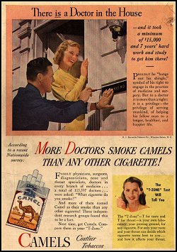 "COPD" includes two very common "diseases" that typically
occur together.
"COPD" includes two very common "diseases" that typically
occur together.

 Emphysema
Lung pathology series
Dr. Warnock's Collection
"Types of emphysema"
Lung pathology series
Dr. Warnock's Collection
Emphysema
Australian Pathology Museum
High-tech gross photos
{20239} emphysema
{20993} emphysema
{29153} emphysema
{38362} emphysema
{38365} emphysema
{49068} "pan-acinar emphysema"
{49069} "centrilobular emphysema"
* The elasticity explanation for emphysema probably was
first stated by Laennec, inventor of
the stethoscope: Lancet 335: 1372, 1990.
 Tobacco smoke augments elastases, inhibits anti-proteases, and
promotes infection with further
release of elastases.
We do not know why some smokers get
much worse emphysema than others.
Tobacco smoke augments elastases, inhibits anti-proteases, and
promotes infection with further
release of elastases.
We do not know why some smokers get
much worse emphysema than others.
 Emphysema patient
Barrel chest
Well-developed arms
That carries you off,
It's the coffin
They carry you off in.
-- Ogden Nash
Chronic bronchitis
Lung pathology series
Dr. Warnock's Collection
* According to one group, the lungs of asthmatics lack T-bet+
lymphocytes which produce gamma-interferon which modulates the immune
response here, and T-bet+ knockout mice have histological
and clinical asthma (NEJM 346: 857, 2002).
Asthma, great pictures
Lung pathology series; follow the arrows
Dr. Warnock's Collection
Asthma
Australian Pathology Museum
High-tech gross photos
{08231} asthma, mucus plugging airways
{08234} asthma, plugs
{29236} asthma, plugs
{07564} asthma, thick basement membrane
{08240} asthma, thick basement membrane, lots
of smooth muscle, lots of epithelial cells lying around
{08243} asthma, thick basement membrane,
mucus plug (trichrome stain)
{09899} Curschmann's spirals
{25999} Curschmann's spirals
{27492} occupational asthma; isocyanates
are infamous
 BOOP
BOOP
Very nice pictures and
discussion by Dr. Epler
* Researchers are presently looking at platelet-derived growth
factor
as a major player in the pathogenesis of this process (Am. J.
Resp.
CCM. 155: 676, 1997). Another group reports finding mast
cells
in the alveoli of BOOP patients, but none in controls,
contradicting
a traditional teaching of histologists: Chest 110: 383,
1996.
Bronchiolitis
Lung pathology series
Dr. Warnock's Collection
Bronchiectasis
Lung pathology series
Dr. Warnock's Collection
{10787} bronchiectasis
{24542} bronchiectasis
{27242} bronchiectasis
{38383} bronchiectasis
{38389} bronchiectasis
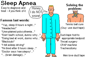
A common (1-5% of adults,
and some kids), serious, long-neglected health problem,
characterized by many episodes of upper
airway obstruction each night.
 * Missionary physician-hero Dr. David
Livingstone ("I presume?") cured himself of sleep apnea by
cutting off his own uvula while in
Africa.
* Missionary physician-hero Dr. David
Livingstone ("I presume?") cured himself of sleep apnea by
cutting off his own uvula while in
Africa. It is
probably the same disease as obstructive sleep apnea, except that
the struggle to breathe is delayed.
* I predicted in these notes in the 1980's that
"Gaisbock's primary idiopathic polycythemia" (long-noted to be
a disease of heavy, middle-aged
men) would prove to be due to sleep apnea. Anyway,
this diagnosis is almost never made any more, and sleep apnea is now
a well-recognized cause of polycythemia. So I think that's the explanation.
Smoker bronchi tend to contain a few H-flu and pneumococci.
Lobar Pneumonia
Australian Pathology Museum
High-tech gross photos
Aspergilloma
Lung pathology series; follow the arrows
Dr. Warnock's Collection
Aspergilloma
Lung pathology series; follow the arrows
Dr. Warnock's Collection
Chronic necrotizing aspergillosis
Lung pathology series; follow the arrows
Dr. Warnock's Collection
Chronic necrotizing aspergillosis
Lung pathology series; follow the arrows
Dr. Warnock's Collection
Aspergillus tracheobronchitis
Lung pathology series
Dr. Warnock's Collection
Cryptococcosis
Photo and mini-review
Brown U.
Bronchopneumonia
Photo and mini-review
Brown U.
{49076} bronchopneumonia
Aspiration pneumonia
Lung pathology series; follow the arrows
Dr. Warnock's Collection
Pneumonia
never mind the exotic bug...
Pittsburgh Illustrated Case
Klebsiella pneumoniae ("Friedlander's pneumonia", named by German pathologist Carl Friedlander whose relationship if any to your lecturer remains unknown) causes lobar pneumonia in deteriorated alcoholics. This gram-negative rod is coated by a thick slimy capsule, and victims cough up sticky slime ("cranberry sauce").
* Staphylococcus (after influenza), H. 'flu, pseudomonas, and
others are notable causes of lobar
pneumonia in those with damaged lung defenses.
Abscessing pneumonia
WebPath Photo
In the era when huge numbers of people died of pneumococcal pneumonia, the classical anatomic pathologists distinguished four successive stages of lobar pneumonia:
1. Hyperemia and edema: the bugs divide like crazy, and the blood vessels dilate and leak fluid in response to injury. "Congestion", given in traditional accounts of pneumonia, is an obvious misnomer.
2. Red hepatization: the inflammation progresses, and the damaged vessels now leak fibrinogen, which forms fibrin meshworks in the alveoli. Some red cells are released by damage to the blood vessels, and polys come in to fight the bacteria. The alveolar exudate becomes "rusty sputum".
3. Gray hepatization: fibrin dominates the picture, while polys and red cells break down ("gray" because hemorrhage is no longer taking place and the red cells have lysed)
4. Resolution: plasmin clears out the fibrin, and the lung returns to normal (hopefully).
COMPLICATION: The pleural surfaces overlying the infection are almost always involved, accounting for the pain of lobar pneumonia. There will be fibrinous adhesions, which may resolve or turn into scars.
COMPLICATION: In Klebsiella, staph, or pseudomonas lobar pneumonia, there is often necrosis and abscess formation, which greatly complicates healing. Necrosis is rare in pneumococcal pneumonia (exception: the slimy type 3). More on this below.
{12641} staph producing abscess
COMPLICATION: If the infection gets really bad in the pleural
space, it will fill with pus
("empyema") and this will need to be drained. (This was the
most helpful
thing that a doctor could do for
pneumococcal pneumonia before penicillin.)
{49077} empyema
COMPLICATION: Sometimes the fibrin in the alveoli mostly
organizes instead, leaving a scar.
{39530} organizing pneumonia; balls of fibrin in
the alveoli
COMPLICATION: The bacteria often spread to other structures
(pericardial sac, meninges)
Today, uncomplicated lobar pneumonias are easily treated with
antibiotics once the etiologic agent
is identified.
{11431} lobar pneumonia
LEGIONNAIRE'S DISEASE (legionellosis, "Pontiac fever", etc.)
This is a special form of bronchopneumonia named for a lethal
outbreak at an American Legion
convention at a hotel in Philadelphia.
The etiologic agent is Legionella pneumophila. You
need special stains (immunostain or silver) to
see it.
The organism is common in standing water, especially in
air-conditioning systems.
Most healthy people merely experience a bad "chest cold", but
in older people who drink and smoke
heavily, it is likely to be fatal if
untreated.
{08179} legionella demonstrated with a silver
stain
NOTE: We are now recognizing that many cases of tough-to-treat
community acquired pneumonias
are due to CHLAMYDIA (Arch. Int. Med. 148: 1425, 1988),
notably the TWAR strain
("Chlamydia pneumoniae", Chest 95: 664, 1989; NEJM
323: 1546, 1990). These infections have
long been recognized in newborns, who acquire the more familiar
sexually-transmitted pathogen
from the birth canal. It's now a notorious, chronic, hard-to-eradicate
infection of the airways (J. Inf. Dis. 182: 1678, 2000; Am. J. Resp.
CCM 164: 536, 2001).
PNEUMOCYSTIS PNEUMONIA
This is a curious lung infection caused by a protozoan (?)
organism, Pneumocystis carinii.
It was originally identified as the cause of plasma-cell
pneumonia in malnourished children in
Europe at the end of World War II.
It is now familiar as a cause of pneumonia in AIDS patients
and people on cancer chemotherapy.
Instead of a plasma-cell pneumonia, these people show no
visible cellular reaction to the infection.
The organism and its cysts grow in the foamy exudate.
* As junior medical students,
we used to go to the airport to
pick up the pentamidine the CDC
shipped in every time we suspected pneumocystis. It was the only
thing that we knew worked. If you spend any time on the oncology service, you will
probably meet
pneumocystis (i.e., you will develop antibodies), but it can't hurt you as long as your T-cells are
working properly.
Future pathologists: You still need silver stains if you want to
identify pneumocystis with confidence. {0456} pneumocystis pneumonia
LUNG ABSCESS
Polys plus necrosis in a confined space (i.e., walled off by
granulation tissue) within the lung.
Mechanisms (after Baby Robbins):
1. Aspiration of bacteria (bad teeth, tonsils), as when drunk
or unconscious.
2. Complication of necrotizing pneumonia (staph, klebsiella,
pseudomonas, legionella) or
bronchiectasis
3. Obstructed bronchus (as, behind a cancer)
4. Infection within a lung cancer itself
5. Septic pulmonary embolus (leg vein infection, endocarditis,
and now an extremely common
problem of IV drug abusers -- Chest 94: 251, 1988)
6. Infarction of a pre-existing infection (as when a pulmonary
embolus hits an area of
bronchopneumonia)
Anaerobic bacteria are often present, and aerobes may also be
involved.
Sooner or later, the enlarging abscess ruptures into an
airway. The patient gets a worse cough and
bad breath, while the radiologist looks for air-fluid levels.
VIRAL AND MYCOPLASMAL PNEUMONIA ("primary atypical pneumonia",
"chest cold", etc.)
A family of infections by micro-organisms smaller than
familiar bacteria, all causing interstitial
pneumonitis.
A while back, a group biopsied a bunch of folks with colds, and
found what you'd expect -- inflammation,
mostly lymphocytes, in the bronchial mucosa (Am. J. Resp. Crit.
Care Med. 151: 879, 1995).
Many annoying chest-colds are caused by Mycoplasma
pneumoniae (check blood for cold
agglutinins).
Viral infection of the lungs can be seen in measles (major
killer worldwide), influenza (all strains),
chicken pox, adenovirus infection (look for "smudge cells"; this can cause
serious pneumonia even in healthy people, Am. J. Med. Sci. 325:
285, 2003).
Respiratory syncytial virus, once considered merely the usual
cause of "bronchiolitis" in toddlers,
is now known to be prevalent and
lethal among older adults. * Interestingly, a vaccine was tried in the late 1960's and
proved to make the disease worse instead of
rendering the kids immune (J. Inf. Dis. 167: 553, 1993).
Puzzle that one out.
Metapneumovirus, a cause of wheezing mostly in youngsters,
was discovered in 2001 (Lancet 360: 1393, 2002; NEJM 350: 443 & 451, 2004).
Lethal influenza infection without staph superinfection presents
diffuse alveolar damage and necrosis along the epithelium of the bronchi
and bronchioles.
Herpes simplex can present as an ulcerative tracheobronchitis
in moderately immunocompromised hosts. Alternatively,
herpes simplex and herpes zoster can
both present as miliary hemorrhagic areas in the lung parenchyma
if immunosuppression is severe. Look for "herpes cells" with
a single intranuclear inclusion surrounded by a clear halo.
(* In contrast to skin and cervix, these viruses seldom
produce multinucleation in the lung.)
The nastiest infectious pneumonitis in the U.S. is the new
hantavirus, from inhaled mouse droppings
("Navajo pneumonia"; anatomic pathology Am. J. Path. 146:
552, 1995; review NEJM 330: 949, 1994). The virus specifically
damages the endothelial cells, producing an extreme pulmonary edema.
Distinctive for this
infection is an abundance of immunoblasts in lung
and peripheral blood.
SARS is a coronavirus: NEJM 348: 1977 & 1995, 2003.
The origin remains unknown; the original claim that it was a zoonosis
from eating wild animals didn't work out (Science 301: 1031, 2003).
The epidemic: Lancet 362: 1353, 2003.
Thankfully only about 1000 people died (follow-up Nat. Med. 9(s):
S-88, 2004).
The pathology (Lancet 361: 1773, 2003; Am. J. Clin. Path. 121: 574, 2004;
Hum. Path. 36: 303, 2005) features:
* The receptor for the virus in the lung is angiotensin converting
enzyme-2 (Nat. Med. 11: 875, 2005).
* Nobody knows how many nasty chest-colds are really zoonotic Q-fever
(Coxiella). Probably underdiagnosed. Pathologists look for "ring / donut granulomas".
"Poker player's pneumonia", the scourge of one town,
was Q-fever traced to a cat's placenta (NEJM
319: 354, 1988).
In most cases of viral pneumonitis
that come to autopsy, the pathologist sees edema and
inflammatory cells, and the process is
confined to the interstitium.
The lungs are heavy but not airless (why?) The inflammatory
infiltrate is mostly lymphs and
macrophages.
In severe cases, an ARDS picture supervenes, with hyaline
membranes.
* Lymphoid interstitial pneumonitis is a dense, polyclonal
("pseudolymphoma") infiltration
of lymphocytes strictly confined to the pulmonary interstitium,
usually in kids with AIDS or
adults with Sjogren's.
Don't confuse it with a virus.
Some patients have a low-grade clonal lymphomas
which look identical; it's a spectrum (Chest 122: 2150, 2002).
{38402} bad viral pneumonia; the lung is nearly
solid from all the inflammatory stuff in the alveolar septa
Most patients recover without treatment. The most worrisome
thing about chest viruses for
healthy adults is that they predispose the lung to bacterial
superinfection.
You're already familiar with Kaposi's virus infection
("Kaposi's sarcoma") of the lung (Radiology
195: 545, 1995).
TUBERCULOSIS ("TB", "the white plague" -- covered twice in
R&F).
Primary tuberculosis occurs when the TB bacillus first infects a person.
A single lesion (the Ghon focus) occurs just under the pleura in the midportion (midway between
apex and base -- the best-ventilated area) of one lung.
The bacilli find their way to the regional lymph nodes, and in a few weeks, granulomas have walled
off the bacilli in both locations. (The combination of lesions in the lung and node is called the Ghon
complex). Viable bacilli remain in the Ghon focus/complex for life.
{08333} TB, lymph node
Much primary TB is asymptomatic, i.e., you discover you're turned your TB skin test.
Progressive
primary tuberculosis is the name given to overwhelming primary infection, which is not so rare
as we used to teach.
The classic dogma is that almost all progressive adult TB represents reactivation of a latent primary
focus. I have never understood why people believed this, and it turns out that it's clearly not true:
NEJM 330: 1697, 1703 & 1710, 1994 (at least in America's slums; confirmed NEJM 346:
1453, 2002; TB in immigrants does often seem to be reactivation). Only in 2002 did we get molecular
proof that one patient's TB had reactivated:
J. Inf. Dis. 185: 401, 2002. Primary or re-activated, the
pathology of bad TB is that of classic "secondary tuberculosis"....
Secondary tuberculosis ("active TB", "postprimary tuberculosis", "adult tuberculosis", "reinfection
tuberculosis", "cavitary tuberculosis") occurs when bacilli escape the original Ghon focus or
more bacilli enter the body from outside.
{08459} cavitary TB
The bacilli may be released from the Ghon focus by invading cancer or "by immunosuppression"
of some sort.
The infection usually reappears at the apex of one or both lungs ("Simon's foci" -- the TB bacilli
actually entered the bloodstream, but grow best in the lung apex where oxygen is most
abundant.) This is one cause of "white pneumonia" ("pneumonia alba"; the other is syphilis).
The better the patient's cell-mediated immunity, the more classic the granulomas.
In other words, the worst TB cases in America today have very few, if any, granulomas! For
example, an AIDS patient can have lungs teeming with "red snappers" with only a sprinkling of
macrophages trying to fight them. (This patient may not even be very sick, but his ability to
transmit the infection is impressive.)
Polys are most abundant when the caseum has eroded into the large airways (why?). Also
remember that TB tends to calcify (handy for radiologists who want to tell it from cancer).
{05949} TB eroding through the chest wall
Arrested TB is secondary TB that has "calcified" and/or been largely replaced by collagen.
{11423} old TB
By contrast, progressive pulmonary tuberculosis spreads throughout the lungs and can produce a
"tuberculous empyema" by involving the pleural cavities.
When a large portion of the lung has undergone caseous necrosis, extension into a large airway
causes all the debris to be coughed up -- exactly what the TB bacillus "wants", since this is how it is
transmitted to other people. The result is a cavity. (* Howler in R&F: "coin lesions" are lone
granulomas, not cavities.)
TB is also prone to infect the larynx and larger airways ("tracheobronchial TB") and, because bacilli
are swallowed, to affect the intestine (* look for Peyer's patches with ulcers having their long axes
perpendicular to the long axis of the bowel). Miliary TB results when many TB bacilli enter the bloodstream but the granulomatous response is
good. It is supposedly named for millet seed (parakeet seed -- * would anyone prefer "milli-",
meaning "thousands" of little granulomas?).
When TB ruptures into a pulmonary artery, there is miliary involvement of
part of a lung, while rupture
into a vein results in miliary involvement of the rest of the body.
When I have fears that I may cease to be
* Keats went to medical school for a year and was
licensed as a physician and surgeon. He quit
to devote his life to writing poetry.
* In this sonnet, he shares his fear that he may die before
had has time to get all his ideas into writing.
* With his medical training, he had good reason to be afraid.
A few days before he wrote the poem,
he had experienced his first episode of hemoptysis. Three years
later he was dead of tuberculosis.
* Also good: Alexander Dumas Jr.'s Camille (TB is
glamorous), Eugene O'Neill's Long Day's
Journey Into Night (TB and drug addiction aren't glamorous),
1970's Italian film A Brief Vacation
(TB as social-political-feminist problem). I didn't understand
Thomas Mann's The Magic Mountain.
Reminder: In most chronic inflammatory diseases of lung,
clubbing of the nails ("Hippocratic
change") often develops because megakaryocytes embolize through
the new vascular channels formed in the lungs.
INTERSTITIAL RESTRICTIVE LUNG DISEASE ("stiff lung";
"fibrosing alveolitis";
"honeycomb lung")
A generic term for longstanding inflammatory damage leading to
fibrosis of the alveolar walls.
Pulmonary compliance decreases, a mechanical barrier to oxygen
exchange comes into existence
("diffusion barrier", "alveolar capillary block"), and pulmonary
blood pressure goes up (leading to
cor pulmonale and death).
It seems to reasonable to think, as "Big Robbins" does, that
the initial stimulus to inflammation is
the presence of an unknown antigen.
Fibrosis finally wipes out groups of alveoli. In most of
these entities, the process is uneven
throughout the pulmonary parenchyma, with some less-involved
airways (especially respiratory
bronchioles) stretched wide by scar contraction -- hence the
radiographic and autopsy diagnosis of
"honeycomb lung". * Regardless of cause, most fibrosing lung diseases are worst
in the lower lobes, where there is ordinarily
less air and more vasculature. (Asbestosis, a pneumoconiosis,
can be an exception -- why?)
* Clinicians hear distinctive dry "velcro
crackles" in pulmonary fibrosis.
The known etiologies of diffuse pulmonary fibrosis:
Rheumatoid lung,
scleroderma lung, and Sjogren's lung: secondary to
autoimmune disease.
Asbestosis, berylliosis, hard metal
(i.e., cobalt & maybe nickel), and rare cases of longstanding
farmer's lung ("hypersensitivity pneumonitis": T-cell
havoc Chest 104: 38, 1993): due to pneumoconiosis
Histiocytosis X ("Langerhans' cell histiocytosis" is a
better name for this family)
Desquamative interstitial pneumonitis: ("DIP") in
addition to fibrosis, the alveoli clog with lipid-
and mucin-laden macrophages. Some cases supposedly turn into
Hamman-Rich syndrome; the
relationship is dubious (Thorax 52: 333, 1997). The
response to steroids in this disease is generally
good.
Nonspecific interstitial pneumonitis
also responds well to glucocorticoids. It is distinguished
from UIP/Hamman-Rich by the uniformity of the histologic changes,
with all septa involved about equally, and no "honeycomb cysts".
Chest 125: 522, 2004.
Radiation lung, amiodarone lung, cytoxan
lung, busulfan lung, bleomycin lung (Am. J.
Path.
147:
352, 1995), and GVH disease lung: fibrosis due to cancer
therapy
Paraquat ingestion -- patients who drink this herbicide
die of rapidly-developing, severe fibrosis of
the lungs. Sarcoid lung: see below
Polymyositis-dermatomyositis: Especially when
antibodies against t-RNA synthetases (anti-Jo, etc.)
are present.
Idiopathic pulmonary hemosiderosis: usually-mild, not-very-serious
illness of young people with recurring microhemorrhages into
the alveolar spaces
* Acute idiopathic pulmonary hemorrhage occurs in babies and can be fatal.
It's presently being worked-out (MMWR 50: 494, June 15, 2001;
Am. J. For. Med. Path. 22: 188, 2001. There was a flap about
about a mold in the homes of these children as the cause of a supposed
epidemic in Cleveland (Pediatrics 99: E5, 1997); I'm undecided.
* Angiosarcoma metastatic to the lung is a hard-to-diagnose
cause of diffuse pulmonary hemorrhage:
Arch. Path. Lab. Med. 125: 1562, 2001.
Wegener's you know. Pulmonary capillaritis has been
described in anti-myeloperoxidase disease
(Am. Rev. Resp. Dis. 146: 1326, 1993; the histopathology
is the same as pulmonary Wegener's) and generally in systemic
vasculitis (fibrinoid precedes neutrophils: Arch. Path.
Lab. Med. 121: 144, 1997).
Am. J. Clin. Path. 104: 7, 1995.
Interstitial fibrosis is one component of ARDS and
"bronchopulmonary dysplasia" (the latter follows
oxygen treatment of neonatal RDS)
Diffuse pulmonary amyloidosis, part of the systemic
disease in a few cases
Lymphangioleiomyomatosis: a rare disease with marked
proliferation of the smooth muscle throughout the lung.
Patients are women in the childbearing
years, and the mainstay of therapy is hormonal manipulation
(Am. J. Med. Sci. 321: 17, 2001).
This is indistinguishable from the lesions of
tuberous sclerosis, and since these usually develop
only in young female TS patients, probably they are the same disease.
Alveolar proteinosis: surfactant and proteinaceous goop
fills the alveoli (same as in acute silicosis) --
listed here by "Robbins", but rarely includes fibrosis.
Most cases are idiopathic, and probably result from
some loss of ability of the type II pneumocytes and/or alveolar
macrophages to dispose of surfactant.
Infectious agents (notably mycobacteria, but a host of others have
been mentioned) are often present;
no one knows whether they interfere with surfactant processing and/or
simply grow well in this culture medium.
* Certain amphophilic drugs can induce alveolar proteinosis, perhaps by
interacting with the soapy surfactant itself.
* Mouse model Am.
J. Path. 146: 1017, 1995.
There is a congenital form with surfactant protein B absent (NEJM
328: 406, 1993).
Regardless of cause, bronchial lavage is the mainstay of therapy.
Cases in which the etiology is totally obscure are called
idiopathic pulmonary fibrosis /
Hamman-Rich syndrome. See below.
* NOTE: The "Loeffler's" family of eosinophilic pneumonias
seldom produce fibrosis.
Clinicians: "Restrictive lung disease" (sort of the opposite
of "obstructive lung disease") exists when
the airways are widely patent but there is a problem ventilating
the lungs. ("Restrictive" and
"obstructive" lung disease give contrasting patterns on
spirometry. Other causes of "restrictive lung
disease" include large pleural effusions or cancers in the
pleural spaces, pneumothorax, chest wall
and spinal column problems, lungs full of tumor, boa constrictor
attack, metastatic calcification,
massive obesity -- don't overlook this one Am. J. Med. 116: 58, 2004-- etc, etc.)
IDIOPATHIC PULMONARY FIBROSIS ("Hamman-Rich syndrome";
"cryptogenic (idiopathic)
fibrosing alveolitis", "usual interstitial pneumonitis" and its kindred:
CMAJ 171: 153, 2004)
Pulmonary fibrosis, most often occurring in middle-aged and older people,
and progressing
to death.
"Usual interstitial pneumonia", or "UIP" is the most common
and most deadly of several different processes in which
pulmonary fibrosis appears. It is recognized by uneven fibrosis
of the alveolar septa themselves, with overlying cuboidalization
of the epithelium.
The alveoli are thickened, though some areas are
always spared. Focal areas of hyperplastic fibroblasts
have overlying type II pneumocytes.
Contraction of the fibrous tissue causes dilation of some of the
air spaces ("honeycomb cysts").
The involved alveoli show obvious chronic inflammation,
but no pathogenic micro-organism or other
etiology has been forthcoming.
Part of the problem seems to be factors produced by
malfunctioning alveolar macrophages and/or
epithelial cells, notably platelet-derived growth factor (PDGF --
NEJM 317: 202, 1987; J. Clin.
Invest. 86: 1055, 1992, interleukin 8 Am. Rev. Resp. Crit.
Care 151: 1604, 1995), the proto-oncogene product which
is the major factor that turns fibroblasts on in normal wound
healing. There are reports of several others, but so far no common
mechanism.
Survival times are improving, with as long as 5 years being
relatively common.
{40688} Hamman-Rich, gross, remember scar is white
Glucocorticoids are notoriously ineffective for UIP, though they work
well for its clinical mimics (desquamative, lymphoid, and nonspecific interstitial pneumonias).
So far, no other medication has been shown to improve survival for UIP.
* There's an accelerated form (?)
called "acute interstitial pneumonia".
It follows a cold, gets worse over days or weeks,
involves only the lungs, and looks like ARDS under the microscope.
It is evidently curable most of the time
with glucocorticoids (Chest 124: 554, 2003).
SARCOIDOSIS ("sarcoid", "Boeck's sarcoid"; * "sarcoid"
literally means "the fleshy disease"): "A
multi-system disorder of unknown etiology characterized by
formation of noncaseating granulomas"
(Baby Robbins) in many organs of the body, with variable clinical
course. Review Lancet 361: 1111, 2003.
Sarcoidosis might even be a reaction pattern rather than one
disease.
Most sarcoid patients are young adults. In the US, women and blacks are
more often affected, but no group is
immune.
There is only a slight familial tendency.
The majority of people with sarcoidosis never become
symptomatic. Those that do are likely to
have:
Cardiac sarcoidosis is rare but lethal, when granulomas in the
AV node cause 3rd-degree heart block
and sudden death (CMAJ 136: 1064, 1987; Thorax 44:
371, 1989). Suspect it whenever a young
person has heart block or * focal echo abnormalities of the
ventricular wall (Br. Heart. J. 57: 256,
1987).
* In one variant of cardiac sarcoidosis, the heart is
massively involved, with minimal involvement of
other organs (Br. Med. J. 292: 1095).
The "cause" of sarcoidosis continues to elude us.
* Mycobacteria, clay dust, and pine pollen have been
suggested, but never demonstrated, as
"antigens". More recently, interferon therapy has apparently
caused "sarcoidosis": Cancer 59: 896,
1987, and it may appear in response to immune reconstitution in patients
receiving HAART for HIV.
The immune disturbance seems central.
There are lots of T4-helper cells at sites of inflammation,
very few in the circulating blood.
Further, the body's B-cells are hyperactive (polyclonal
gammopathy) -- it seems likely they are
getting bad advice from the T4-cells.
Lymph nodes draining a cancer occasionally exhibit a
morphologic reaction identical to sarcoidosis
(Cancer 68: 1845, 1991) that may stay around after the
cancer is cured (Chest 98: 1300, 1990).
* Sarcoidosis is infamous for popping up in its victims'
tattoos (Arch. Derm. 141: 869, 2005).
Sarcoid granulomas are sharply circumscribed, tend to occur
adjacent to
lymphatic vessels, usually
do not caseate,
and contain giant cells. (You may
hear that foreign-body giant cells are most characteristic of
sarcoidosis, but actually Langhans cells
are just as common.)
{08756} sarcoid in the lung
* "Asteroid bodies" (probably wreckage from the cytoskeleton)
and "Schaumann's conchoid bodies"
(laminated calcium and iron, refractile) can turn up inside the
giant cells, but have no diagnostic
utility.
The diagnosis of sarcoidosis is usually made by finding
noncaseating granulomas on biopsy of some
organ, in the absence of infectious organisms, foreign body, or
other explanation.
* Every writer on sarcoid seems to have a favorite organ to
biopsy first. Right scalene node is
standard, but you'll hear about biopsy of the lip (minor salivary
gland), liver, bone marrow, and even
fine-needle aspiration of the spleen, all in a search of
noncaseating granulomas.
Serum angiotensin converting enzyme (ACE) is a useful adjunct
test (elevated more often than not), to make the diagnosis and
monitor disease activity. You'll hear about the Kveim test, which involves taking
ground-up spleen from someone
with sarcoidosis and injecting it into the dermis. If a
granuloma forms, the living patient supposedly has
sarcoidosis (80% sensitive, 95% specific). Long considered outdated, there is now a resurgence
of interest in the material, especially among
researchers who need to decide who really does
and does not have the disease
(Clin. Chest Med. 18:799, 1997;
Mount Sinai Journal of Medicine 63:335, 1996;
J, Imm. 154: 1450, 1995). To date, nobody knows what's in Kveim
reagent, except that it's supposedly not bacteria: AJRCCM 159 1981, 1999.
Making the diagnosis is important, because immunosuppression
using a few weeks on glucocorticoids is often helpful.
Longer therapy is much more dubious; the side-effects
of the drugs are noxious, and you're not going to clear up scar tissue.
* Sarcoidosis is a common, lethal disease in horses.
Oddly, amyloidosis almost never complicates sarcoidosis (Thorax
43: 422, 1988).
* Treatment of cutaneous sarcoidosis using tetracycline: Impressive.
Is this really a bacterial disease after all? Arch. Derm. 137: 69, 2001.
* "Giant cell interstitial pneumonitis" is a recognizable reaction
pattern almost always caused by inhalation of hard metal dust.
GOODPASTURE'S DISEASE: Antibodies against the basement
membranes of lung and kidneys --
type II immune injury.
Pulmonary hemorrhage is seldom severe, but a few patients do
exsanguinate or drown in blood.
More about this under "Kidney" -- these patients die of renal
involvement.
Not all of these patients have renal involvement clinically:
Thorax 46: 68, 1991.
{24848} Goodpasture's, lung
EOSINOPHILIC PNEUMONIAS ("Loeffler's", etc.)
A grab-bag of illnesses ("idiopathic", drug-related, and
caused by worms) that feature many
eosinophils in the blood and sputum and in the pulmonary alveolar
walls. Patients usually have
pulmonary infiltrates and increased eosinophils in the blood.
In the U.S., the disease is most often due to aspergillus
colonizing the airways of an asthmatic. But
there are many other causes, including ascaris worms migrating
through the lungs, and plenty of
"idiopathic" cases.
In "tropical eosinophilia", microfilaria worms are trapped in
the lungs and break down, producing a
similar syndrome.
* While we are on the subject of worms in the lungs, remember
that dirofilariasis, the dog
heartworm, is prone to die in human lungs and produce a "coin
lesion" simulating lung cancer. I
suspect this accounts for a few cases of "surgically cured
squamous cell carcinoma of the lung."
Among drugs, the best-known offender is nitrofurantoin.
Some of these patients will have Loeffler's eosinophilic
endocarditis.
* Churg-Strauss vasculitis may feature an
"eosinophilic pneumonia" -- future
pathologists, look hard for granulomas before making your
diagnosis of "idiopathic Loeffler's" (Am. J. Clin. Path. 114: 767, 2000).
Do not confuse these with eosinophilic granuloma, a
member of the "histiocytosis X" family of
quasi-neoplastic proliferations of Langerhans histiocytes, seen
mostly in older smokers.
LIPID PNEUMONIA
{38464} lipid pneumonia
Exogenous lipid pneumonia: the lung's reaction to
aspirated oil.
Causes include oily nose drops, mineral-oil laxatives, and forcing
children to take cod-liver oil (the latter
must have been an important killer of children, whose pitiful deaths
were due to "pneumonia"). Don't
give this stuff to your patients.
The more unsaturated the oil, the worse the inflammation.
Mineral oil disease is usually mild.
Polyunsaturated vegetable oils are worst.
The pathologist sees yellow patches grossly. The microscopic
picture features lipid-laden
macrophages and fibroblasts laying down scar tissue.
* "Fire eater's pneumonia" is actually due to aspiration of
petroleum products, though it mimics an infection clinically
(Chest 124: 398, 2003).
Endogenous lipid pneumonia ("golden pneumonia"): buildup of surfactant (in
macrophages) behind an obstructed major
airway (i.e., lung cancer) or many small airways
("bronchiolitis obliterans" family), or in amiodarone lung
(interferes with surfactant processing: Chest 112: 1068, 1997).
* MISCELLANEOUS LESIONS
Bronchocentric granulomatosis: A reaction pattern, most
often seen with infection by
Aspergillus fungus, in which the bronchi break down into
caseating granulomas.
The cystic adenomatoid lesion is a fortunately rare and
dangerous hamartoma seen in infants.
A bronchopulmonary sequestrum is a portion of lung
tissue, inside or outside the healthy lung, with
airways unconnected to the main bronchial tree, and therefore
unaerated. It is prone to become
infected, or to cause problems because of its mass.
Pulmonary interlobar sequestrum is a birth defect in
which part of the lung is supplied by a branch
of the aorta instead of by the pulmonary artery. This is only a
problem later when it gets infected.
Several types of congenital cysts of the respiratory tree also
occur and can cause problems
(hemorrhage, infection, rupture, etc.) at any age.
Remember that most lung diseases will tend to be exacerbated
by gastric reflux
Lung abscesses
WebPath
Suppurative pleuritis
Not quite empyema
WebPath
{11732} lobar pneumonia
{11733} lobar pneumonia
{11734} streptococci in lobar pneumonia,
gram stain
{12464} lobar pneumonia
{17565} lobar pneumonia
{17567} lobar pneumonia
Pneumocystis
Lung pathology series, great photos
Dr. Warnock's Collection
Pneumocystis
Photo and mini-review
Brown U.
Viral lung infection
Lymphocytes
WebPath Photo
Lymphocytic interstitial pneumonia
Pittsburgh Pathology Cases
Kaposi's in the lung
Lung pathology series
Dr. Warnock's Collection
You are already familiar with how TB causes tissue injury.
(Click here for a review.)
Tuberculosis
Very large Ghon focus
WebPath Photo
{08336} TB, lymph node
{10230} TB in the lung, good cavity
{21151} disseminated, miliary TB in liver
TB
WebPath Photo
Tuberculosis; white pneumonia
Lung pathology series; follow the arrows
Dr. Warnock's Collection
{08187} TB, kidney
{08188} TB, lung
{08190} TB, lung
{08193} TB, lung
Before my pen has gleaned my teeming brain,
Before high-piled books, in charactery,
Hold like rich garners the full-ripened grain;
When I behold, upon the night's starred face,
Huge cloudy symbols of a high romance,
And think that I may never live to trace
Their shadows, with the magic hand of chance;
And when I feel, fair creature of an hour,
That I shall never look upon thee more,
Never have relish in the faery power
Of unreflecting love! -- then on the shore
Of the wide world I stand alone, and think
Till Love and Fame to nothingness do sink.
TB granulomas
WebPath Photo
Pulmonary tuberculosis
Photo and mini-review
Brown U.
Interstitial Lung Disease
Notes on processing tissue; great photos
Dr. Warnock's Collection
* The actual histopathology is very complex.
There is always some inflammation.
There may also be
nodules of smooth muscle, vascular changes, etc., etc.
In many cases, the honeycombing is worst just under the pleura.
Elastin and proteoglycans also accumulate.
Honeycomb Lung
Great gross photos
Dr. Warnock's Collection
The histopathology in rheumatoid lung is widely variable:
Chest 127: 2019, 2005.
 Rheumatoid lung, capillaritis
Lung pathology series
Dr. Warnock's Collection
Asbestosis
Photo and mini-review
Brown U.
Desquamative interstitial pneumonitia
Lung pathology series
Dr. Warnock's Collection
* Some people are super-sensitive to the fibrosing
effects of bleomycin. This is now yielding up its secrets,
thanks to a mouse model of bleomycin sensitivity with
scrambled pulmonary interstitial matrix remodelling (Am. J. Path. 152:
821, 1998).
Despite the selective indignation,
nobody got sick from smoking marijuana sprayed with
paraquat.
Wegener's granulomatosis
Lung pathology series; follow the arrows
Dr. Warnock's Collection
Wegener's granulomatosis
Lung pathology series; follow the arrows
Dr. Warnock's Collection
Wegener's granulomatosis
Lung pathology series; follow the arrows
Dr. Warnock's Collection
Wegener's granulomatosis
Lung pathology series; follow the arrows
Dr. Warnock's Collection
Wegener's with capillaritis
Lung pathology series; follow the arrows
Dr. Warnock's Collection
Busulfan lung
Lung pathology series
Dr. Warnock's Collection
Nodular amyloidosis
Lung pathology series; follow the arrows
Dr. Warnock's Collection
Lymphanioleiomyomatosis
Lung pathology series
Dr. Warnock's Collection
Pulmonary lymphangiomyomatosis
Great photos
Pittsburgh Pathology Cases
Patients have shortness of breath and may note that they cough
up "white jello".
Alveolar proteinosis from silicosis
Lung pathology series
Dr. Warnock's Collection
Amyloid and proteinosis
Lung pathology series
Dr. Warnock's Collection
Fibrosing alveolitis
Lung pathology series
Dr. Warnock's Collection
Usual interstitial pneumonitis
Lung pathology series
Dr. Warnock's Collection
Restrictive Lung Disease
Queen's U., Kingston
Thanks Dr. Boag
{40685} Hamman-Rich, histology, the alveoli are slits
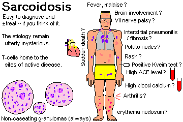
{11417} sarcoid in a node, asteroid body
{11464} sarcoid, odd stain
{11774} sarcoid, lymph node
{12376} lupus pernio
{10976} sarcoid granuloma
{10979} sarcoid granuloma
{14427} sarcoid granuloma
{15474} brain sarcoid
{20220} sarcoid, lymph node
{23380} sarcoid, granulomas
{34913} sarcoid, lung
{35945} sarcoid, muscle
{38416} gross, sarcoid
{42030} sarcoid, marrow
* For following the course of severe
disease, the gallium-67 lung scan
seems to be preferred nowadays (over sed rate, lung function
tests, ACE).
* Thalidomide for sarcoidosis: Chest 122: 227, 2002.
Also watch infliximab and pentoxifylline.
{29104} Goodpasture's, lung, iron stain
{29581} Goodpasture's
{29584} Goodpasture's
Lupus and Goodpasture's
Lung pathology series
Dr. Warnock's Collection
Remember that intra-alveolar hemorrhage may not produce
massive hemoptysis, and instead mimic pneumonia (why?)
You can also see intra-alveolar pulmonary hemorrhage with
lupus (Chest 118: 1083, 2000; Arch. Path. Lab. Med. 125: 475, 2001), small-vessel polyarteritis
or cryoglobulin vasculitis, abciximab
(anti-platelet antibody) therapy (Chest 120: 126, 2001),
crack cocaine (don't miss this one, Doc: Chest 121: 1231, 2002)
and so forth.
Future
pathologists: All forms of "histiocytosis X" feature "Birbeck
tennis-racket granules" and the
histiocytes of eosinophilic granuloma show "coffee bean nuclei".
Since "eosinophilic granuloma" occurs just under the pleura,
it's blamed for causing pneumothorax.
Histiocytosis X with Birbeck granules
Lung pathology series
Dr. Warnock's Collection
Eosinophilic granuloma
Lung pathology series, follow the arrows
Dr. Warnock's Collection
Lipid pneumonia
Endogenous, golden surfactant pneumonia
WebPath
Lipid pneumonia
Lung pathology series
Dr. Warnock's Collection
Amiodarone lung
Lung pathology series
Dr. Warnock's Collection
A similar histologic pattern has been reported in "hot tub lung",
in which immunocompetent people get an overwhelming infection with
Mycobacterium avium complex (Am. J. Clin. Path. 115: 755, 2001;
Arch. Int. Med. 163: 845, 2003).
Cystic adenomatoid malformation
Virtual Hospital
The vast majority of primary malignant neoplasms of the lungs are some form of BRONCHOGENIC CARCINOMA. Easy review Lancet 355: 479, 2000.
In 2005, the US had around 184,800 new cases of bronchogenic carcinoma (102,420 men, 82,380 women) -- CA, in loc. There were around 168,140 deaths.
Bronchogenic carcinoma is the leading cancer killer of US men, although age-adjusted rates have been dropping for men since 1980 (NEJM 321: 1197, 1989), and the total absolute number is now decreasing; the total percent of people dying of lung cancer is way down.
It surpassed breast cancer as the leading cancer killer of women in 1985. The rates in women have just started to drop (2004).
Most lung cancers are silent until they've become inoperable. Those lucky patients whose cancers are of operable subtype and are detected very early have a reasonably good prognosis (maybe 60% long survival with stage I, 40% stage II: Ann. Thorac. Surg. 60: 466, 1995).
Bronchogenic carcinoma is a disease of older people, and is unusual under age 30. The incidence increases with age and "pack-years smoked".
Lung cancer exhibits Nowell's law in action, with mutations in the surrounding "normal" epithelium. Prognosticating lung cancer using genetic markers has been underway since the mid-1990's, and centers now look at around 100 genes (Am. J. Resp. CCM. 170: 167, 2004); clinical decisions are not yet based on the genetic profile, but probably will be within a decade.
 Risk factors are well-established (Chest 103-S1: 20-S,
1993)
Risk factors are well-established (Chest 103-S1: 20-S,
1993)
Cigaret and cigar smoking: This is the overriding risk factor.
The danger is proportional to the amount of smoke inhaled every day and the duration of smoking, and is measured in "pack-years." After ten pack-years the increased risk is measurable, and after fifteen pack-years, there is clear-and-present danger. After quitting, the risk decreases slowly and approaches normal after ten or fifteen years. (Cigar smokers inhale less smoke overall than cigaret smokers, but contrary to what you may hear, their risk for lung cancer is still greatly increased.)
"Passive smoking" seems to be of some importance, but much less than active smoking (Am. J. Pub. Health 82: 1525, 1992). Pipe smoking and tobacco chewing are much less risky than cigaret smoking, at least as far as the lungs are concerned.
The other risk factors are synergistic (i.e., more than just additive) with cigaret smoking.
* By the way, former smokers filling out research forms will often say they never smoked. Remember this when you're evaluating research articles (Chest 101: 19, 1992).
Radiation: uranium miners (powerful effect, all histologies Cancer 89: 2613, 2000), atomic bomb survivors, after radiation therapy (mild effect; Cancer 71: 3054, 1993), radon in homes ("if you say so....": NEJM 330: 159, 1994.)
* During the 1980's and early 1990's, there was a hoopla over radon from the ground building up in our energy-efficient homes. The claim, advanced by the Environmental Protection Agency, was that it caused 5-20 thousand lung cancers yearly, multiplicative with tobacco smoking. Was it just junk science? I wasn't the only one to say so... Lancet 337: 1329, 1991; Arch. Int. Med. 151: 674, 1991 (AMA council report). It does seem possible that smokers are at increased risk if there's radon in their homes. Most of the epidemiologic studies have come back negative, one controversial study found a negative correlation (as if radon protected you: Health Phys. 72: 623, 1997), the last big paper to find a link was Am. J. Pub. Health 89: 1042, 1999 and even the authors admit it's really weak, and even Br. J. Cancer 84: 134, 2001, which did a study of radon and lung-cancer in nonsmoking men, couldn't quite get a statistically significant correlation. There seems to be agreement that non-smokers aren't at serious risk, and that smokers can protect themselves much less expensively by stopping smoking (Am. J. Pub. Health 88: 811, 1998).
As you would expect, the nucleic acid damage from radiation is more random than in tobacco smokers. See Lancet 339: 576, 1992.
Asbestos inhalation: Notorious cause of lung cancer. More than half of asbestos workers who smoke have died of lung cancer (more: Br. Med. J. 306: 1503, 1993).
Certain metal dusts (nickel, silver, chromate ion as in dyestuffs)
Coal tar fumes, carbon black (Lancet 358: 562, 2001) and other chemicals (* notably vinyl chloride, chloromethyl ethers) Occupational lung cancer: Mayo Clin. Proc. 68: 183. 1993.
Urban pollution: a minor risk factor compared with the others, with which it may be synergistic (JAMA 287: 1132, 2002)
* In one study which generated a hoopla, the investigators failed to take into account the fact that people living near the steel mill smoked much, much more than did their rural counterparts. When somebody noticed this, the "cancer risk" from the steel mill disappeared (Arch. Env. Health 48: 184, 1993).
In the poor nations, living in a coal-heated dwelling that's not ventilated clearly causes a great deal of lung cancer (Lancet 362: 849, 2003; JNCI 94: 826, 2002).
* I think that silica / sand exposure, made "official" in 1997 by the International Agency for Research on Cancer, is a crock. The numbers are very soft and there's no credible mechanism. See Am. J. Epidemiol. 153: 695, 2001.
 * Pigeon keeping: Possible promoter, but not a
mega-risk factor (Br. Med. J. 305: 986 & 989, 1992).
A 1992 flap about keeping a canary or parakeet being a risk factor for
lung cancer
was laid to rest by a
bunch of negative studies (Br.
Med. J. 313: 1218, 1996).
* Pigeon keeping: Possible promoter, but not a
mega-risk factor (Br. Med. J. 305: 986 & 989, 1992).
A 1992 flap about keeping a canary or parakeet being a risk factor for
lung cancer
was laid to rest by a
bunch of negative studies (Br.
Med. J. 313: 1218, 1996).
* Right there is a hoopla about carotenoids (there are at least five kinds) as protecting people (smokers, non-smokers) from lung cancer. Given that a high level of blood carotenoids strongly suggests an overall healthy-lifestyle, and that today's "pop wisdom" is that vegetables protect from cancer, it's hard to do reliable studies. Protection by dietary carotenoids: Am. J. Clin. Nutr. 72: 990, 2000; Am. J. Ep. 156: 536, 2002 (about a whopping 15%, and based on self-reports); no protection Canc. Causes Contr. 13: 231, 2002; more confusion Br. J. Cancer 84: 728, 2001 (something's going on but it can't all be the tomato sauce).
NOTE: We know who is at risk. The traditional wisdom is that screening (routine x-rays, exfoliative cytology) is a waste of time and money (Ann. Int. Med. 111: 232 & 239, 1989). With today's improved low-radiation scanning, we may be putting all smokers into the machine yearly: Cancer 89(S-11): 2474, 2000, curing 60-80% of the early cancers we find.
Classification scheme:
The classification was updated in 1999, but most bronchogenic carcinomas still get assigned to one of the four light-microscopic categories of the old WHO classification (1967):
Tumor Type "Features" (find one....)
Squamous Cell...keratin, squamous pearls, "bridges" (desmosomes); EM: tonofilaments, diffuse keratin; IP: high MW keratin
Adenocarcinoma... glands, papillae, mucin; EM: microvilli, secretory granules, gland formation; IP: low MW keratin, CEA
* Other adenocarcinoma subtypes exist and except as noted below are of no known significance.
Large Cell Undifferentiated... none of the above, plenty of cytoplasm
Small Cell Undifferentiated... "small blue cells" (i.e., very little cytoplasm to stain pink); EM: "neurosecretory" (dense-core, APUD) granules, fine chromatin, little cytoplasm; IP: neuron-specific enolase, bombesin
These categories (especially the first three--"squamous", "adeno", and "large cell undifferentiated") are difficult to distinguish unless the tumor is very well-differentiated. (* The new WHO system ignores tiny areas with "squamous" or "adeno" features taking up less than 10% of a resected tumor; so the number of "large cell undifferentiated" tumors will increase). The treatment (surgery, palliative radiation, maybe some chemotherapy) and prognosis (bad) are the same anyway.
"Subtypes exist, of course" but making the distinctions is not all that useful. Reported relative frequencies vary widely. For this course, remember that bronchogenic carcinomas of all four types are about equally common. Adenocarcinoma the most common if you are asked on somebody's exam (won't be mine). Squamous cell carcinoma used to be easily the commonest, now it's probably a close race between adenocarcinoma (listed as "most common" in a few recent reviews; "because smoke from filtered cigarets goes deeper into the lungs") and small-cell ("increasing in frequency").
The trend still seems to be "men get more squamous cells, women get more adenocarcinomas" (Cancer 69: 86, 1992; Cancer 103: 2566, 2005; we may suppose us very macho men cannot inhale our non-filtered cigs as deeply as the ladies inhale their filtered cigs).
EM and immunohistochemistry have further complicated things.
Clinicians consider it most important that the pathologist distinguish small cell undifferentiated carcinoma ("small cell" carcinoma) from the others.
"Small cell" carcinoma is not a surgical disease, and usually had a nice initial response to chemotherapy. See below.
The other types are treated surgically if feasible, radiated ("spot-welded") as needed, and seldom respond well to chemotherapy. My own favorite article from 1998 was BMJ 317: 771, 1998: People who had been given chemotherapy for advanced non-oat-cell carcinoma of the lung would NOT have accepted it if they'd been told the truth beforehand. And a comparison of four expensive, horrific protocols which showed that none gave better than 20% transient responses ended with the frank admission that it's not right to do this (NEJM 346: 92, 2002).
* Adjuvant chemotherapy after surgery does seem to help some (NEJM 350: 353, 2004).
* Managed-care comes to the pathology lab. Appropriate handling of lung cancer specimens: Arch. Path. Lab. Med. 119: 695, 1995.
|
|
SQUAMOUS CELL CARCINOMA ("Epidermoid carcinoma")
|
Squamous Cell Lung Cancer Australian Pathology Museum High-tech gross photos
|
|
|
{17520} squamous cell carcinoma of lung,
large mass at hilum
{20994} squamous cell carcinoma of lung,
find the cavity
{20996} squamous cell carcinoma of lung,
gross
{17525} squamous cell carcinoma of lung,
poorly differentiated
{15401} tonofilaments
{15402} desmosome
Tobacco smoking is the major risk factor (90-99%, estimates vary).
Tumors arise anywhere in the bronchi, often near hilum. Large squamous cell carcinomas tend to cavitate. Often hemorrhage into the cavity and out the mouth is the final event (exsanguination, drowning in blood).
Generally the pathologist can find squamous metaplasia and dysplasia (and even carcinoma in situ) in the bronchi surrounding a squamous carcinoma. ("Nowell's law" triumphant.)
Squamous cell carcinomas very commonly produce a parathyroid-hormone-like substance that may cause elevated serum calcium even in the absence of bone metastases -- "humoral hypercalcemia of malignancy". (Production of other ectopic hormones is unusual.)
The protein is PTHrP, parathormone-related peptide, required for proper development of teeth and bones (Proc. Nat. Acad. Sci. 95: 11846, 1998).
The epidermal growth factor receptor, coded by the erbB proto-oncogene, is much over-expressed in this particular cancer (Nature 307: 521, 1984), but adenocarcinomas and large-cell-undifferentiated carcinomas will also stain some for this. Oat-cell is usually negative, and this is another helpful distinction.
If the tumor is found when small, and is well-differentiated, the surgical cure rate is reasonably good. Squamous differentiation still remains an independent "good" prognositic sign in a bad disease: Ann. Thor. Surg. 77: 1173, 2004.
* Spontaneous regression of this dread cancer is very rare but happens: Chest 94: 701, 1988.
ADENOCARCINOMA
Adenocarcinoma of the lung was once thought not to be related to tobacco smoking (Acta Path. Microbiol. Scand. S-157: 1, 1962.) Nowadays 75-90% of these patients in recent studies have been smokers.
Given similar risk factors, women supposedly get more adenocarcinomas and men get more of the other types.
Most adenocarcinomas arise in the periphery beneath the pleura.
|
|
{20997} peripheral adenocarcinoma
Adenocarcinomas have been said to arise in scars in the lung,
including old TB, old infarcts, and physical wounds.
These are called "Yokoo tumors", and Dr. Yokoo taught me in residency.
I used to think that the
the cancer produces the fibrosis, but I've seen several
adenocarcinomas at autopsy in non-smokers and these have always
been in the setting of scars of known etiology.
The histology correlates with the prognosis for small tumors
(Cancer 61: 2083, 1988), while large
tumors are very lethal.
Surfactant apoprotein stain is now available and should be
handy in telling this tumor from
metastatic disease. Other special
stains: Am. J. Clin. Path. 92: 150, 1990.
Oddly, about 20% of lung adenocarcinomas arise
multicentrically (Chest 95: 151, 1989).
* Molecular biologists again: Mutational activation of the
K-ras proto-oncogene (at "hot spot" 12) is
involved in many smoking- and occasional non-smoking-related
adenocarcinomas, in contrast to
other lung cancers.
About 10% (figures vary) of adenocarcinomas are of the
BRONCHIOLO-ALVEOLAR
CARCINOMA subtype, considered to include cancers of the bronchial
goblet cells ("mucin-positive") as well as type II
pneumocytes and Clara cells ("mucin-negative"; includes the "sclerosing"
sub-subtype).
These malignant cells grow along the alveolar septal framework
and (if "mucin positive") secrete mucus into the alveoli,
producing "consolidation." This cancer does not invade, but kills
by growing as a single layer of thick cells covering over the respiratory membranes.
This type of cancer is getting to be more common (Cancer
68: 1973, 1991). WHO-1999 separates it from other adenocarcinomas.
Before 1980 it was thought to be non-cigaret-related; newer
studies show a relationship (but less
than any of the four "major" types.
* Pathologists distinguish primary bronchioloalveolar cell
carcinomas from metastatic colon cancer (which can look identical)
because only the latter stains for the villin antigen; this is not 100%.
New technique: Cdx2 stain usually does not stain bronchiolo-alveolar carcinomas,
but usually does stain colon metastases (Am. J. Clin. Path. 122: 421, 2004).
Beyond this, bronchioloalveolar carciomas are a genetically and immunologically
heterogeneous lot: Arch. Path. Lab. Med. 128: 406, 2004.
* Future pathologists: the term for this pattern of growth
along a surface without invasion is "lepidic".
{11454} bronchiolo-alveolar carcinoma; very
small cells growing along the alveolar septa
The tumor is slow-growing but very lethal unless
it's a small resectable lesion (Ann. Thor. Surg. 71:
971, 2001). A few may be benign, but no one knows how to recognize these.
* Jaagziekte, an infectious disease of South African &
other sheep, resembles this tumor
histologically and clinically, but there is no good evidence of
transmissibility in humans.
* Cigaret-smoking beagles, an animal model for human lung
cancer, get mostly bronchioloalveolar
carcinoma, and so do scleroderma patients (Arch. Pathol. Lab.
Med. 108: 7, 1984.)
LARGE CELL UNDIFFERENTIATED CARCINOMA ("hundred-day cancer")
Most are related to tobacco smoking (90-99%, estimates vary.)
These tumors arise anywhere in
lungs. Cures are rare, and death comes quickly.
Obviously these tumors are mostly occupied with growth
and invasion rather than looking like they did in health.
There are no useful
tumor markers (hPTH-like substance may be produced) or other
clinical behaviors that really show
this is a distinct entity.
SMALL-CELL UNDIFFERENTIATED CARCINOMA ("OAT CELL")
(Lancet 345: 1285, 1995; treating it Chest 112(4S):
251-S, 1997.)
{11428} oat cell carcinoma, spreading along
the bronchi
The "cell of origin" is the
Kulchitsky (APUD) cell of the bronchial
epithelium.
Almost all (99+%) tumors are related to tobacco smoking
(rarely, just uranium mining).
Small cell carcinomas arise anywhere in the lung, most often
near the hilum, and quickly spread
along bronchi.
This cancer is infamous for early and widespread metastases
and rapid death of the patient.
(Surgery is usually considered futile; a few
heroes are trying it again, however.)
The tumor is composed of small (2x size of a lymphocyte) cells
with very little cytoplasm ("small
blue cells", "oat cells"), with many mitoses and usually a lot of
necrosis.
Future pathologists: The tumor cells stain positive for
neuron-specific enolase (NSE) and
bombesin/GRP. Like the K-cells from which they derive, "small cell" carcinoma is also known for secreting a lot of
different substances.
Among these are ACTH, hADH, neurophysin, neuron specific enolase, bombesin (and/or the
closely-related gastrin-releasing peptide),
* bradykinin, * calcitonin, * growth hormone (hGH), * histamine, * hLH,
* lipotropin (finding this suggests that elevated ACTH is not of
pituitary origin: J. Clin. Endo. Metab. 86: 2997, 2001),
* oxytocin,
* prolactin, * somatostatin, * several different enzymes, etc., etc.
(Bombesin/GRP is the cancer's
autocrine growth factor).
IMPORTANT: Of these, the ones most likely to be detected are hADH
(hyponatremia / water intoxication)
and ACTH (Cushing's syndrome). Each syndrome occurs in 5-10% of
"small-cell" patients (and ectopic
secretion of the hormone is much commoner); they are seldom very
serious but help make the diagnosis.
PTH-like hormone secretion and hypercalcemia are not typical
of small-cell tumor patients.
The molecular biology is small cell carcinoma was worked out in the
early 1980's.
One of the first genetic signatures discovered in cancer
is the deletion at 3p21 in oat cell carcinomas.
Actually, some material is lost here in most lung cancers.
At least many small cell carcinomas have amplification (many
extra copies) of exactly one member
of the myc family of proto-oncogenes. This is more common
in relapsed or super-aggressive
small-cell carcinoma cases.
* Rb is mutated more often than not. There's a lot more known
about the molecular genetics of oat cell and non-oat-cell lung cancers,
but little has found clinical usefulness. The surrounding, morphologically
normal epithelium bears many of the same mutations.
Oat cell's initial response to chemotherapy is usually very good.
Most cases relapse and die, but occasional
cures of the early disease are now being claimed (Chest 123: 259-s, 2003;
two percent of all patients alive at 5 years Cancer 89: 523, 2000).
Second primaries and chemotherapy-induced leukemias may
still occur and kill the patient.
* Future pathologists: (1) On biopsy, it's often tough to tell
crushed oat cells from crushed lymphocytes, especially if your
biopsy instrument is dull.
Most helpful is a common leukocyte antigen (Semin.
Onc. 20: 153, 1993). (2) On fine needle aspiration,
there is a tendency to mistake oat-cell carcinoma for non-oat-cell carcinoma.
and carcinoids for oat-cell carcinoma.
How not to make these errors: Arch. Path. Lab. Med. 129: 614 & 619, 2005.
PREINVASIVE LESIONS
* Autofluorescence bronchoscopy is now in use to find the squamous in-situ lesions. A majority
will turn invasive (Chest 117: 1572, 2000).
CLINICAL MANIFESTATIONS OF BRONCHOGENIC CARCINOMA
The onset is notoriously subtle. Patients present with cough,
chest pain, shortness of breath, and/or (especially) weight
loss. The disease is most often unresectable when the patient
comes to the physician.
Extension of the tumor in and near the chest causes many
problems:
Metastatic disease:
It is very common for lung cancer to present as a brain tumor.
Oat-cell is infamous for this, but any common type can do this.
{18764} brain "mets"
Bronchogenic carcinoma often involves the brain, bones, liver,
adrenals (why?), kidneys, heart,
pleura, and skin; no organ is immune.
{18774} adrenal "mets", this
may well have produced adrenal insufficiency
Extrapulmonary, nonmetastatic manifestations of bronchogenic
carcinoma are frequently seen.
They include:
"Oat cell carcinoma causes
autoantibodies against the nervous system because the cancer
cells are of neural crest origin and the
body is fighting them." This is probably mostly true.
Most histologically low-grade lung carcinomas are
typical carcinoids
(Chest 119: 1647, 2001), from Kulchitsky cells.
The only known risk factor for these tumors is MEN-I. Tobacco smoking is not a risk factor.
Typical carcinoids are located in the large bronchi, exhibit a pushing
border and are quite vascular (can
hemorrhage after biopsy.)
Typical carcinoid by light microscopy (benign-appearing
cuboidal cells in rows), immunoperoxidase (chromogranin-positive, neuron-specific-enolase positive),
and electron
microscopy (APUD granules).
Carcinoid syndrome is seldom produced by these lesions. Only about 10%
metastasize, and even these tend to be indolent.
WHO-1999 distinguishes two other neuroendocrine (K-cell derived)
lung cancers. Intermediate neuroendocrine carcinomas
are neuron-specific-enolase positive but
can be chromogranin-negative, and have much larger cells
than do classic oat-cell carcinomas. These are almost all fatal.
It may not be worth distinguishing them from other large-cell carcinomas:
Exp. Mol. Path. 70: 179 2001. OTHER TUMORS
* Tumorlets ("chemodectomas"): little meningioma-like things that are
probably hyperplastic chemoreceptor cells.
* Alveolar adenoma is a benign, multicystic tumor of
older women. The cell of origin is the surfactant-producing pneumocyte.
Lymphomas (including "lymphomatoid granulomatosis") are
cancers, really-clonal (Am. J. Clin.
Path. 103: 341, 1995). Pseudolymphomas (lymphoid
hyperplasias -- "Arthur Godfrey's cancer")
may turn into lymphomas in some cases. * Ask a
hemato-pathologist about lymphomatoid
granulomatosis.
Pulmonary hamartoma: A growing nodule of cartilage,
with clefts lined by pneumocytes. Future
radiologists: recognize the familiar calcified, popcorn-shaped mass.
If the radiologist can't call it, then it must be removed for
fear of missing a cancer.
{38506} lung hamartoma, gross
* Pulmonary blastoma: A very rare, mixed epithelial and
mesenchymal ("carcinosarcoma") cancer
that resembles fetal lung. It occurs at any age, and there are
no risk factors.
Metastases to the lung are common in many (if not most)
carcinomas and sarcomas. * Taking out
lung metastases of sarcoma has long been popular, and nowadays
the statistics are quite good for
long-term survival (Clin. Orth. Rel. Res. 310: 188, 1995).
Pulmonary lymphatic carcinomatosis (misnomer:
"lymphangitic spread"): Seen in disseminated
malignancy. Tumor plugs the pulmonary lymphatics, leading to
pulmonary edema and rapid death.
{21001} pulmonary lymphatic carcinomatosis (white
strings)
Adenocarcinoma
Photo and mini-review
Brown U.
Bronchioloalveolar carcinoma
Freshly-opened at autopsy
WebPath
Atypical adenomatous hyperplasia
Forme fruste of bronchioloalveolar
cell carcinoma?
Bronchioloalveolar cell carcinoma
Various cell types can give rise to it
Dr. Warnock's Collection
{12608} bronchiolo-alveolar carcinoma; the
lung is solidified by mucus in the alveoli
* Future pathologists only: Here's a popular system
for subdividing the small (2 cm or less) adenocarcinomas
that you will get as surgical specimens (Cancer 75 2844, 1995):
Large cell and adenocarcinomas
Lung pathology series
Dr. Warnock's Collection
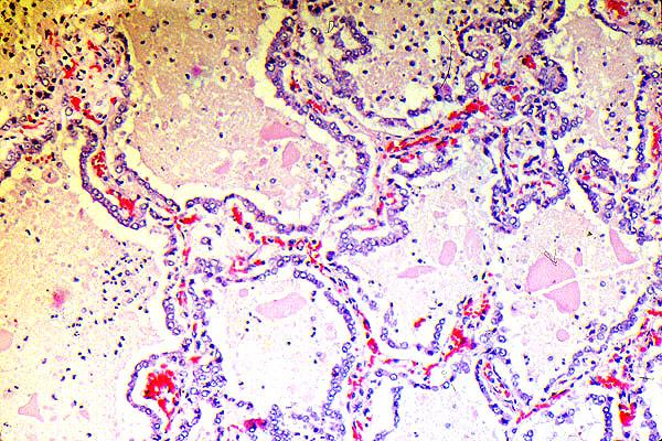 Bronchiolo-Alveolar Carcinoma
High magnification
KU Collection
{10475} oat cell carcinoma, growing
down between the cartilage rings
{10435} oat cell carcinoma
{11693} oat cell carcinoma
{12578} oat cell carcinoma
{14076} oat cells, spinal fluid
{12581} oat cells in sputum
{38521} oat cell
{38527} oat cell
{09082} oat cell carcinoma, electron
micrograph showing neurosecretory granules
EM shows oval nuclei, finely dispersed chromatin, scanty
cytoplasm, usually a few neurosecretory
(dense-core, APUD) granules, sometimes squamous and/or "adeno"
features.
The World Health Organization (J. Clin. Path. 54: 257, 2001)
now recognizes three "in situ" lesions.
If there is carcinoma in situ at the resection margin, a stump recurrence
is likely. If it's down in the glands, it's near-certain (Chest 128:
1736, 2005).
{18778} bone "mets", this hurts
* Preoperative staging of lung cancer has long been a challenge; watch for
fluorodeoxyglucose
PET scanning to become the mainstay (NEJM 343: 254, 2000; Radiology
212: 803, 1999).
Nowadays around 40%
of non-oat I-A tumors are apparently cured surgically.
 BRONCHIAL CARCINOIDS
Atypical carcinoids stain for chromogranin but have anaplasia
and mitotic figures and often necrosis; about half of these
eventually metastasize and some cause death.
* Proposed reclassification of carcinoids of the lung:
Am. J. Clin. Path. 116: S65, 2001.
The truly hard-core pathologist can use a gene probe for chromogranin mRNA: Cancer 82: 468, 1998.
Carcinoid and oat cell
Lung pathology series
Dr. Warnock's Collection
Bronchial hamartoma
Lung pathology series
Dr. Warnock's Collection
 Cancer metastatic to lungs
"Cannonball metastases"
KU Collection
UPPER AIRWAY, LARYNX AND TRACHEA
|
|
Congenital anomalies: The most important is the various forms of tracheo-esophageal fistulas. The newborn chokes and turns blue while eating. Mild forms are easy to treat (* celebrated in the original M*A*S*H). In the severe forms, the esophagus begins in a blind pouch, and all food ends up in the airways.
Sinusitis
Edema (allergy, infection) around the outlets of the sinuses sets up a vicious cycle, with non-drainage followed by bacterial infection followed by additional obstruction.
The most serious complication is infection of the cavernous sinus and nearby structures. Although sinus trouble is common, serious complications are rare.
|
|
Infections of the throat and larynx are common:
The common cold and viral laryngitis require no description.
Epiglottitis ("croup") usually results from infection with H. 'flu B. Airway obstruction is the major problem.
Laryngeal papillomatosis is a life-threatening infestation of the larynx with HPV (* usually strain 1). This time, warts are no laughing matter.
Trauma most often results today from intubation (grisly
article: Chest 96: 877, 1989). Forensic
pathologists look for fractures of the hyoid bone in suspected
strangling.
Laryngeal papilloma
Lung pathology series
Dr. Warnock's Collection
Foreign bodies in the trachea can cause sudden death, especially in toddlers and drunken adults. I've seen a peach section, a roll of ham, and a denture, and a friend lost his two-year-old daughter to a peanut (keep these and popcorn away from toddlers). The "cafe coronary" is usually due to poorly-chewed beef.
Laryngeal nodules ("Justin Timberlake's disease") are benign fibroepithelial polyps on the vocal cords of those who use their voices a lot -- drill sergeants, singers, teachers. They present as hoarseness and are easy to remove.
Angiofibroma of the nasopharynx is usually a teenaged boy's
tumor. This big white nuisance lesion is best left alone,
since it bleeds copiously if biopsied.
Nasopharyngeal carcinoma, usually poorly-differentiated
squamous, is rampant in China and seems
to be caused by a combination of Epstein-Barr virus (herpes 4)
and pickled fish
and pickled vegetables. How herpes 4
transforms cells: NEJM 333: 693 & 742, 1995.
* Cancer of the trachea is rare and is usually adenoid
cystic carcinoma or mucoepidermoid
carcinoma, arising from tracheal glands that are homologous to
salivary glands. (These can appear on the bronchi too.)
Dysplasia of the laryngeal epithelium: Pre-cancerous,
more or less aggressive depending on how
mean it looks. Cancer 75: 457, 1995.
TB of the true vocal cords is transmissible by speaking,
not just coughing.
Cancer of the larynx is very common, and is almost
invariably a squamous cell carcinoma due to
cigaret smoking. Ethanol abuse is also a risk factor; nobody
knows why. There is usually some nearby carcinoma
in situ.
Cancers confined to the true vocal cords (or the region just
below them) are usually easy to cure
using radiation, saving the voice. Cancers above the true vocal
cords usually require laryngectomy.
Angiofibromas
Student case
Galveston
Mucoepidermoid carcinoma and adenoid cystic carcinoma
Lung pathology series
Dr. Warnock's Collection
Metaplasia in a smoker's larynx.
Precedes dysplasia / cancer.
WebPath Photo
{11696} laryngeal squamous cell carcinoma, supraglottic
* Subclassifying cancers of the larynx for wiser therapy: Ann. Ot. Rhin. Laryng. 104: 587, 1995.
PLEURAL DISEASES
"Devil's grip", or pleurodynia, is a complication of coxsackievirus infection in children who are obviously sick with an acute febrile illness.
Pleural effusion -- fluid in the pleural space -- is a ubiquitous problem in clinical medicine. (Future radiologists: Before you can see it on x-ray, there must be about 700 mL -- a lot of fluid).
Each side can actually hold about 4000 mL if the lung is completely collapsed. Effusions cause atelectasis and restrict lung movement, you can tap them for diagnostic or therapeutic purposes, you can prevent their buildup by sclerosing the pleural space, etc.
Transudates: congestive heart failure, nephrotic syndrome, cirrhosis. All other pleural effusions will probably be exudates.
Hydrothorax: pleural effusion that is a transudate or serous exudate.
Hemothorax: a pleural effusion that is blood
Pyothorax or empyema: a pleural effusion that is
pus
Chylothorax, from injury to the thoracic duct
Hemothorax
Urbana Atlas of Pathology
Pneumothorax: Air in the pleural space, i.e., the lung
has collapsed.
This can result from a wound to the chest wall (why?), or from
a tear in the visceral pleura. The
latter can happen in healthy people ("spontaneous pneumothorax")
or from tumor or injury
(iatrogenic, pink puffer).
* Catamenial pneumothorax accompanies menstruation, usually in multiparous
women. The remedy is to do pleurodesis with talc during the menstrual period.
Pneumothorax is most serious ("tension pneumothorax") if a
valve-like flap on the visceral pleura
lets air into the chest wall but not out.
Chylothorax
WebPath
|
|
|
Pleural plaques of dense collagen are harmless but are a marker for asbestos exposure. They have nothing to do with smoking.
Benign mesothelioma is a pedunculated nubbin made of fibrous tissue. It has nothing to do with asbestos.
 Malignant mesothelioma ("Steve McQueen's disease"):
cancer of the pleural mesothelium
Malignant mesothelioma ("Steve McQueen's disease"):
cancer of the pleural mesothelium
This extremely lethal cancer almost always results from asbestos exposure.
* Turkey has some asbestos-rich soils and in these regions, blown dust may explain the abundance of asbestos fibers in the lungs and the migh prevalence of mesotheliomas. See Env. Health Perspect. 108: 1047, 2000.
The usual picture is that of a biphasic tumor (why?); histologic subtyping into "epithelioid" and "sarcomatoid" variants is of no use in prognosticating survival (Thorax 44: 496, 1989). Distinguishing mesothelioma from look-alike cancers is difficult. Tips for future pathologists:
Mesothelioma
Electron micrographs
VCU Pathology
As a matter of fact, telling mesothelioma from reactive hyperplasia of the mesothelium can be difficult, in biopsies or effusions.
Pathologists can now tell it from metastatic disease most of the time by staining for keratins of various MW's -- a wide range are present in mesothelioma. Autopsy is still helpful in confirming the diagnosis.
|
|
|
|
|
|
|
|
Oddly, a few percent of mesotheliomas produce insulin-like molecules that cause hypoglycemia.
Treating mesothelioma: Chest 107(S6): 332-S, 1995. Don't get your hopes up for a cure.
* An ultra-inbred community in Turkey in which 50% of the villagers die of mesothelioma, with only a dubious link to environmental carcinogens: Lancet 357: 444, 2001.
Rule: The parietal pleura is very sensitive to pain, far more so than the lung or any but the largest airways. If chest pain accompanies respiratory movements, the cause is probably inflammation or irritation of the parietal pleura. You may have experienced the "stitch", caused by a wrinkle and relieved by super-inflating your lungs.
{27597} mesothelioma, gross
CILIARY DYSKINESIA SYNDROMES (Am. J. Resp. CCM. 151: 1559, 1995).
Too big a topic for the end of "Respiratory", but includes Kartagener's (no dynein arms) and several others. Patients have:
We will be glad to help you make the diagnosis by electron microscopy.
A transgenic mouse with Kartagener's: Nature 354: 306, 1991.
* The Fellowship of those who bear the Mark of Pain. Who are the members of this fellowship? Those who have learned by experience what physical pain and bodily anguish mean, belong together all the world over; they are united by a secret bond. Praise God! One and all they know the horrors of suffering to which man can be exposed, and one and all they know the longing to be free from pain. He who has been delivered from pain must not think he is now free again, and at liberty to take life up just as it was before, entirely forgetful of the past. He is now a "man whose eyes are open" with regard to pain and anguish, and he must help to overcome those two enemies (so far as human power can control them) and to bring to others the deliverance which he has himself enjoyed. The man who, with a doctor's help, has been pulled through a severe illness, must aid in providing a helper such as he had himself, for those who otherwise could not have one. He who has been saved by an operation from death or torturing pain, must do his part to make it possible for the kindly anaesthetic and the helpful knife to begin their work, where death and torturing pain still rule unhindered. The mother who owes to medical aid that the child still belongs to her, and not to the cold earth, must help, so that the poor mother who has never seen a doctor may be spared what she has been spared. Where a man's death agony might have been terrible, but could fortunately be made tolerable by a doctor's skill, those who stood around his deathbed must help, that others, too, may enjoy that same consolation when they lose their dear ones.
Such is the Fellowship of those who bear the Mark of Pain.
-- Albert Schweitzer, M.D., Ph.D.
On the Edge of the Primeval Forest
* SLICE OF LIFE REVIEW
{11512} lung, normal
{11739} lung, normal
{14900} epiglottis, normal
{14901} olfactory epithelium, normal
{14902} olfactory epithelium, normal
{14903} trachea (false & true vocal cords)
{14904} trachea (false & true vocal cords)
{14905} trachea, normal
{14906} respiratory epithelium (of trachea)
{14907} respiratory epithelium (of trachea)
{14908} bronchus, normal
{14909} bronchus, normal
{14910} bronchiole, normal lung
{14911} bronchiole, normal lung
{14912} respiratory epithelium, normal
{14913} respiratory bronchiole, normal
{14914} respiratory bronchiole, normal
{14915} alveolar duct, normal
{14916} alveolar duct, normal
{14917} alveolus, normal
{14918} alveolus, normal
{15145} trachea
{15147} trachea
{15148} lung
{15149} lung
{15150} lung, normal
{15151} lung, normal
{15152} lung
{15154} pneumocytes, type ii lung
{15289} trachea, normal
{15290} trachea, normal
{15291} epiglottis, normal
{15292} epiglottis, normal
{15293} epiglottis, normal
{15294} trachea, * seromucous glands
{15295} trachea, normal
{15297} lung, normal
{15298} bronchiole, respiratory bronchiole
{15299} pneumocyte, type ii
{15300} bronchus, normal
{15302} bronchiole, normal
{15303} bronchus, * seromucous gland
{15763} lung, normal
{15764} lung, normal
{15765} lung, normal
{15766} lung, normal
{15767} lung, normal
{15768} lung, normal
{15791} larynx, normal
{15792} larynx, normal
{17521} bronchus, normal
{17566} lung, normal
{20898} trachea
{20899} trachea, seromucous gland
{20900} cilia, tracheal epithelial cells
{20902} epiglottis
{20903} epiglottis
{20904} trachea
{20907} trachea, epithelium
{20909} alveolus, lung
{20910} lung, respiratory duct
{20911} lung, terminal bronchiole ?
{20912} lung, terminal bronchiole ?
{20913} bronchus, lung
{25654} lung, normal
{27215} lung, normal
{27218} lung, normal
{27221} lung, normal
{27224} lung, normal
{27227} lung, normal
{27468} lung, normal
{29168} bronchitis, chronic with normal to compare
{37604} lung, normal
{37619} bronchus, normal
{41517} lung, normal cast of capillary bed
{46452} xerogram laryngeal, norma
{15289} trachea, normal
NOTE: You may not share Mr. Housmann's "scientific" nihilism. I'm always surprised and pleased when such folks at least pay lip service to basic human goodness.


| Visitors to www.pathguy.com reset Jan. 30, 2005: |
Ed says, "This world would be a sorry place if
people like me who call ourselves Christians
didn't try to act as good as
other
good people
."
Prayer Request
 Teaching Pathology
PathMax -- Shawn E. Cowper MD's
pathology education links
Ed's Autopsy Page
Notes for Good Lecturers
Small Group Teaching
Socratic
Teaching
Preventing "F"'s
Classroom Control
"I Hate Histology!"
Ed's Physiology Challenge
Pathology Identification
Keys ("Kansas City Field Guide to Pathology")
Ed's Basic Science
Trivia Quiz -- have a chuckle!
Rudolf
Virchow on Pathology Education -- humor
Curriculum Position Paper -- humor
The Pathology Blues
 Ed's Pathology Review for USMLE I
![]()
![]()
 | Pathological Chess |
 |
Taser Video 83.4 MB 7:26 min |

)window.location='http://edcenter.med.cornell.edu/Pathology_Images/342.gif') Pulmonary edema
Pulmonary edema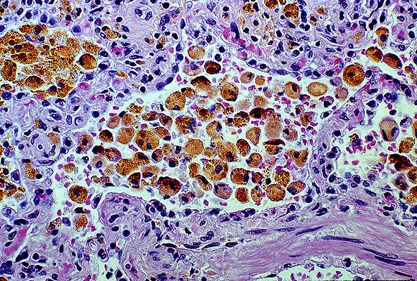 Heart Failure Cells
)window.location='http://edcenter.med.cornell.edu/Pathology_Images/341.gif') Pulmonary infarct, healing
Pulmonary infarct, healing)window.location='http://edcenter.med.cornell.edu/Pathology_Images/312.gif') Pulmonary infarcts
Pulmonary infarcts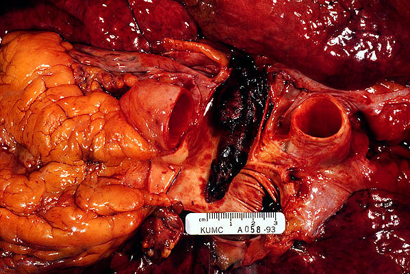 Pulmonary thromboembolus
)window.location='http://edcenter.med.cornell.edu/Pathology_Images/309.gif') Saddle embolus
Saddle embolus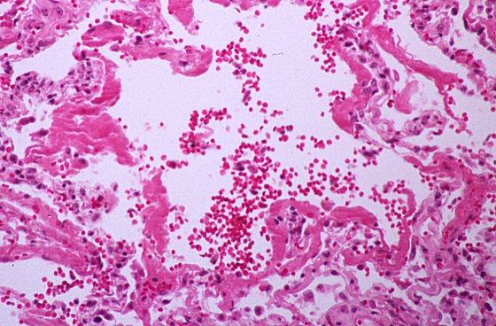 ARDS
ARDS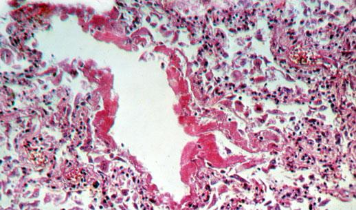 ARDS
ARDS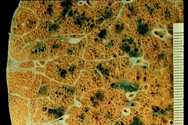 Centrilobular emphysema
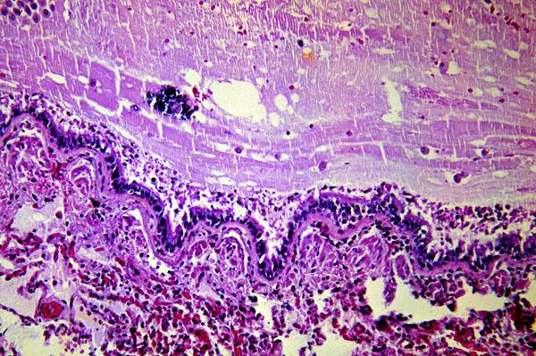 Asthma
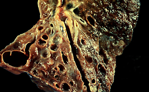 Cystic fibrosis
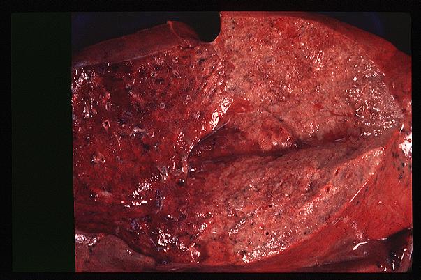 Lobar pneumonia
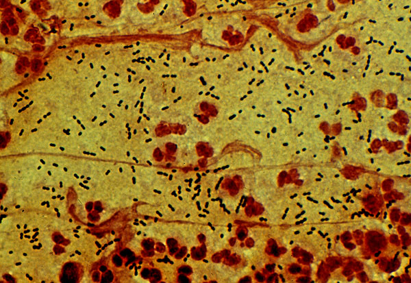 Pneumococcal pneumonia
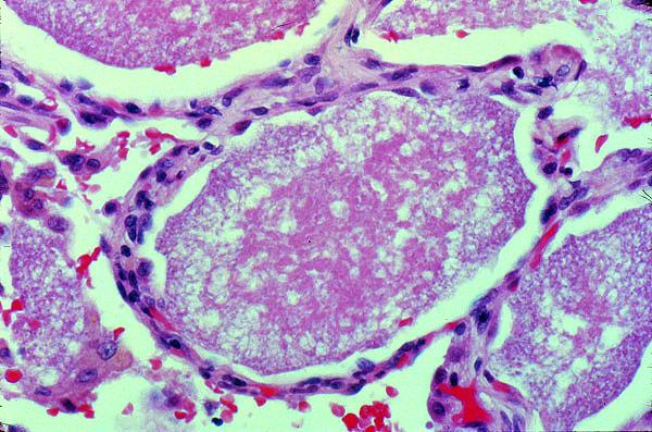 Pneumocystis carinii
 Tuberculosis
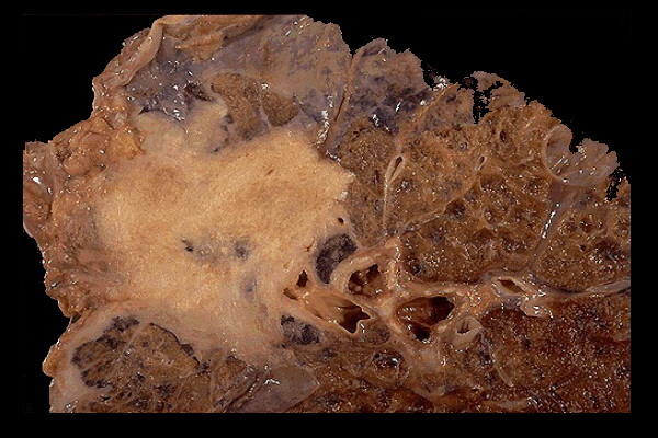 Lung cancer
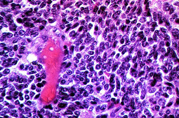 Oat cell carcinoma
 Laryngeal cancer, fatal
)window.location='http://www.mdchoice.com/photo/img/img0102.jpg') Tension pneumothorax
Tension pneumothorax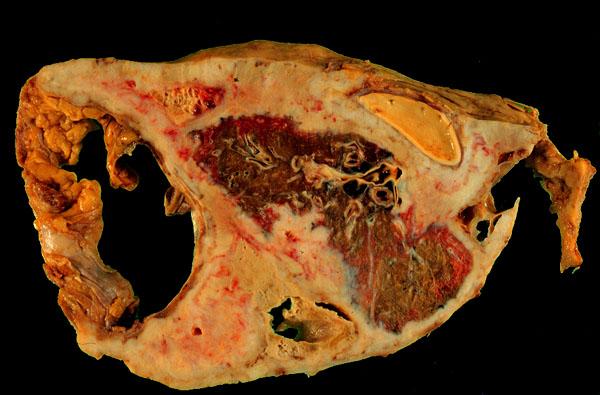 Mesothelioma
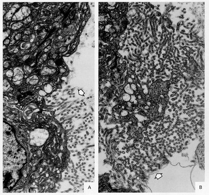 Elongated microvilli
)window.location='http://www.path.queensu.ca/present/boag/resp/images/lgcames1.jpg') Mesothelioma
Mesothelioma