Ed Friedlander, M.D., Pathologist
scalpel_blade@yahoo.com


Cyberfriends: The help you're looking for is probably here.
Welcome to Ed's Pathology Notes, placed here originally for the convenience of medical students at my school. You need to check the accuracy of any information, from any source, against other credible sources. I cannot diagnose or treat over the web, I cannot comment on the health care you have already received, and these notes cannot substitute for your own doctor's care. I am good at helping people find resources and answers. If you need me, send me an E-mail at scalpel_blade@yahoo.com Your confidentiality is completely respected.
 DoctorGeorge.com is a larger, full-time service.
There is also a fee site at myphysicians.com,
and another at www.afraidtoask.com.
DoctorGeorge.com is a larger, full-time service.
There is also a fee site at myphysicians.com,
and another at www.afraidtoask.com.
Translate this page automatically
 |
With one of four large boxes of "Pathguy" replies. |
 I'm still doing my best to answer
everybody.
Sometimes I get backlogged,
sometimes my E-mail crashes, and sometimes my
literature search software crashes. If you've not heard
from me in a week, post me again. I send my most
challenging questions to the medical student pathology
interest group, minus the name, but with your E-mail
where you can receive a reply.
I'm still doing my best to answer
everybody.
Sometimes I get backlogged,
sometimes my E-mail crashes, and sometimes my
literature search software crashes. If you've not heard
from me in a week, post me again. I send my most
challenging questions to the medical student pathology
interest group, minus the name, but with your E-mail
where you can receive a reply.
Numbers in {curly braces} are from the magnificent Slice of Life videodisk. No medical student should be without access to this wonderful resource. Someday you may be able to access these pictures directly from this page.
Also:
Medmark Pathology -- massive listing of pathology sites
Freely have you received, freely give. -- Matthew 10:8. My
site receives an enormous amount of traffic, and I'm
handling about 200 requests for information weekly, all
as a public service.
Pathology's modern founder,
Rudolf
Virchow M.D., left a legacy
of realism and social conscience for the discipline. I am
a mainstream Christian, a man of science, and a proponent of
common sense and common kindness. I am an outspoken enemy
of all the make-believe and bunk that interfere with
peoples' health, reasonable freedom, and happiness. I
talk and write straight, and without apology.
Throughout these notes, I am speaking only
for myself, and not for any employer, organization,
or associate.
Special thanks to my friend and colleague,
Charles Wheeler M.D.,
pathologist and former Kansas City mayor. Thanks also
to the real Patch
Adams M.D., who wrote me encouragement when we were both
beginning our unusual medical careers.
If you're a private individual who's
enjoyed this site, and want to say, "Thank you, Ed!", then
what I'd like best is a contribution to the Episcopalian home for
abandoned, neglected, and abused kids in Nevada:
My home page
Especially if you're looking for
information on a disease with a name
that you know, here are a couple of
great places for you to go right now
and use Medline, which will
allow you to find every relevant
current scientific publication.
You owe it to yourself to learn to
use this invaluable internet resource.
Not only will you find some information
immediately, but you'll have references
to journal articles that you can obtain
by interlibrary loan, plus the names of
the world's foremost experts and their
institutions.
Alternative (complementary) medicine has made real progress since my
generally-unfavorable 1983 review linked below. If you are
interested in complementary medicine, then I would urge you
to visit my new
Alternative Medicine page.
If you are looking for something on complementary
medicine, please go first to
the American
Association of Naturopathic Physicians.
And for your enjoyment... here are some of my old pathology
exams
for medical school undergraduates.
I cannot examine every claim that my correspondents
share with me. Sometimes the independent thinkers
prove to be correct, and paradigms shift as a result.
You also know that extraordinary claims require
extraordinary evidence. When a discovery proves to
square with the observable world, scientists make
reputations by confirming it, and corporations
are soon making profits from it. When a
decades-old claim by a "persecuted genius"
finds no acceptance from mainstream science,
it probably failed some basic experimental tests designed
to eliminate self-deception. If you ask me about
something like this, I will simply invite you to
do some tests yourself, perhaps as a high-school
science project. Who knows? Perhaps
it'll be you who makes the next great discovery!
Our world is full of people who have found peace, fulfillment, and friendship
by suspending their own reasoning and
simply accepting a single authority that seems wise and good.
I've learned that they leave the movements when, and only when, they
discover they have been maliciously deceived.
In the meantime, nothing that I can say or do will
convince such people that I am a decent human being. I no longer
answer my crank mail.
This site is my hobby, and I presently have no sponsor.
This page was last updated February 6, 2006.
During the ten years my site has been online, it's proved to be
one of the most popular of all internet sites for undergraduate
physician and allied-health education. It is so well-known
that I'm not worried about borrowers.
I never refuse requests from colleagues for permission to
adapt or duplicate it for their own courses... and many do.
So, fellow-teachers,
help yourselves. Don't sell it for a profit, don't use it for a bad purpose,
and at some time in your course, mention me as author and KCUMB as my institution. Drop me a note about
your successes. And special
thanks to everyone who's helped and encouraged me, and especially the
people at KCUMB
for making it possible, and my teaching assistants over the years.
Whatever you're looking for on the web, I hope you find it,
here or elsewhere. Health and friendship!
We have two ends with a common link;
--Author unknown
QUIZBANK GI tract (all)
 I am presently adding clickable links to
images in these notes. Let me know about good online
sources in addition to these:
I am presently adding clickable links to
images in these notes. Let me know about good online
sources in addition to these:
Pathology Education Instructional Resource -- U. of Alabama; includes a digital library
Houston Pathology -- loads of great pictures for student doctors
Pathopic -- Swiss site; great resource for the truly hard-core
Syracuse -- pathology cases
Walter Reed -- surgical cases
Alabama's Interactive Pathology Lab
"Companion to Big Robbins" -- very little here yet
Alberta
Pathology Images --hard-core!
Cornell
Image Collection -- great site
Bristol Biomedical
Image Archive
EMBBS Clinical
Photo Library
Chilean Image Bank -- General Pathology -- en Español
Chilean Image Bank -- Systemic Pathology -- en Español
Connecticut
Virtual Pathology Museum
Australian
Interactive Pathology Museum
Semmelweis U.,
Budapest -- enormous pathology photo collection
Iowa Skin
Pathology
Loyola
Dermatology
History of Medicine -- National Library of Medicine
KU
Pathology Home
Page -- friends of mine
The Medical Algorithms Project -- not so much pathology, but worth a visit
National Museum of Health & Medicine -- Armed Forces Institute of Pathology
Telmeds -- brilliant site by the medical students of Panama (Spanish language)
U of
Iowa Dermatology Images
U Wash
Cytogenetics Image Gallery
Urbana
Atlas of Pathology -- great site
Visible
Human Project at NLM
WebPath:
Internet Pathology
Laboratory -- great site My team:
Ed Lulo's Pathology Gallery
Bryan Lee's Pathology Museum
Dino Laporte: Pathology Museum
Tom Demark: Pathology Museum
Dan Hammoudi's Site
Claude Roofian's Site
Pathology Handout -- Korean student-generated site; I am pleased to permit their use of my cartoons
Estimating the Time of Death -- computer program right on a webpage
Pathology Field Guide -- recognizing anatomic lesions, no pictures

St.
Jude's Ranch for Children
I've spent time there and they are good. Write "Thanks
Ed" on your check.
PO Box 60100
Boulder City, NV 89006--0100
More of my notes
My medical students
Clinical
Queries -- PubMed from the National Institutes of Health.
Take your questions here first.
HealthWorld
Yahoo! Medline lists other sites that may work well for you

We comply with the
HONcode standard for health trust worthy
information:
verify
here. 

![]()

With one we sit, with one we think.
Success depends on which we use;
Heads we win, tails we lose!
 Georgetown Med School
GI Pathology
Lots of ulcerative colitis
Gastrointestinal System I
Great pathology images
Indiana Med School
Gastrointestinal System II
Great pathology images
Indiana Med School
Gastrointestinal Pathology
Virginia Commonwealth U.
Great pictures
Tulane Pathology Course
Great for this unit
Exact links are always changing
Gastrointestinal Diseases
Mark W. Braun, M.D.
Photomicrographs
GI Tract Ia
Introductory Pathology Course
University of Texas, Houston
GI Tract Ib
Introductory Pathology Course
University of Texas, Houston
GI Tract II
Introductory Pathology Course
University of Texas, Houston
KCUMB students: Questions for this system
Esophagus and Stomach GI Tract 1-187
Small Bowel GI Tract 188-256
Large Bowel GI Tract 257-280, 322-332
Colon Cancer GI Tract 281-321
Hepatobiliary I Liver 1-86
Hepatobiliary II Liver 87-172
Pancreas Pancreas Do 'em all
 Consider this mastery material.
Be sure you can use the following terms correctly, and tell how and where they apply to the gut.
achalasia
atresia
diverticulum
erosion
fistula
hematemesis
hematochezia
hernia
melena
polyp
pseudo-diverticulum
reflux
stenosis
tenesmus
ulcer
Be sure you can recognize each of the kinds of lesions presented on the videodisc!
INTRODUCTION
Disease of the gut troubles most people at some time during their lives, and claims the lives of many people.
This easy unit focuses on problems with the alimentary canal. Conceptually, the material presents no serious problems. Some of the "why"'s are only now being clarified, and several common problems have complex causes. The human gut withstands considerable abuse. For a review of how to injure the gastroduodenal mucosa, see J. Clin. Gastroent. 13(S1): S1, 1991.
A few terms for review now:
Polyp: Any bump on the gut that sticks up above the inner surface into the lumen.
Hernia: Presence of a portion of an organ in a body space where it doesn't belong. Incarcerated hernia: One that can't be reduced, i.e., put back in its proper place. Strangulated hernia: Where venous drainage is compromised and infarction is imminent / present.
Erosion: A portion of the epithelium
mucosa has been lost due to necrosis, but there has been no loss of the
underlying connective tissue. In the stomach, as the term is generally
used, there may be loss of some
connective tissue but sparing the muscularis mucosae.
Ulcer: A portion of the epithelium and some of the underlying
connective tissue has been lost due to necrosis. In the stomach, an "ulcer"
is typically diagnosed only if the muscularis mucosae is lost.
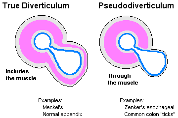
Diverticulum: An outpouching of all the layers of the wall of a hollow organ. Pseudo-diverticulum:
Outpouching of mucosa through a defect in the muscularis propria.
Paracrine / endocrine cells ("Kulchitsky cells", "enterochromaffin", "neurosecretory", "argentaffin /
argyrophil", formerly "APUD" cells) are found individually all along the gut, and various ones
produce various peptide hormones of known and unknown significance. (You met these in the lung
when we studied oat cell carcinoma and bronchial carcinoid.)
)window.location='http://www.mdchoice.com/photo/img/img0016.jpg') Strangulated umbilical hernia
Strangulated umbilical hernia
EMBBS
In the GI tract in particular, "atypia" and "dysplasia" are probably interchangeable terms.
The common cancers of the GI tract are carcinomas. Often the first physical finding is Virchow's sentinel lymph node, where the thoracic duct joins the left internal jugular vein.
ESOPHAGUS
|
|
The "gullet" begins at the cricophagyngeus (the short "upper esophageal sphincter" starts here) and ends at the diaphragmatic hiatus or thereabouts ("lower esophageal sphincter"). Solid food may hang up at either site, or where the esophagus passes behind the left main-stem bronchus or between enlarged hilar nodes. Neither sphincter is fool-proof (or even anatomic). Considerable coordination is required for a proper swallow (the big word for "swallowing" is "deglutition"). Most of the time, the esophagus performs its simple but important task well.
You remember that the upper esophagus (i.e., the first inch or so) has mostly skeletal muscle (and is thus subject to diseases of nerve and skeletal muscle), the middle esophagus (i.e., maybe another inch) has both skeletal and smooth muscle, and the distal esophagus (i.e., most of the esophags) has mostly smooth muscle. Unlike most of the rest of the gut, the esophagus has no serosa to help limit the spread of rips or cancers.
Problems with the esophagus manifest as difficulty and/or pain on swallowing, and/or problems with regurgitation.
Birth defects of the esophagus are relatively common.
Agenesis of some or all of the esophagus is uncommon. Atresia of the esophagus, as elsewhere, is failure of a normally-hollow organ to develop its lumen. An atretic esophagus is represented, over part or all of its length, by a fibromuscular cord without a lumen. Less severely, portions of the esophagus may be congenitally stenotic (i.e., too narrow).
{20073} esophageal atresia, from behind. Lungs at the sides, stomach at the bottom
Tracheo-esophageal fistulas of several varieties are common neonatal surgical problems. The proximal esophagus may enter the trachea or bronchus, producing coughing upon feeding (* original movie of M* A* S* H). The proximal esophagus may end blindly and the distal esophagus arise from a large airway, preventing feeding and causing the stomach to fill with air (the most common version). Or there may simply be a window between the two organs.
Throat problems: A little bit higher than the real esophagus, but worth mentioning here:
Globus hystericus ("globus syndrome", "globus pharyngeus"), a "lump in the throat", is spasm of the back of the throat and perhaps the upper esophagus. It creates the feeling of a mass in the hypopharynx. Sometimes the cause is organic (i.e., a reflex from something wrong nearby, perhaps reflux from the esophagus spilling into the larynx); often it's psychosomatic. Psychiatrists attribute it to "swallowed tears", and the cure is to cry. (Despite its banal nature, the sensation of a mass may frighten a patient. "Spontaneous cures of non-biopsy-proven throat cancer" sound like globus.)
Zenker's "pulsion" diverticulum: Adjacent to the cricophagyngeus muscle. A hiding-place for last week's spaghetti and last month's pills (so that's why they didn't work....) The smell alone may create a serious social problem.
* An epiphrenic diverticulum, also of mysterious origin, occurs just above the diaphragm. It can harbor large amounts of fluid that are disgorged back to the mouth shortly after the patient goes to bed.
Achalasia means "failure of a sphincter to relax when it should". Usually, this means the lower esophageal sphincter.
In the U.S., the problem is usually idiopathic failure of the lower sphincter to relax. The etiology is just now becoming clarified; the cause is usually inflammation of the myenteric plexus (Am. J. Surg. Path. 24: 1153, 2000; Histopathology 35: 445, 1999; Gastroenterology 111: 648, 1996), with eventual nerve damage, but nobody knows why this happens. In other cases, the ganglion cells are simply lost instead; no one knows why.
The esophagus remains filled with food. The patient (typically a young adult) will notice regurgitation and bad breath. The situation may become really nasty as more and more food accumulates in a mega-esophagus. Fortunately, most patients are cured by a single endoscopic dilatation of the sphincter; there's also "botox", or laparoscopic myotomy and partial fundoplication for the hard cases (Am. J. Surg. 181: 471, 2001.)
Untreated, achalasia is a life-threatening problem (carcinoma, aspiration pneumonia).
* Pseudo-achalasia usually results from cancer obliterating the myenteric plexus (Am. J. Surg. Path. 26: 784, 2002).
You remember that Chagas' disease (T. cruzi) is an important cause of mega-esophagus worldwide.
Glycogen plaques are acanthotic (i.e., thick spiny layer) epithelium with extra glycogen. The most common lesion of the esophagus, by far, and exactly as serious as freckles. You'll see it whenever you do endoscopies.
 {15553} glycogen acanthosis of esophagus, gross
Webs are contracted, localized fibrous scars that form little shelves ("ledges") that may obstruct the lumen.
* No one knows quite what to make of a supposed association between upper esophageal webs, iron deficiency anemia, and a risk for squamous cell carcinoma in the proximal esophagus. Whether or not this really exists, it's called "Plummer-Vinson syndrome". Current thinking is that somehow iron deficiency causes the webs to form just beyond the circoid (Am. J. Gastro. 97: 190, 2002).
Schatzki's ring is a washer-shaped partially-obstructing fibrous ring at the squamo-columnar junction just above the gastroesophageal junction. It is a radiologist's delight and may be seen in any adult; long a mystery, cases that occur in the absence of reflux may be due to pill enlodgement (Am. J. Surg. 158: 563, 1989). A-ring is the same, at the gastro-esophageal junction.
A big chunk of solid food (i.e., poorly-chewed beef) may hang up on a web or ring. In bad cases, softer food may have trouble negotiating the obstruction.
{09433} Schatzki ring
{09434} Schatzki ring
{09435} web
Hiatus hernia is said to be present whenever a portion of the stomach pooches up through the diaphragmatic hiatus. Said to be present in up to 10% of adults (typically the overweight), they are most often sub-clinical.
Sliding hiatus hernia (the usual kind) is present when a short (congenital, fibrous scarring from years of reflux, diaphragm pulled low by obesity) pulls the proximal stomach into the chest. As the esophagus contracts during swallowing, radiologists watch the stomach slide further up through the diaphragm. As with "reflux esophagitis", the distal esophagus is likely to become inflamed and damaged as a result of exposure to pepsin and acid.
Para-esophageal ("rolling") hiatus hernia (the less common kind) is present when a portion of the stomach rolls up through the diaphragm alongside an esophagus of normal length. This is typically a non-problem, but the herniated portion of stomach may become strangulated.
Future pathologists: Don't expect to see either type of hernia (or, for that matter, a Schatzki's ring, or an intestinal hernia) after death, when the muscles of the body relax / go into rigor mortis).
Traction diverticula, illustrated without comment in "Big Robbins", probably result from scar contraction in the mediastinum (i.e., TB). They are uncommon.
* Aphthous ulcers, the familiar painful white "canker sores" that most people have experienced on the oral mucosa, may be a serious problem in the esophagus for people with HIV infection. No one knows why. Thalidomide for this problem: J. Inf. Dis. 180: 61, 1999.
Gastroesophageal reflux (GERD, formerly, "peptic esophagitis") is the common esophagal problem, the result of an incompetent lower esophageal sphincter.
The sphincter is inflamed, scars, and becomes further damaged. The epithelium keeps getting digested and regrowing, with much more opportunity to select for mutated cells. Pathophysiology Am. J. Med. Sci. 326, 274, 2003.
Update for clinicians: JAMA 287: 1972 & 1982, 2002. The correlation between clinical symptoms and endoscopy is remarkably poor. Only about half of the patients with severe "GERD" even have heartburn (Dig. Dis. Sci. 48: 2237, 2003.
The cause of common "reflux" remains obscure. Mechanical problems (including overweight and
sliding hiatus hernia) must contribute, and the problem is more severe if there's "excess stomach acid
/ pepsin / bile" or the gastric contents stays for some reason within the esophagus. Other things that
irritate the esophagus (swallowing spicy food, alcohol (Gut 34: 727, 1993, others), very hot
beverages, and/or tobacco juice) will not help either. Pregnancy, benzodiazepines, intubation, and
tobacco use are all implicated as well. Lying flat makes matters worse (tip: try propping the head of
the bed up on cinder blocks).
The diagnosis of GERD is made clinically on endoscopy
(Dig. Dis. Sci. 45: 217, 2000),
but pathology may be obtained for confirmation.
Pathologists diagnose reflux based on the following criteria (older review: Gast. Clin. N.A. 19: 631,
1990; update Am. J. Surg. Path. 20(S1): S-31, 1996):
Future pathologists: Be sure you've got that specimen properly oriented, and consider asking the
clinician to send it up on cardboard;
Why the thickness of the basal cell layer? In reflux, the surface cells get digested and the basal cells
are multiplying overtime to replace them.
 Peptic esophagitis ulcers
WebPath Photo
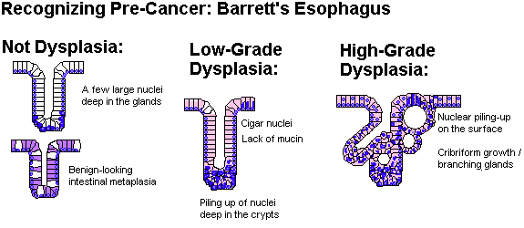
If replacement of the normal squamous epithelium by a columnar epithelium has occurred, you may have a Barrett's esophagus. All about Barrett's: Gastroenterology 122: 1569, 2002; NEJM 346: 836, 2002; Med. Clin. N.A. 86: 1423, 2002.
Today's definitions of Barrett's usually require goblet cells, and for the truly hardcore, the non-goblet cells are both absorptive and secretory. Older definitions of Barrett's allowed any columnar epithelium type.
Ignore hamartomas of gastric-type mucosa; cancer probably only occurs if there is metplasia with goblet cells, so perhaps one day Barrett's will be redefined as limited to this.
There are at least 2 million "Barrett's" patients in the U.S. In Sweden, 1.6% of the population is affected, with alcohol and tobacco being risk factors (Gastroent. 129: 1825, 2005).
As you'd expect ("Nowell's law triumphant"), finding a Barrett's esophagus means there's been some hits on the genome, and the genetically damaged cells have had a chance to overgrow the area because of repeated healing from reflux. This is a fertile breeding ground for adenocarcinoma of the esophagus.
The molecular biology of transformation to cancer is now fairly clear: Lancet 360: 1587, 2002.
* Anti-reflux therapy may be helpful, but don't count on it to reverse the process. This includes the new, popular procedure of laparoscopic fundoplication (Ann. Thor. Surg. 77: 393, 2004).
In today's cost-conscious era, it seems reasonable to screen adults who complain of heartburn once for Barrett's, with follow-up if and only if there is dysplasia (numbers Ann. Int. Med. 138: 176, 2003).
More onNowell's law: Arch. Surg. 132: 728, 1997. Deciding on therapy (lasers, electrocoagulation, mechanically removing the mucosa, surgery) based on how bad the dysplasia is: Br. J. Surg. 84: 760, 1997, Am. J. Med. 111 S 8A: 147A, 2001; lasers see Gut 51: 776, 2002. Some pathologists recommend waiting to resect until there is "superficial adenocarcinoma", but the truth is that pathologists can't seem to distinguish this reliably from high-grade dysplasia (Gut 51: 671, 2002).
Future pathologists: You'll look at dysplasias from Barrett's biopsies frequently, and there are likely to be further refinements that will help you let the surgeons know when to operate (laser, scrape, etc). All Barrett's have some hyperchromasia of the nuclei in the lower portions of the glands. Low-grade dysplasia features loss of mucus production, stratified nuclei in the crypts, and elongated nuclei in parallel. High-grade dysplasia features stratification on the surface, branching glands, and/or cribriform stuff in the crypts. Be careful about calling "severe dysplasia" if there is inflammation; it might be better to treat the reflux and repeat the biopsy. One major criterion that says "operate" is loss of the basal orientation of the nuclei, i.e., this is more than just the kind of atypia that one finds in an adenomatous polyp. Grading update: J. Clin. Path. 55: 910, 2002. Biomarkers Mayo Clin. Proc. 76: 438, 2001. Does screening really save enough lives to be worthwhile? It's still a tough call (Am. J. Gastro. 98: 1931, 2003; Gastroenterology 127: 310, 2004 from KU).
* HCA, a tumor marker, to help spot the aggressive dysplasias: Am. J. Clin. Path. 122: 747, 2004.
{15428} Barrett's esophagus (note tan columnar, rather than white squamous, mucosa)
{15429} Barrett's esophagus
{15427} acute reflux esophagitis (one heck of a case of heartburn;
stomach is on the left)
Reflux leads to fibrosis (scarring, shortening, perhaps narrowing) of the esophagus, difficulty on swallowing ("dysphagia"), pain in swallowing ("odynophagia"), retrosternal pain ("heartburn") and/or slow GI bleeding leading to iron-deficiency. In severe cases, massive GI bleeding (hematemesis, melena) can occur.
Other noteworthy causes of esophagitis include candida, CMV, herpes, radiation, generalized diseases of stratified squamous epithelium (* pemphigus, other) or keeping a stomach tube down for more than a few minutes. All the above can give some nasty ulcers.
You also remember the esophageal changes ("rubber hose") in scleroderma and graft-vs.-host disease.
{11096} candida esophagitis (scrapes off, unlike glycogen acanthosis)
{11099} candida esophagitis
{49128} candida esophagitis
|
|
{19449} herpes, histology
* Future pathologists: Herpes of the esophagus is often relatively devoid of good herpes-inclusion
cells. Look instead for clusters of macrophages (Hum. Path. 22: 541, 1991).
Drinking lye (Drano, etc., an extremely painful and unreliable method for would-be suicides) or
some other caustic substance leads to corrosive esophagitis, strictures, etc., etc.
{11772} lye burn of stomach
Perforated esophagus can result from swallowing the wrong
thing. Chicken bones are infamous (AJFMP 19: 166, 1998) --
these can pierce the heart or cause other dreadful problems.
Lacerated esophagus usually results from heavy-duty vomiting during which the esophagus fails to
relax (alcohol abuse, pregnancy, post-anesthesia; "Mallory-Weiss syndrome"). Endoscopists and
pathologists see little longitudinal tears, usually in the distal esophagus. They are a problem only if
bleeding is massive, or if the esophagus actually ruptures ("Boerhaave's syndrome"; at these sites of
rupture, the muscularis mucosae is apparently absent: Am. J. Surg. 158: 420, 1989).
{15420} lacerated esophagus
Esophageal varices are dilatations of the esophago-gastric venous plexus. These result from portal
vein hypertension from any cause, as the blood from the stomach and intestines seeks the low-pressure pathway back to the
heart.
You won't know varices are there until they bleed. And they bleed massively when their attenuated
overlying mucosa is rubbed away, or they just pop from pressure. This is the fast way out of life for
many problem drinkers.
{38629} varices, gross
Portal hypertension patients will also commonly exhibit hemorrhoids and dilated veins around the
umbilicus ("caput medusae", why?) Remember that portal hypertension will greatly accelerate GI
bleeding from non-variceal causes (gastritis, peptic esophagitis, ulcer, Mallory-Weiss) as well.
Learn now: The causes of portal hypertension....
Benign tumors of the esophagus: Banal, and relatively uncommon. For example, fibrovascular
hamartomas: AJR 166: 781, 1996.
Carcinoma of the esophagus strikes around 8000 people each years and kills most of them.
Squamous cell carcinoma has historically been the most common esophageal cancer
in the United States, but is becoming less common.
Most patients are males (more than 4:1), black men are at higher risk than others, and in the U.S., the
large majority are both smokers (cigarets, cigars) and drinkers.
Old lye strictures are also frequent sites for esophageal cancer, as is the Chagas-disease ridden mega-esophagus (Digestion
47: 138, 1990).
The epidemic of highly-aggressive squamous cell carcinoma in Mainland China (Cancer 73: 2027, 1994;
Cancer 74: 573, 1994) has been attributed to aspergillus fungus contamination, nitrosamines,
vitamin deficiency, zinc deficiency, molybdenum deficiency, ethnic teas, and ethnic delicacies that
are pickled (i.e., rendered rich in certain fungi).
* The South Carolina lowlands have a great excess of squamous cell carcinoma of the esophagus,
and these cancers have a distinctively high rate of p53 mutations, suggesting some chemical in the
environment (J. Thor. Card. Surg. 108: 148, 1994). Still a mystery: South. Med. 95: 900, 2002.
In West Kenya, it is a common disease of teens and young adults (Lancet 360: 462, 2002).
Any portion of the esophagus can be involved; the middle third is slightly more common than the
others.
Like most squamous cell carcinomas, esophageal cancer arises in squamous dysplasia (update Gut 54:
187, 2005), is often multifocal (Cancer 73: 2687, 1994), grows as a
fungating lesion (less often, just an ulcer), and produces symptoms (dysphagia, food "sticking") only
late. Because of its location, the later stages of this disease are particularly cruel.
* Biomarkers: Surgery 127: 552, 2000.
* A claim from the 1990's that many esophageal carciomas
contained HPV remains unconfirmed.
* Honesty or the impact of managed care? The surgeons finally
tell it like it is: radiation and chemotherapy for cancer of the
esophagus have little or no impact beyond making life more miserable
for these patients (Arch. Surg. 133: 722, 1998).
{15423} carcinoma of the esophagus, exophytic growth
Adenocarcinoma is now almost as common as squamous
cell carcinoma, and is probably increasing in frequency.
It arises most often arising in a Barrett's
esophagus. Again, the male predominance is marked (more than 6:1)
and as you'd expect, symptomatic reflux is a strong risk
factor (NEJM 340: 825, 1999). Tobaco and obesity are risk factors,
alcohol consumption is not.
It's basically the same lesion as
cancer of the gastric cardia: Br. J. Surg. 529: 529, 1999.
The longer the Barrett's region
(Gut 33: 1155, 1992), the higher the risk, and smoking also increases risk: Cancer 72: 1155, 1993
(big study). Histopathology of Barrett's cancer: Hum. Path. 19: 942, 1988.
Most of the current work on esophageal carcinoma focuses on early, accurate diagnosis and surgical
treatment (Br. J. Surg. 76: 759, 1989; how to cut Ann. Surg. 219: 475, 1994; J. Am. Col. Surg.
178: 363, 1994). This will be of special interest to those of you who do primary care in rural areas;
you are likely to do your own endoscopy. Both types of cancers often arise multifocally (Br. J.
Surg. 75: 531, 1988). Since normal esophageal epithelium contains glycogen, you can help
demarcate dysplastic squamous epithelium using Lugol's iodine solution (J. Surg. Onc. 50: 149,
1992).
Other tumors of the esophagus are banal (little leiomyomas) or curiosities (sarcomas; melanomas,
Am. J. Clin. Path. 92: 802, 1989, others).
{49132} leiomyoma, gross
Histopathology and cell-of-origin of cancers of the esophagus: Cancer 75(S6): 1440, 1995.
* Esophageal angina: Spasm simulating myocardial infarct (including "crushing chest pain radiating
to the left arm and jaw", etc., etc.) Once said to be common, we hear little about this now.
{11889} stomach with biscuit
Most stomach problems start with the mucosa. Review of the healthy mucosa:
Surface, pits ("crypts" / "foveolae") in all areas... Tall surface mucous cells (make "neutral mucus")
Deep in pits, all areas... Neck cells (reserve cells for both above and below; "the proliferative zone")
Cardiac glands... More neck-type cells
Gastric glands... Parietal cells (eosinophilic, packed with mitochondria; make acid and intrinsic factor);
Chief cells (pale, granular)
Pyloric glands... Neck-type cells; G-cells (gastrin producers)
In 1999, Lancet 354: 134, 1999
published a short report on
biotech-enhanced potatoes thickening the mucosa of the stomach
and elongating the crypts of rats to which they were fed.
A small-sample study with a conclusion that didn't make sense...
and the small differences could easily be explained by differences
in the angles produced by hand-cutting and hand-embedding, as happens on human biopsies
routinely. Did the author compare the heights of the villi to the
depths of the crypts for control purposes? Or for that matter,
the relative thickening of crypt layer and gland layer?
Of course not! Your instructor wrote to Lancet (who turned down my letter)
and to the author (Arpad Pusztai,
who had gone on TV and made inflammatory statements
about the public being used as guinea pigs;
he didn't respond either and is now a leading antibiotechnology activist
and celebrated "persecuted genius").
Lancet ended up admitting (May 29, 1999) that
it knew the paper was junk science when it was published but that if it
hadn't published it, it would have been smeared for "suppressing
information" by
anti-biotechnology militants. I am not making any of this up.
Regrettably the
"environmental movement" thrives on this kind of stuff.
Greenpeace USA's website
(2005) is still citing the paper as proof of the dangers of
bioengineered food ("damaged immune systems and stunted growth of vital organs").
* The mucosa protects itself with a host of protease-inhibitors, phospholipids, glycoproteins, etc., etc.;
neutrophils and lots of other things damage it (Dig. Dis. Sci. 39: 138, 1994), etc., etc. You'll go
crazy trying to keep them all straight.
* Curiosities
It's not rare to find a bit of ectopic pancreas.
You may also find lung, complete with bronchus -- the famous "pulmonary sequestrum." It's harmless.
Dieulafoy's malformation is an extra-large artery running along the
mucosa of the lesser curvature. It can cause severe bleeding.
Intestinal metaplasia (review Gut 52: 1, 2003):
A common finding. Most often it's caused by helicobacter; you might see it
in autoimmune atrophic gastritis with achlorhydria (perhaps due to the associated bacterial
colonization); bile reflux and previous radiation. An experienced endoscopist can spot areas of intestinal metaplasia
by their slightly more whitish color.
Intestinal metaplasia is clearly the precursor lesion for "intestinal type" stomach cancer.
* Some pathologists distinguish subtypes of worsening severity: Type I: Straight crypts, regular architecture, mature enterocytes, Paneth cells, and goblet cells, neutral
mucin
{15432} type I intestinal metaplasia (right)
Type II: Mild distortion of glands, few enterocytes or Paneth cells, many mucin-producers and goblet cells,
neutral mucin, * carboxymucin
Type III: Variable degrees of glandular distortion, cells less differentiated, mostly sulfo-mucin ("colonic differentiation").
Any kind of stomach cancer can harbor any kind of mucin. In case
somebody (not me) asks:
Sulfomucin... Very acid... Intestine; if in stomach, the epithelium is atrophic-metaplastic and is likely to turn
nasty (?)
Carboxymucin... Acid... Intestine (also called "acid sialomucin")
Neutral mucin,,, Neutral... Stomach (also called "neutral sialomucin").
Future pathologists: Use alcian blue to stain the acid mucins!
I am almost sorry to have to add that newer work indicates that all three types may represent
either multistep-mutations-of-carcinogenesis or just tissue regeneration (Cancer 74: 556, 1994;
the distinction is of no importance (J. Clin. Path. 54: 679, 2001).
This is sad because for the past decade, we pathologists have been honing our
color-vision to distinguish normal gastric neck cells from "small-intestinal type"
intestinal metaplasia, etc., etc. * Stay tuned for prognosticating "intestinal metaplasia", judging whether it is reversible
upon eradicating helicobacter (consensus is now that this is the norm: Gut 54:
1536, 2005), based on gene studies (Gut 52: 1, 2003)
and/or immunostaining and/or repeat endoscopy (Dig. Dis. Sci. 45: 1754, 2000). "Das-1 positive is premalignant": Gut 52: 80, 2003).
Birth defects
Diaphragmatic hernias result from failure of the diaphragm to form properly. Portions of stomach,
intestines, and other organs end up in the chest.
{15607} diaphragmatic hernia, left leaf never formed
It is more common in boys, and Turner's XO is also a risk.
* A claim that some of
these kids lack nitric oxide synthetase (NEJM 330: 969, 1994) has failed to find
additional support (Clin. Genet. 53: 421, 1998).
Often impressive. Causes include beer chug-a-lugging, bowel obstruction, misplaced endotracheal
tubes, gastroparesis (think of diabetic autonomic neuropathy),
and (in the dead) inept CPR attempts. (Amateurs first blow air into the stomach, then rupture
it by pressing in the wrong place. See Ann. Int. Med. 30: 343, 1997.)
{49141} gastric dilatation
Bezoars
These are swallowed goodies that remain permanently in the stomach.
Hairballs (trichobezoars) are seen in people who enjoy nibbling their long hair ("Rapunzel
syndrome"). Or ask a pet cat. Review Mayo Clin. Proc. 73: 653, 1998.
Phytobezoars may be chunks of ill-chewed vegetable matter, or (worst) persimmon remnants. The
latter contain a substance that, complexed with acid, turns into cement and may require surgery.
Things that interfere with gastric emptying (diabetes, other dysautonomias, post-vagotomy, anti-cholinergic medicines)
enhance one's ability to personally experience a bezoar.
{49150} bezoar
Acute gastritis: Acute damage to the gastric mucosa from any cause.
If there is necrosis of any epithelial cells, it is "erosive gastritis" --
you remember that an erosion is inflammation plus necrosis of an epithelium without necrosis
of the underlying connective tissue (at least yet).
Pathogenetic mechanisms include:
"Big Robbins" listed, or could have listed, these important causes:
You can figure out for yourself what the mechanisms might be in each case. Anatomically, you may
see anything from mild edema and a few polys to bloody sloughing of chunks of the upper mucosa,
and symptoms can range from "upset stomach" to vomiting blood by the pint.
Autoimmune ("fundic", "chronic active", "diffuse atrophic", "type A") chronic gastritis is an autoimmune process
that attacks primarily the fundic glands.
You'll see loss of mucous secretion, striking shortening of the glands, and usually loss of the parietal cells.
Patients usually (60+%) have autoantibodies against parietal cell H+/K+ ATPase (and usually others against intrinsic
factor). This is the usual cause of the achlorhydria (i.e., diminished or no stomach acid) and
Addisonian pernicious anemia.
Around 10% of these patients go on to develop stomach cancer.
Many of these patients have other autoimmune endocrine diseases as well. The
three to remember are Addison's disease of the adrenals, Hashimoto's disease of the thyroid (Arch. Int. Med. 159: 1726, 1999), and
insulin-dependent diabetes.
* Future pathologists: Since there is no acid and no feedback,
the G-cells undergo hyperplasia in the antrum. They are a single layer of clear cells. This is a breeding-ground
for carcinoids supposedly.
{15426} "atrophic gastritis", gross
Helicobacter gastritis is a serious problem worldwide.
The vast majority of the old "unexplained chronic gastritis" ("type B gastritis")
cases are caused by helicobacter. You'll have no trouble recognizing
the familiar wiggly creatures on the surface of (often obviously damaged)
gastric mucosa. Giemsa, immune, or silver stains show them to advantage.
Helicobacter flourishes in the stomach because it cleaves urea to ammonia
under acid conditions (Science 287: 482, 2000).
Helicobacter's virulence factor, CagA protein, actually gets inoculated
into the stomach cells (Science 287: 1497, 2000). Nobody knows
yet exactly what it does to cells after it enters, though it deregulates
at least one growth control gene (J. Inf. Dis. 187: 334, 2003).
The other virulence
factor, VacA (vacuolizing cytotoxin A) is even more mysterious.
There's a chronic infiltrate with both lymphocytes and neutrophils.
Lymphoid follicles in the mucosa suggest "Helicobacter".
Pyloric metaplasia advances from the antral area into the fundus,
perhaps decreasing acid production, and the fundic glands may atrophy as well.
You may see intestinal metaplasia,
especially near any ulcers that may be present.
Acid production may be increased, decreased, or normal.
When the fundus is involved, the fundic glands become shallow
("patchy atrophic gastritis", to be distinguished from autoimmune
atrophic gastritis), and typically exhibit intestinal
metaplasia (i.e., enterocytes, goblet cells; it's especially pre-malignant -- type III -- when they are
filled with "acid mucus/sulfomucus" as in the real intestine) and/or antral metaplasia (neck cells, G-cells). In advanced autoimmune
gastritis only, the parietal cells are completely gone.
In any portion, the rugae are likely to flatten and vanish. The process begins at the surface and work
downward into the glands. Lesions of various stages are present simultaneously in different parts of
the stomach. Eventually, the surface will come to exhibit at least some intestinal metaplasia and
probably some degree of dysplasia.
Helicobacter gastrits involving most of the stomach is the precursor lesion
to the epidemic stomach cancers seen in much of the world.
Removing Helicobacter cures this illness (J. Clin. Path. 4: 22, 1994).
Most people with significant
duodenitis (i.e., polys) have Helicobacter gastritis (Am. J. Clin. Path. 90: 711, 1988, kids Am. J.
Clin. Path. 102: 188, 1994 & Dig. Dis. Sci. 39: 1488, 1994), etc., etc.
{11728} Helicobacter in gastritis (look close)
Other forms of "gastritis":
Menetrier's disease ("idiopathic hypertrophic gastritis";
"enlarged fold gastitis"): Idiopathic hyperplasia of the surface
mucous cells, with corresponding atrophy o the glands. Makes for some big folds, and a lot of protein loss in the excessive mucus. Intestinal
metaplasia and neoplasia may form (nice case: NEJM 1/14/88).
Menetrier's is caused (at least sometimes) by Helicobacter, and resolves when you
clear the bacteria (Gut 35: 701, 1994). Growth factors producing
the huge folds: Gut 39: 787, 1996.
{19486} Menetrier's, gross
* Hypertrophic hypersecretory gastropathy: Idiopathic hyperplasia of the stomach glands themselves,
with excessive numbers of parietal and chief cells.
Zollinger-Ellison syndrome: Gastrinoma (often in the pancreas) causing hyperplasia of the gastric
glands. Makes for a very upset, ulcerated stomach.
* Granulomatous gastritis: Sarcoid, Crohn's, idiopathic (for the latter see Dig. Dis. Sci. 39: 1649,
1994). All are non-caseating, of course.
Stress ulcers (the usual "acute erosions") are small (less than 1 cm) areas of loss of some (or all) the mucosa. Note
that nobody really knows where "acute gastritis" leaves off and "stress ulcers" begin; probably
they're part and parcel of the same reaction pattern.
Nomenclature: If the patient has burns, they are "Curling's ulcers" (* think of a hot curling iron). If
the patient has intracranial trauma, they are "Cushing's ulcers" (attributed to vagal stimulation of
gastric acid secretion, named for famous neurosurgeon Harvey Cushing).
Their pathogenesis constitutes a major mystery of medicine. Except in Cushing's ulcers,
hyperacidity does not seem to be the problem. Most of the factors that produce "acute gastritis" can
also help produce stress ulcers. Some workers now favor catecholamine effect (i.e., ischemia)
and/or some glucocorticoid effect.
Pre-pyloric erosions are due to stress (Scan. J. Gastr. 24: 522, 1989).
{15425} "stress ulcer", gross
* A sweet-and-simple new test for the integrity of your gastric mucosa: swallow some sucrose and
test for sucrose in the blood! Lancet 343: 998, 1994.
{10471} benign stomach peptic ulcer
Ulcers resulting from the digestive action of acid-pepsin ("no acid, no ulcer") on the gastric mucosa.
A common problem, somewhat more common in men, usually subclinical, prone to remit and
relapse ("once an ulcer, always an ulcer"). "Ulcers" can occur anywhere along the GI tract.
Frequency:
80% duodenum
19% stomach
1% elsewhere
The vast majority of both gastric and duodenal ulcer patients have Helicobacter on board (Gut 35:
19, 1994), and eliminating the creature eliminates the disease.
Other risk factors for ulcer include being under physical or emotional stress (maybe; Gastroent. 99:
1628, 1990), smoking, taking aspirin or NSAID's, boozing, a family history, blood type O (* blood group
factors, notably Lewis B, mediate attachment of Helicobacter: Science 262: 1892, 1993;
a factor BabA binds to blood group O), having a
job that requires you to be physically active (Gut 32: 983, 1991), * non-secretor status (ask a blood
banker or criminologist, these people keep Lewis B on board), hypercalcemia from any cause
(enhances gastrin secretion), cirrhosis and emphysema (make life stressful), and having a stomach
tube down (Surgery 111: 274, 1992). None of these are overwhelmingly powerful. (You will get
an ulcer, and probably several, if you develop a gastrinoma.)
Ask a gastroenterologist about the differences among ulcer patients.
Those with duodenal ulcers tend to secrete acid too abundantly when stimulated, and to empty their
stomachs too readily.
Gastric ulcer types tend to have low-normal levels of acid, and tend either to have chronic gastritis
or take lots of aspirin or other substances noxious to the stomach. People with longstanding gastric
ulcers almost all have "chronic gastritis" in the antrum, and intestinal metaplasia near the ulcer.
* Whether or not Helicobacter is on board, gastric ulcers seem to be preceded by abnormalities of the
phospholipids in the gastric epithelium, with loss of their normal protective function. This may be a
common pathway by which Helicobacter, atrophic gastritis, cirrhosis, certain animal models, and
others produce ulcer (Gastroent. 107: 362, 1994).
Most (almost all) patients with duodenal ulcers, and a majority of those with gastric ulcers, now are
known to have Helicobacter on board, and the bug is thought to be an important part of the
pathogenesis.
In the duodenum, there appears to be a vicious cycle between Helicobacter infection and
antral
metaplasia (J. Clin. Path. 43: 981, 1990, Am. J. Clin. Path. 102: 188, 1994) that permits the bugs
to thrive. This is not surprising, considering the abrupt appearance and disappearance of ulcers.
Antral metaplasia is a common finding in normal duodenum (Am. J. Clin. Path. 90: 711, 1988), and
perhaps this how duodenal ulcers begin.
The rare Helicobacter-negative duodenal ulcer patient may have Crohn's, Zollinger-Ellison, taking
steroids or NSAID's (review Gut 35: 891, 1994; nobody knows how it works), or just be unlucky
(Gut 34: 762, 1993; Am. J. Med. 91: 15, 1991).
Peptic ulcers are usually single, and most are less then 3 cm across. They look sharply punched-out
(as you'd expect, rolled borders suggest malignancy). However, they may penetrate deeply. The
base is always keep clean by digestive juice. Gastric ulcers are usually on the lesser curvature, and
the favorite site is near or in the antrum. Duodenal ulcers are usually in the first portion, but may be
anywhere. As scar contracts, the mucosal folds radiate from the ulcer.
Peptic ulcers may cause pain (relieved when food or antacid neutralizes stomach acid, recurring
after a meal stimulates stomach acid), hemorrhage, perforate (call the surgeon!) and/or cause
fibrosis leading to obstruction (notably of the pylorus). We believe they do not undergo malignant
transformation ("cancers often ulcerate, but ulcers seldom/never cancerate").
Future surgeons: You may "CHOP" out the ulcer if it is chronic, hemorrhaging uncontrollably,
obstructing the gastric outlet, or perforated.
Benign tumors
Hyperplastic ("inflammatory") polyps are extremely common and thought to result from exuberant
regeneration of the mucosal epithelium. They usually look like multiple little rice grains, but may
be larger. Microscopically, they are composed of dilated glands lined with pit-type cells.
The big ones (over 1.5 cm) can turn cancerous (Dig. Dis. Sci. 41: 377, 1996).
{09292} hyperplastic polyps of stomach
* Inflammatory fibroud polyp: is a mass of vessels and stroma
looking like granulation tissue, plus a lot of eosinophils.
* Juvenile polyps resemble those we'll meet later in the colon.
Adenomatous polyps ("neoplastic polyps", "adenomas") tend to be pedunculated, with villi and/or
crypts, and the current definitions require
some dysplasia. They are premalignant, there is a link to syndromes
and a
familial tendency, and maybe half would turn into cancer if left alone (but who wants to do that
study?)
{09333} adenomatous polyp
{09299} leiomyoblastoma
* Subclassifying them: Ann. Surg. 242: 64, 2005. Those resulting
from atrophic gastritis almost never kill, other types are more aggressive.
Gastric adenocarcinoma (common "stomach cancer"; Lancet 362: 305, 2003; Ann. Surg. 241: 27, 2005)
While its prevalence in the U.S. has decreased strikingly in recent decades, this continues to be an
important cancer killer in every country. Around 15,000 people per year die of stomach cancer in
the U.S. Rates in the rest of the world are also declining. Risk factors include:
Diet and environment are evidently much more important than ethnic
background. First-generation immigrants
have the risk of their home countries; second-generation immigrants the risk of their new countries
(Ann. Surg. 241: 27, 2005).
There are two major types of gastric adenocarcinoma, with different cells of origin.
(1) Diffuse infiltrative gastric adenocarcinoma
arises from the neck cell.
Helicobacter, autoimmune gastritis, and intestinal metaplasia are not
risk factors.
The frequency of this cancer is actually increasing dramatically
in the US (CDC: Arch. Path. Lab.
Med. 128: 765, 2004). No one knows why.
* Hereditary diffuse gastric adenocarcinoma, an anti-oncogene
deletion syndrome, is caused by mutated
E-cadherin: Gut 53: 814, 2004. Signet-ring cancers
begin at the body-antral junction, and are already invading the upper mucosa
long
before they are visible on endoscopy. Prophylactic gastrectomy
seems like a good idea.
(2) Intestinal type gastric adenocarcinoma
typically arises in the setting of longstanding "atrophic gastritis" or other
"chronic gastritis", usually in the presence of helicobacter and/or autoimmune gastritis and/or
intestinal metaplasia and/or
bile reflux. How we discovered
the link to Helicobacter: Am. J. Clin. Path. 100: 236, 1993); Cancer
73: 2691, 1994; Cancer 75: 2203, 1995; Dig. Dis. Sci. 41: 950, 1996.
Does eradicating helicobacter prevent progression of intestinal metaplasia to cancer?
Yes! (Gut 53: 1244, 2004. No! JAMA 291: 187, 2004.
* News: Around half of stomach cancers arising in the gastric cardia (but not elsewhere) probably
arise from Barrett's esophagus (Arch. Surg. 129: 609, 1994).
* Some cancers, espsecially after partial gastrectomy,
are rich in lymphocytes and these are almost always packed with Epstein-Barr
virus (Cancer 74: 805, 1994). Similar tumors are common
in the Far East regardless of previous surgical procedures.
Stomach cancers "with lymphoid stroma"
are less aggressive than common gastric cancers (Arch.
Surg. 129: 615, 1994), even if they are not the result of
previous surgery, and regardless of Epstein-Barr positivity (the virus is usually
present). Histopathology 36: 252, 2000; molecular biology
Gastroent. 121: 612, 2001; Am. J. Path. 161: 1207, 2002;
Am. J. Clin. Path. 121: 237, 2004.)
The gross is the usual for a cancer (a cauliflower, an ulcer, or diffuse invasion). The histology is
what you'd expect. In either case, you may see invasive glands, papillae, signet-ring cells, mucus
lakes, desmoplasia, and most anything else, though when you see signet ring cell
invasion, it is usually the "diffuse" type rather than the "intestinal" type.
Terms: linitis plastica: "leather bottle" stomach from a
diffusely-infiltrating, desmoplastic cancer. Krukenberg tumor: Drop-metastases causing
enlargement of the ovaries. Sister Mary Joseph's node: metastasis to the umbilicus (named for the
Mayo brothers' scrub nurse). Rectal shelf ("of Blumer"): Drop-metastases to the lowest place on the peritoneum
(remember the "pouch of Douglas"?)
* Grading stomach cancer is based on whether the cancer makes tubules and/or mucus (Goseki I-IV): Gut 35: 758, 1994;
some studies claim this gives no useful prognostic information beyond the stage;
Cancer 88: 2114 & 2426, 2000 did find it to be of some help.
Symptoms are also what you'd expect -- nausea, vomiting,
early satiety, GI bleeding. As you'd also expect, there are usually
no sumptoms until it's too late. (If you have an extra cauliflower
in you stomach, you'd never know it.) There is so much stomach cancer in Japan that people get
endoscoped routinely in search of it, and there are many cures (unlike in the US). Biopsy all stomach ulcers you see -- even a cancer may shrink
on a regimen of H2-blockers and antacids.
{17511} Stomach cancer, exophytic
Most gastric adenocarcinomas are probably preceded by high-grade carcinoma-in-situ/dysplasia (see
Arch. Surg. 140: 644, 2005; dysplasia usually invades the mucosa soon,
and until it penetrates the muscularis propria, it's
called "early gastric cancer" (EGC). It can stay in the mucosa for a long time (Arch. Path. Lab. Med. 114: 1046,
1990). Some diffuse-infiltrating cancers may start de novo, without
dysplasia. {15443} early stomach cancer
Detected late (and it still usually is), stomach cancer has a generally poor prognosis.
{15441} non-metaplastic dysplasia
The mainstay of therapy for stomach cancer is surgery. Chemotherapy after surgery: Lancet 343:
1122 (yes!) & 1309 (no!), 1994.
* Serum tumor markers: CA 19-9, CA 72-4. Am. J. Surg. 169: 595, 1995.
Stomach lymphomas (and other GI lymphomas) are less common than carcinomas but have a better
prognosis. Since they tend to be bulky, patients present with obstruction.
Western ("American") lymphomas are usually familiar B-cell lymphomas. Helicobacter seems to be
the big risk factor: J. Clin. Path. 47: 437, 1994, J. Clin. Path. 47: 436, 1994; Lancet 345: 26, 723,
724 & 798, 1995 (no surprise, since Helicobacter makes lymphoid follicles grow).
* Mediterranean lymphomas occur especially around the Near-East, and usually feature
plasmacytoid differentiation. A subset is alpha-heavy chain disease.
* Sprue-associated lymphoma exhibits T-cell markers (review Gut 49: 804, 2001).
* "Pseudolymphoma" of the stomach is an old term that has no meaning
in light of today's molecular diagnostic techniques (Cancer 79:
1656, 1997). You can't always tell severe chronic inflammation from true lymphoma
just on histopathology.
SMALL INTESTINE
The whole nine yards! -- Ed
The would-be small-bowel pathologist must be able to recognize three distinct
patterns of "villous atrophy".
Birth defects
You'll learn about exotic malrotation syndromes and reduplication anomalies on "pediatric surgery".
Atresia and stenosis of portions of the gut probably result from poor vascular flow during
embryogenesis.
{53768} duodenal atresia
Pancreatic choristomas are little curiosities that may be biopsied to rule out a tumor.
Diverticula are not uncommon in the duodenum, where they may fill with bacteria that get first
pickings on foodstuffs, but they are uncommon farther down.
{18709} diverticulosis, duodenum
Cancer of the ampulla of Vater is rare and presents as if it were
an early cancer of the head of the pancreas. It's often curable with the Whipple procedure.
More about this later.
Persistence of the omphalomesenteric duct as Meckel's diverticulum (2% of people, two inches long,
two feet proximal to the ileocecal valve, 2 types of choristomas -- antral and pancreatic,
2x2 complications -- painful ulcers, bleeding ulcers, "appendicitis", volvulus.)
{10157} Meckel's
Vascular disease of the bowel
Despite its abundant vascular supply, the bowel often suffers damage from lack of blood.
Transmural infarction
Severe, abrupt compromise of the mesenteric circulation will cause necrosis of the bowel. This is
always hemorrhagic (why?)
The small intestine is much more vulnerable, since the colon can receive some accessory supply and
drainage from its retroperitoneal portions. Even so, the process must be severe, since there's great
collateral circulation throughout the bowel.
Causes of bowel infarcts (transmural or less):
(Obviously this will be acute only when there is some superimposed problem, i.e., thrombosis,
rupture into a plaque, shock, congestive heart failure)
After 3-4 days, the gangrenous bowel will perforate. Ischemic necrosis of the bowel is often missed
clinically, to everyone's eventual dismay.
Future pathologists: As in any red infarct, the vessels will be dilated and engorged with blood by the
time you get the specimen. Mucosal infarction ("hemorrhagic gastroenteropathy")
When the hypoperfusion of the gut is less severe, only the inner portion of the bowel will die, and the
serosa will remain intact.
Usually this results from shock. Remember that digitalis, norepinephrine, and dopamine ("great for
keeping the kidneys open") all shunt blood away from the gut. Further, many long-distance runners
have considerable experience with GI bleeding because of diversion of blood from guts to muscles.
Patients have pain, bleeding, etc. As long as the muscularis propria is spared, the lesions are
completely reversible.
{15515} ischemic enteritis, gross
Chronic ischemia will cause fibrosis and narrowing of the affected portions of bowel. These people
may have "intestinal angina" after meals.
Bowel infections
You'll learn more about these in "Micro". Remember that extensive inflammation of the bowel from
any cause produces diarrhea (why?) and protein loss (why?)
Remember that the common diarrheal illnesses (nasty E. coli, viruses) can and do kill people,
particularly children. E. coli strain O157:H8
of course produces the toxin and a shigella-like illness.
Morphologists:
* TB produces ulcers with their long axes perpendicular to the long axis of the bowel (in contrast to
the eschar-filled ulcers of typhoid, in which they run parallel.) Popular trivia question.
* Yersinia enterocolitica and Campylobacter jejuni produce granulomas with stellate microabscesses.
Salmonella (typhoid, paratyphoid) infections attract primarily macrophages. Look for
erythrophagocytosis.
Cholera -- no anatomic pathology apart from the fluid shifts
Mycobacterium avium-intracellulare -- packed with macrophages filled with acid-fast mycobacteria
Cryptosporidiosis is newly recognized as an important cause of diarrhea. Healthy people can shake
the infection; it will linger in the immunodeficient.
* Intestinal spirochetosis just means a layer of anaerobic spirochetes
(Serpulina pilosicoli, Brachyspira aalborgi, maybe others) overlying
a slightly damaged surface epithelium, usually in the distal colon and rectum.
See J. Clin. Microb. 36: 261, 1998; J. Clin. Microb. 37: 2093, 1999;
Am. J. Clin. Path. 120: 828, 2003.
Causes abdominal pain and/or diarrhea.
Gourmets:
Pea-soup stool: Typhoid, salmonellosis, other.
Current-jelly stool: Inflammatory bowel disease, dysentery (shigella, amoebas);
intussusception
Rice-water stool: Cholera, other enterotoxins
Making the diagnosis:
Gram stain: Staphylococcal colitis (i.e., you've taken too many antibiotics
and killed off the friendly commensals)
Smear for polys: Invasive bacteria (shigella, salmonella, yersinia, campylobacter, enteroinvasive E.
coli), Crohn's, ulcerative colitis
Fresh mounts: Amoebas, worms, giardia (for this, try a duodenal aspirate), cholera
Electron microscope: Viruses.
Biopsy: CMV, herpes.
Blood culture: Best way to find typhoid
Stool culture: Invasive bacteria (shigella, salmonella, yersinia, campylobacter)
Acid-fast: TB (good luck), atypical mycobacteria, cryptosporidiosis (easy), isospora
{09826} acid-fast stain, atypical mycobacteria
Serology: Typhoid, extra-intestinal amebiasis
Toxin assay: Pseudomembranous enterocolitis for C. difficile
Classic male gay bowel syndrome / proctitis was caused by any of a host of infectious agents,
including amoebas, campylobacter, echovirus, giardia, gonococci, herpes, shigella, salmonella, and
yersinia. With changing life-styles, the problem has become less common.
A systemic disease where ulcers and fibrosis, often with granulomas, affect portions of the
alimentary canal.
The morphology of Crohn's is distinctive.
Transmural involvement (i.e., all three layers) of the gut is the rule.
The mucosa shows ulcers, which begin as pinpoint lesions ("aphthae")
and coalesce into longitudinal, serpentine fissures and cobble-stoning. Later in the disease,
or after surgery, these are likely to penetrate deep through the gut wall.
They will either perforate or form fistulas skin or other loops of bowel. Healing produces fibrosis of
the mucosa.
The wall is edematous and rubbery, progressing to fibrosis ("garden-hose"). The thickening of the
wall narrows the lumen (radiologists: "string sign").
The serosa becomes dusky gray and fibrous, and the fat tends to "creep around" the mesentery ("fat
wrapping", which helps the surgeon recognize the diseased segments; see Br. J. Surg. 79: 955,
1992).
In any layer, you will see a lymphoid infiltrate. You may see sarcoid-like granulomas (this is the
only way, on mucosal biopsy of the large bowel, to prove the disease isn't ulcerative colitis instead).
In the small bowel, around 50% of cases have granulomas. In the colon, they are usually present
("granulomatous colitis").
Most impressive, these lesions are sharply-demarcated from normal regions ("skip lesions").
The most common site of involvement is the terminal ileum.
Since any portion of the gut can be involved, these patients are prone to aphthae in the mouth,
fibrosis ("sclerosing cholangitis") of the bile ducts, perianal abscesses, and fissures and fistulas the
anus.
The healing epithelium in Crohn's disease may exhibit atypia (dysplasia), but cancer is uncommon
(supposedly 2% or so; it behaves as in ulcerative colitis Gut 35: 950, 1994; one new study considers
careful screening to be in order: Ann. Surg. 223: 186, 1996); colonoscopy
does seem to save lives (Gut 44: 580, 1999).
Despite the well-known anatomic pathology, the etiology remains obscure.
There is an obvious disturbance involving the immune system of the gut.
Alterations in the lymphocyte populations are very different from
normal, even in fistulas (Gut 53: 1314, 2004), and a tremendous
literature exists on immunologic abnormalities but with no unifying
mechanism yet clear.
Despite decades of trying, and many positive reports,
there's no agreement as to whether mycobacteria are usually
present in the lesions. The newest claim that they are present in the bloodstream
of many Crohn's patients but no controls is Lancet 364: 1039, 2004.
Claims from the early 1990's that
anti-mycobacterial therapy was very helpful in Crohn's didn't hold up.
One point against a link is that anti-TNF-α medications are notorious
for allowing ordinary TB to disseminate; there has been no such
dissemination of mycobacteria in Crohn's patients treated with these drugs.
However, some problem with immune response to the normal gut flora
is probably involved. Remember that the disease is usually worse in the terminal
ileum (i.e., where the bugs really start to be numerous); there's also some
experimental evidence including animal models.
One susceptability gene for familial's Crohn's
has been located; NOD2/CARD15 (Gastroenterology 122: 867, 2002;
Am. J. Gastro. 97: 3095, 2002; Lancet 359: 1661, 2002).
This is some sort of an immune modulator, but exactly how it works is not clear.
Since giving an elemental diet helps (Dig. Dis. Sci. 42: 408,
1997), it's inviting to think that the local T-cells are angry
with something in common food.
Blacks seldom get Crohn's disease, while it is very common in some Jewish ethnic groups. The
disease is more common in the developed world, and has been increasing considerably in frequency
over the past few decades.
* Most interestingly, the more hygienic your parents kept their house, the more likely you are to get
Crohn's (Lancet 343: 766, 1994).
Before relapses, mucosal
levels of alpha-TNF and interleukin 1beta rise;
watch for specific therapies directed against these (Lancet 353:
459, 1999).
Crohn's disease is a particular miserable one to have.
Patients are likely to suffer from a variety of systemic problems also seen in other kinds of
inflammatory enteropathy. These include arthritis ("enteropathic arthropathy"), uveitis, erythema
nodosum, and (if HLA-B27 positive) ankylosing spondylitis.
If you touch the bowel during surgery, a lesion will often develop at the site. Try not to operate on
these people (though sometimes you must, for obstruction); the procedure is likely to make the process worse.
The monoclonals have revolutionized treatment of Crohn's.
In 1999, I predicted the remarkable responses to
natalizumab ("Antegren", "Tysabri"), the
α4 integrin inhibitor,
for both ulcerative colitis and Crohn's (Gastroenterology 121:
268, 2001 and 122:
1592, 2002); NEJM 348: 24, 2003. Right or wrong, the medication
was withdrawn in February 2005 after two cases (only one tissue-confirmed)
of progressive multifocal leukoencephalopathy. * Dwight Eisenhower was operated for Crohn's during his presidency.
{09825} Crohn's disease, gross
Carcinoid tumors: Slightly
malignant cancers arising from the endocrine (paracrine, Kulchitsky,
enterochromaffin, APUD, neuroendocrine, neurosecretory, chromaffin, etc., etc.) cells of the gut or
elsewhere. Review Lancet 352: 352, 1998; NEJM 340: 858, 1999.
The histology looks like a non-secreting adenoma. The cells are monotonous, and the architecture is
pretty. Depending on the site, carcinoids tend to secrete various hormones (check "Big Robbins" for
the list, which is long).
You'll see neurosecretory granules, if you stain for them or use EM.
* Future pathologists: They'll often concentrate at the bottoms of cells.
Synaptophysin is a good stain to use.
Carcinoids are very common in the appendix (1 in 300 autopsies). Here, and in the rectum, they
have very limited ability to metastasize.
Carcinoids elsewhere may be more aggressive.
When the liver cannot detoxify the secretory
products of the carcinoid (i.e., it is not in the portal system, or there are liver metastases), patients are
prone to symptoms.
The classic carcinoid syndrome (blamed in part on serotonin) involves (1) wheezing; (2) flushing;
(3) fibrosis of the right-sided endocardium (lung detoxifies whatever does this).
* The truth is that nobody knows which tumor product
causes the fibrosis of the right-sided endocardium, though serotonin is once
again a "usual suspect".
Prolonged survival is common in carcinoid disease, even with liver metastases. (Surgeons: You
may de-bulk them.)
Future clinicians: Suspecting a carcinoid?
Check the urine for the serotonin metabolite, 5-HIAA (5-hydroxy-indole-acetic acid).
Even better... if you really think this might be carcinoid,
an isotope scan using 111-In-pentetreotide (an octreotide analogue)
will light up carcinoids (good for other neuroendocrine tumors too; in high doses
it can used for radiotherapy).
{19521} carcinoid, appendix, gross
* Future pathologists:
Argentaffin: Capable of reducing silver (and thus having the granules stain black) without a separate
reducing agent.
Argyrophil: Capable of reducing silver (and thus having the granules stain black) so long as a
separate reducing agent is supplied. All argentaffin cells are also argyrophil.
Chromogranin is a great immunostain for carcinoid, adrenal medulla, and generally for
"neuroendocrine" stuff.
Foregut carcinoids are almost never argyrophil. (NOTE: This includes lung, duodenal, and biliary
carcinoids. These are not usually hormonally active.) All about gastric carcinoids: Cancer 73:
2053, 1994.
Midgut carcinoids are almost always argyrophil. (NOTE: This includes small intestinal,
appendiceal, and most colonic carcinoids. These are usually hormonally active.)
Hindgut carcinoids are variable. (NOTE: Descending colon and rectum.)
Malabsorption is a common clinical problem. Regardless of the cause, the effects of malabsorption are unpleasant.
Undigested food produces diarrhea. This is especially nasty when some substance is broken down
into constituents but not absorbed (mucosal problems, disaccharidase deficiency).
If there is a shortage of bile, stools lack their normal brown color and look like clay instead. If there
is malabsorption of fat ("steatorrhea"), stools are extra-stinky, greasy, and buoyant enough to resist
flushing, and absorbing vitamins A, D, E, and K becomes a problem.
People with diseases involving the terminal ileum are prone to become deficient in B12 and folic
acid. There may be loss of trace minerals. (Most famous: Lack of zinc produces acrodermatitis
enteropathica).
"On the plus side" for most Americans, getting and staying trim is no longer a problem....
Celiac sprue (review from Mayo's: Am. J. Clin. Nutr.
69: 354, 1999; also NEJM 346: 180, 2002;
Lancet 362: 383, 2003; Gastroenterology 128(S4: S-19 and S-74, 2005)
A very common disease caused by an idiosyncratic reaction to gliadin, a protein in the gluten of
wheat, rye, and barley. (Oats are okay: Br. Med. J. 313: 1300, 1996;
NEJM 337: 1884, 1997).
The epitope is known (Nat. Med. 6: 337, 2000).
Possibly gliadin binds to the enterocyte, creating a
hybrid antigen to which the immune system responds.
There's a lot of sprue around; don't hesitate to order
anti-reticulin antibody / endomysial antibodies
assays. BMJ 381: 164, 1999.
The histology is typical. The villi shrink and then
disappear along the small bowel, and the crypts deepen. Electron
microscopists: The microvilli vanish, too.
The mature enterocytes are being destroyed
by apoptosis (Am. J. Clin. Path. 115: 494, 2001),
and the reserve cells are trying to replenish them.
Not surprisingly, activated cytotoxic killer-T cells invade the epithelium itself (Am. J. Path. 148:
1351, 1996). Nowadays, this finding is key to the diagnosis, especially
if the villi are not completely atrophic.
Future pathologists: In the fully-expressed disease, the villi are gone, but you
can still suggest a diagnosis of gluten enteropathy if the villi are obviously too short and the crypts are obviously
hyperplastic.
Occasionally an identical histologic pattern appears as part of a self-limited
illness with malabsorption ("probably a virus": J. Clin. Path. 121: 546, 2004).
Patients present with malabsorption, and an abnormally smooth gut mucosa (often lacking even the
normal folds) is seen on endoscopy (Gut 31: 1080, 1988; NEJM 319: 741, 1988). The diagnosis is
clinched when remission occurs following compliance with a gluten-free diet.
Around 10% of these people eventually get aggressive primary T-cell
lymphoma (less often, carcinoma) of the gut if
they're not properly treated.
Many of these patients also have "dermatitis herpetiformis", and conversely, most "dermatitis
herpetiformis" patients have celiac disease, at least on biopsy.
{15513} celiac sprue, small bowel; no villi
In tropical sprue, probably an infectious disease caused by
one or more unknown bacteria, the
crypts are hyperplastic, and the villi are usually short but seldom
completely gone. (* Tip for future pathologists: Look for eosinophils!) These patients
remit on long-term tetracycline, B12 and folic acid supplementation.
Infection by the newly-characterized Tropherhyma whippli bacillus (NEJM 327: 293, 1992; NEJM
332: 390, 1995), a close kin to the actinomyces.
The bug has been observed for decades as PAS-positive rods inside histiocytes in the gut and
elsewhere. Antibiotic treatment has long been known to eliminate the organism.
The disease is much more common in men (more than 10:1). No one knows why some men can't shake
this bug. In Whipple's disease, there doesn't seem to be any inflammatory response to the presence
of millions of the little creatures.
In addition to malabsorption, the organism may involve lymph nodes (harmless), joints,
endocardium (J. Inf. Dis. 190: 935, 2004) and/or brain (J. Neurosurg. 101:
336, 2004). Systemic Whipple's is easy to miss: Arch. Path. Lab. Med. 127: 1619, 2003.
Once uniformly fatal, Whipple's disease is now easily treated with long-term antibiotic therapy.
(* Tetracycline, nowadays trimethoprim-sulfamethoxazole Dig. Dis. Sci. 39: 1642, 1994 or new
cephalosporins).
The bug was finally grown, in cell culture, in 1997. The trick is
to add interleukin 4, which the bug evokes in vivo (Lancet 350:
12262, 1997; more NEJM 342: 620, 2000; JAMA 285: 1039, 2001).
{06100} Whipple's disease
Disaccharidase deficiency
Many adults lose their intestinal lactase (less often, other disaccharidases) as they get older. People
of North European extraction tend to hold onto it longest; Blacks tend to lose it early. Rarely, the
deficiency is present at birth.
Since lactose cannot be broken down, diarrhea results upon taking milk products.
Anyone who's recovering from any inflammatory disease of the gut is prone to be lactase deficient,
at least temporarily.
If you are lactase deficient and still like your dairy foods, exogenous lactase (for your milk) and
special ice cream are available. Mmm.
* Future researchers: The normal gut flora breaks lactose down into products including hydrogen, so
some people make the diagnosis by measuring breath hydrogen. (No, this isn't bad enough to cause
problems at birthday parties....) I suggest taking a history and making a therapeutic trial instead.
Abetalipoproteinemia
Difficulty making apolipoprotein B, which is necessary to absorb fat. Patients also lack
chylomicrons, VLDL's, LDL's, and have scrambled lipid content in cell membranes (look for "spiny
red cells", or "acanthocytes"). Pathologists see enterocytes loaded with dietary fat that has
nowhere to go. Rare, but tends to pop up on exams because we understand the molecular biology.
Mild cases might be "longevity genes" and a reason for some people just having low LDL's.
Intestinal obstruction
One of the most unpleasant, slowest ways to die. There are a variety of causes.
You may hear of the non-mechanical problems being called "paralytic ileus". Unless the bowel is
paralyzed, obstruction produces severe crampy-colicky pain.
Hernias result when gut enters a defect in the wall of the peritoneal cavity. Most often, this is in a
man's inguinal ring, though there are a variety of other possible locations.
Adhesions are fibrous bands that follow peritonitis from any cause (most typically, peritoneal
irritation during surgery). These can snare loops of bowel and require re-entry into the abdomen.
{40680} adhesions
Intussusception is a telescoping of the bowel into itself, often because of a bump in the wall (big
Peyer's patch, polyp, Meckel's) and the gut mistakes it for dinner.
Most common in kids, you can see it at any age. Future radiologists: Here's one you can sometimes
both diagnose and cure with a barium enema!
* Philologists: The trapped part is the intussusceptum. The enclosing part is the intussuscipiens.
{09832} intussusception
Volvulus: Twisting of a loop of bowel 360 degrees around its stalk (or Meckel's). Rare, but don't miss it.
When it occurs in the colon, the lowest-level medical student is given the privilege of untwisting it,
while everyone else stands way back (why?)
{07210} volvulus
{24441} meconium ileus and volvulus (cystic fibrosis)
Fecal impaction
A rectum (usual location) full of hard, immobile feces. No joke. You'll learn the techniques of
"rapid digitalization" and "3H enema" ("high, hot, heck-of-a-lot") to remove these in the clinic.
An under-recognized lesion is "stercoraceous ulcers", pressure decubiti from fecal impaction. A
common autopsy finding in hospitals where bowel care is sometimes neglected, and an entry route
for "idiopathic sepsis".
Small bowel biopsy can be performed using standard endoscopy, or
a swallowed device ("Watson capsule", "Storz capsule") that is
monitored on fluoroscopy and
then retrieved (Scand. J. Gastro. 36: 1230, 2001).
Wireless capsule endoscopy is an exciting new technology.
The patient swallows a capsule containing a tiny video camera,
a transmitter, and an antenna. Using it to confirm the diagnosis of
Crohn's: Gut 52: 390, 2003.
APPENDIX
There's less "mystery" to our having the appendix than "Big Robbins" would have you believe.
Many animals have a large pouch here that they need for digestion. We've still got it, but it's no
asset. (* Darwin's world; the imperfections of nature tell stories of our origins. Pseudoscience fans:
Ignore what you've heard about the appendix being essential for the developing immunity of the
unborn child. It's only one of many sites where B-cells can mature.)
A familiar, common surgical problem, mostly of young people, caused (in most cases, we think) by
obstruction of the lumen (usually a fecalith or hyperplastic lymphoid tissue, less often a tumor,
gallstone, or
maybe pinworms).
Probably impaction by a fecalith is most important, since appendicitis is rare in countries with high-bulk eating habits.
{20088} appendicitis with fecalith
As mucus continues to be secreted behind the obstruction, the bacteria get the upper hand and the
organ begins to swell.
As the osmotic pressure within the pus builds up to tremendous levels, the appendix becomes
gangrenous (vascular compromise) and/or bursts (nasty, you'll get peritonitis or worse).
Early symptoms are a poorly-defined belly-ache (typically referred to the umbilicus) and no appetite
("Would you like some ice cream?" "NO!!"), with maybe fever and leukocytosis. Later, when the
appendix presses against peritoneum, the pain becomes knife-like and peritoneal signs appear. Call
a surgeon.
The pathology is what you'd expect. Early, there's the obvious hyperemia. Later, there's fibrin on
the outside, pus on the inside. At the end, there's gangrene. The weakened wall may rupture,
spreading the infection.
{13283} acute appendicitis, gross
Future surgeons: If fewer than 20% of your operations for "appendicitis" don't reveal a normal
appendix, your index of suspicion is low. Once in there, you may find mesenteric adenitis (Yersinia
enterocolitica), an inflamed Meckel's, the GI 'flu, gonococcal salpingitis, etc., etc.
* I got a chuckle from a surgeon's claim (Lancet 347: 1076, 1996) that a "histologically
normal appendix" removed at surgery for suspected appendicitis often exhibits widespread immune
activation. The "immune activation" looks like the result of overstaining the sections. Mucocele of the appendix is an appendix transformed into a bloated, tense-walled bag of mucus.
The cause may be (1) a hyperplastic polyp of the appendix; (2) a mucin-producing adenoma; (3) a
mucinous cystadenocarcinoma.
Of these:
Carcinoid is the common tumor of the appendix. It is a very low-grade malignancy.
COLON AND RECTUM
Colon Exhibit
{49202} melanosis coli from cascara laxatives, the
terminal ileum is normal color
Terms:
Tenesmus is the continuous, painful urge to defecate, whether or not you really need to do so. Any
inflammatory problem in the anus or rectum is likely to lead to tenesmus, which is very unpleasant.
Dysentery is the passage of bloody-mucoid stools, usually not voluminous, usually painful.
Birth defects
Malrotation may first become apparent when the inflamed appendix isn't where it should be.
Reduplication may produce double-barreled portions of the colon or rectum. One kid in 5000 has
an imperforate anus (failure of the cloacal diaphragm to open;
there are several varieties, including those with fistulas to nearby organs).
{49199} imperforate anus ("atresia of the anus")
* There are normally some white cells (mostly lymphocyte, plasma cells,
and some eosinophils) in the colonic mucosa, but they are not so numerous
as in the small bowel, and if they extend all the way to the muscularis mucosae,
there is probably some real inflammation here.
* Acute typhlitis today describes bacterial ulcers of the cecum,
a potentially deadly hazard in those who lack neutrophils.
* Diversion colitis results from depriving mucosal cells of the
short-chain fatty acids they need to get from the fecal stream.
This can happen when a diverting
colostomy is performed proximal to a portion of bowel; we treat it with
fatty acid enemas.
Hirschsprung's disease ("congenital megacolon"; * 7 consonants in a row) results from failure of
Auerbach's and Meissner's plexuses to develop over a stretch of bowel, usually rectosigmoid.
If the entire colon is involved, it's aganglionosis.
This isn't rare. Around 1 in 6000 people suffer from Hirschsprung's.
Future pathologists: You will make the diagnosis on rectal suction biopsy,
which samples the mucosa without any need to get the ganglion cell layer itself.
In the absence of proper
ganglia, the unmyelinated nerve fibers in the mucosa are thicker
and more abundant (Arch. Path. Lab. Med. 122:
721, 1998), and distinguishing this from normal is usually easy.
{20090} aganglionic megacolon
* A Hirschsprung's gene has been found to be an allele at the RET receptor (MEN-IIb locus, for GI
tract ganglioneuromas.) See Nature 367: 319, 1994.
* Intestinal neuronal dysplasia is a Hirschsprung's variant
with an okay Auerbach's plexus but
lots of giant ganglia in the submucosa instead of the hypertrophic nerve trunks
and excessive adrenergic and cholinergic fibers seen in the submucosa
in
Hirschsprung's. See Gut 48: 671, 2001.
Slow-transit chronic constipation ("colonic inertia") might perhaps be
a forme fruste of IND, with malformations of the ganglionic
plexus. One report: Eur. J. Gastro. 12: 755, 2000.
For an adult, there are many causes of megacolon, including damage to the nerve tissues from
inflammatory disease. Pretty much the only thing that destroys ganglion cells is Chagas' disease.
{49197} acquired megacolon
Diverticular disease ("ticks") This common problem is the result of the mucosa pooching out through the weak spots where
vessels penetrate the muscularis propria 120 degrees
on either side of the mesentery.
At least a few diverticula are present in most older people eating a "western diet". The cause, of
course, is high intra-lumenal pressures necessary to propel these peoples' hard little feces. As you
would expect from the etiology, diverticula are most numerous distally.
Purists: Note these are really "pseudo-diverticula".
Usually asymptomatic (the bowel complaints are a result of the high intraluminal pressures),
uncomplicated diverticulosis may result in copious bleeding if a feeder artery gets nicked.
{42227} colonic diverticulosis, gross (inflated specimen)
Diverticulitis ("left-sided appendicitis") results from one diverticulum getting infected, probably
when a fecalith occludes its mouth and lets bacteria proliferate.
Suppuration may result, and nearby diverticula may become involved when bacteria spread and
edema narrows their mouths.
The process usually subsides, but surgery may be required. Severe diverticulitis may cause scarring
and chronic problems.
Functional bowel syndrome ("spastic colon") is cramping, constipation, and/or diarrhea without a
good anatomic correlate.
Long attributed to "emotional causes", and then to "inadequate fiber in the diet", at least part of the
problem is uncoordinated peristalsis, resulting in a portion of the bowel trying to push feces through
a distal portion that is clamped shut ("segmentation").
This may also be a mechanism underlying diverticular disease.
* "The mind and the gut": The "emotional causes" business may reflect the common experience that
anxiety speeds transit time, and depression slows it; some enterprising psychiatrists
actually
quantitated this in 1996, and found that anxious psych patients had a 14 hour transit time, depressed
psych patients had a 49 hour transit time, and controls had a 42 hour transit time. (Br. Med. J.
1996).
* Future pathologists: Ignore anything anybody tells you are
determining post-mortem interval from how far dinner (eaten at
a known time) has passed
along the gut.
* Amoebas probably are not an important cause of functional bowl syndrome symptoms (Lancet
349: 89, 1997).
* Type 3 serotonin receptor blocker (alosetron) for spastic colon:
Lancet 355: 1035, 2000. The medicine was approved too fast and
killed people by producing ischemic colitis: Lancet 357: 1544, 2001.
It's now back, but be careful.
Hemorrhoids are dilated perianal venous plexi.
The causes are (1) constipation and straining at stool; (2) venous stasis of pregnancy; (3) portal
hypertension.
Rectal problems attributed to hemorrhoids may simply be due to constipation. However, a
thrombosed or twisted-infarcted hemorrhoid can be very painful. Bleeding hemorrhoids in portal
hypertension can be fatal (Dr. Livingstone, of "I presume" fame, died this way; he may have had
portal hypertension due to schistosomiasis).
Angiodysplasia of the colon is a dilatation of vessels, usually in the cecum of older people, that
can bleed slowly or copiously.
Angiographers see this lesion more readily than do pathologists, since the lesion collapses on
removal from the body.
{08088} angiodysplasia of cecum; the glands
are normal
Idiopathic ulcerative colitis (Lancet 359: 331, 2002).
An inflammatory disease of unknown etiology but predictable pathology.
The disease begins in the rectum and may extend up as far as the cecum. The pathology is
continuous, without the "skip lesions" of Crohn's. (* Well, sometimes
it's patchy, but usually it's continuous up from the rectum.
Am. J. Gastro. 92: 1247, 1997; J. Clin. Path. 49: 319, 1997.)
Involvement of the terminal ileum is called "backwash ileitis". Otherwise, the small bowel is spared.
There may be systemic problems, most notably enteropathic arthritis, erythema multiforme, and
uveitis, as well as sclerosing cholangitis and even the dread, necrotizing skin lesion "pyoderma
gangrenosum".
* An interesting finding is that, if your assay is super-sensitive, a large majority of ulcerative
colitis and/or primary sclerosing cholangitis patients have small amounts of anti-neutrophil
cytoplasmic antibodies on board (Am. J. Clin. Path. 99: 277, 1993). These antibodies seem to be
directed against lactoferrin, cathepsin, lysozyme, and/or beta-glucuronidase, but not proteinase 3
(Wegener autoantigen) or myeloperoxidase (polyarteritis autoantigen).
* Yet another interesting new finding: In ulcerative colitis,
soluble fas (apoptosis trigger) ligand in the feces
very likely induces apoptosis of the colonic epithelial cells,
breaking down the barrier for germs to do their damage (Gastroent. 113:
160, 1997).
The pathology is inflammation (predominantly plasmacytic), hemorrhage and tissue loss. Except in
the most severe cases, the damage is strictly limited to the mucosa.
Look for "crypt abscesses", accumulations of neutrophils in the dilated bases of surviving colonic
crypts. These are fun to find, and are usually abundant in ulcerative colitis, but not pathognomonic
of anything.
As the ulcers coalesce, the surviving mucosa sticks up as pseudopolyps. These may grow long
and look like worms. Or they may tunnel under a "bridge" of intact mucosa.
In the most severe cases (as with any severe, extensive inflammatory colonic disease), "toxic
megacolon" may develop, with the bowel wall paralyzed and the lumen filled with incredibly nasty
stuff. Call a surgeon.
In milder cases, lesions in various stages of healing are common.
The cause remains obscure.
Current work is consistent with the idea that some bacterial product in the gut of specially-vulnerable people damages the
colonic epithelium, shutting down metabolism of fatty acids (Gut 35:
73, 1994; Lancet 343: 35, 1994). The best candidate right now is
a fusobacterium that produces butyric acid (Gut 52: 79, 2003).
The cells become hyper-permeable, and eventually gut products
reach the lamina propria, resulting in inflammation. See Dig. Dis. 10: 17, 1992; Disease-a-Month
37: 605, 1991; Dis. Col. Rect. 31: 482, 1988.
Whatever the cause, the disease is increasing in prevalence in the U.S. The disease waxes and
wanes, though the talk about "stress" as a precipitator hasn't held up.
Smoking is supposed to make the disease less severe. It's not worth it though.
* The 1997 media hype about measles vaccine causing ulcerative colitis
(refuted): Lancet 350: 764, 1997.
As you would expect, patients are troubled with bloody diarrhea, and may have systemic symptoms.
The most serious complication is the development of adenocarcinoma of the colon.
Gastroenterologists follow patients with this disease, and if the mucosal cells begin to look
dysplastic, colectomy is probably advisable (Lancet 343: 71, 1994). The cancer risk continues even
when the disease is quiescent (Gastroent. 103: 431, 1992), and approaches 100% in thirty years.
Even "low grade dysplasia on flat epithelium", rumored to be less aggressive,
has a 50% chance of going malignant in five years (Gastroenterology 125: 1311, 2003).
* Future pathologists: "Special malignant stains" to detect dysplasias: Intracytoplasmic sucrase
(Hum. Path. 23: 774, 1992); TAG (Int. J. Cancer 43: 810, 1989). Dysplastic cells have p53 hit:
Gastroent. 107: 369, 1994, and src hit: J. Clin. Inv. 93: 509, 1994.
In any case, removal of the colon at any time cures ulcerative colitis.
If the sulfa and the steroid enemas fail, and you elect "no surgery", consider cyclosporine by vein
(NEJM 330: 1841, 1996). Newer remedies include epidermal growth factor per rectum (NEJM 349: 350, 2003).
Right now, a pathologist's call of "low grade dysplasia" probably does not
justify colectomy, and pathologists can't agree among ourselves whether it is present
in particular specimens: Gut 52: 1127, 2003.
Future clinicians beware: Shigellosis and amebiasis can mimic ulcerative colitis pretty well.
Future pathologists:
Here is the inevitable chart comparing "Crohn's" and
"Ulcerative Colitis"...
Pseudomembranous colitis: Clostridium difficile. Review: NEJM 334: 257, 1994.
Necrotizing enterocolitis
A serious gut problem in babies, especially bottle-fed preemies. The etiology is unclear but is
probably infectious.
Inflammation and necrosis of the terminal ileum, cecum, and ascending colon produce a medical and
surgical emergency.
Collagenous colitis (see
Gut 44: 881, 1999; histopathology Am. J. Gastro. 98: 340, 2003)
A newly-recognized illness of older adults (mostly women) featuring a dense band of collagen under
the surface mucosa of the colon.
The etiology is obscure, but patients often have evidence of autoimmunity. The collagenous band is
often accompanied by a chronic inflammatory infiltrate. (The cases I've seen remind me of the
infiltrate you see in active scleroderma.)
Patients have difficulty absorbing water and electrolytes from the colonic contents. It would seem
reasonable that the collagenous band simply interferes with their reabsorption.
Future endoscopy artists: The colon may look normal. Biopsy anyway. Future pathologists:
Beware, the collagenous band can be seen in ulcerative colitis and in Crohn's.
* Apparently NSAIDS can cause these people to get superimposed ulcers:
Am. J. Gastro. 98: 1834, 2003.
{08103} collagenous colitis (thin band under
the surface mucosa; the normal colonic glands are cut at an angle)
* A similar lesion in the small bowel is an uncommon cause of malabsorption ("collagenous sprue").
Benign colon polyps * "Big Robbins'" distinction between "neoplastic" and "non-neoplastic" seeks artificial in light of
Nowell's law. No one questions that the premalignant lesions (tubular adenomas, villous
adenomas)
are true neoplasms.
Hyperplastic polyps are the most common lumps and bumps in the colon. Most older people have
some.
Grossly, they usually look like rice grains on the colonic mucosa.
Histologically, they look like colonic mucosa with cells of variable height in the crypts creating a
cute scalloped appearance.
* Some splitters call them serrated adenomas
if they have visible nucleoli. The sessile serrated adenoma,
considered premalignant, features branching or L-shaped glands
or mitotic figures high in the glands (Am. J. Clin. Path. 124:
380, 2005).
Cronkhite-Canada syndrome, an obscure, non-inherited polyposis syndrome
with unknown genetics (i.e. probably post-zygotic mutation),
seems to feature a progression of these to cancer (Digestion 69:
57, 2004.)
The significance of a hyperplastic polyp to a person's health is zero. All but the largest ones
get left alone. {15516} hyperplastic colon polyp, histology
Juvenile hamartomatous polyps ("retention polyp") occur in infants and toddlers, usually in the
rectum. They cause rectal bleeding and sometimes iron deficiency.
Grossly, these are pedunculated masses. Microscopically, you'll see colonic crypts that are mostly
dilated and filled with mucus. Their loose connective tissue and constant exposure to feces means
that they are almost always inflamed.
These polyps auto-amputate and get passed in the feces, which is convenient.
Unfortunate victims of "juvenile polyposis syndrome", probably mild anti-oncogene deletion
syndrome, have lots of these little hamartomas and a somewhat increased cancer risk.
{15540} juvenile polyp protruding through anus
Peutz-Jeghers hamartomatous polyps
These are large, firm polyps with a tree-like central
structure rich in smooth muscle.
These polyps occur multiply in the Peutz-Jegher anti-oncogene deletion syndrome, and may appear
anywhere in the gut. (Physical diagnosticians: Look for distinctive freckles on the lips, palms, and
genitals.) Contrary to classic teaching, Peutz-Jegher's patients are at increased risk for colon cancer
and other cancers; the gene (STK11/LKB1 is now linked to various GI cancers
Am. J. Path. 154: 1835, 1999). Why these seldom turn cancerous
("stuck at first base") is still mysterious despite the existence of a mouse model:
Nature 419: 127, 2002.
{09342} Peutz-Jeghers polyp Inflammatory polyps: Unfortunate, archaic term for pseudopolyps.
Real adenomas
These are true neoplasms of the gut, composed of benign-but-dysplastic cells arranged as crypts
("tubes") or villi.
All forms of real adenoma are most common in the rectosigmoid, but may occur anywhere in the
colon. All are most common in people with the colon-cancer-linked anti-oncogene deletion
syndromes (which are common).
* They probably begin as "aberrant crypt foci", which an endoscopist
can spot with methylene blue dye, and which may harbor dysplasia.
See NEJM 339: 1500, 1998.
One person in three will get at least one colon polyp (Science 262: 1734, 1993).
The histology helps name the adenoma. They may be:
(1) tubular (75%)
(2) villous, or
(3) mixed tubulo-villous.
Lesions with less than 20-25% villi are called tubular adenomas. Lesions with more than50% villi are called villous
adenomas. Villous areas are much more likely to turn malignant, and at least a third of villous
adenomas harbor at least carcinoma-in-situ.
Any of the three types of polyps may be (1) pedunculated (i.e., have a stalk, most typical of tubular
adenomas, making them look like little raspberries), or (2) sessile (i.e., be flat, most typical of
villous adenomas).
Large sessile villous adenomas, in addition to being fertile breeding ground for cancer
("Reaganomas"), can bleed. They can also leak mucus, protein, potassium and water into the bowel,
making a person sick.
The dysplastic cells of either type of adenoma are tall, crowded, with slight pseudo-stratification of
nuclei.
{15519} normal colon vs. dysplasia in an adenoma
When the cells themselves start piling up, showing frequent mitotic figures,
showing obvious nucleoli, and/or forming glands
within glands, carcinoma has probably supervened. The diagnosis is tricky, and if glands-within-glands are present, the
tendency today is to diagnose cancer. Lesions can metastasize when the
cancer invades through the muscularis mucosae.
{15547} colon polyp (a villous adenoma)
Familial polyposis coli syndromes
This includes defects in the APC locus (the common variety), in which literally thousands of polyps
develop in the colon early in life, and the more severe Gardner's syndrome (in which polyposis coli
is accompanied by minor birth defects and a risk for other tumors, notably of mesenchymal origin;
same locus). These are both autosomal-dominant anti-oncogene deletion syndrome.
Patients with one of these syndromes have a 100% chance of eventually having one or more polyps
turn malignant. Early colectomy is a good idea.
Turcot's syndrome is features with colon polyps and brain
tumors. The genes may be the APC locus (gives mostly medulloblastomas),
one of the Lynch loci (gives mostly glioblastomas;
see below; PMS2 Oncogene 19: 1719, 2000) or both (NEJM 332:
839, 1995).
Colorectal cancer is the second leading cancer killer of U.S. men, and the third of U.S. women. It is
almost always an adenocarcinoma, and death from colorectal cancer is almost entirely preventable.
This cancer often arises in one of the benign neoplastic colonic polyps cited above; only recently
have we started focusing on the large minority of these tumors that arise de-novo (Cancer 75(S6):
1534, 1995). The over-riding risk factors seem to be (1) mild hereditary defects in anti-oncogenes
lost in colon cancer, and (2) years of eating the typical western diet
(well maybe).
Despite much pontificating, nobody really knows why diet is a risk (if it really is);
the "red meat" business doesn't
hold up in at least one study, which includes a "political incorrectness" disclaimer: Am. J. Pub.
Health 84: 856, 1994. More recently, even the received wisdom that
low-fiber diets place you at risk (which several previous studies have failed to demonstrate)
flunked a major test: NEJM 340: 169, 1999 (also NEJM 342: 1156, 2000).
HNPCC genes ("hereditary non-polyposis colon cancer", Lynch's syndrome)
cloned: Nature 366:
722, 1994. They turn out to be the genes for repair of DNA mismatches
(review the "Neoplasia"
handouts if you like.) One person in 200 is born with one copy knocked out, and is at risk (upon the
knocking-out of the second copy in a colonic epithelial cell) for cancers
of the colon (* also ovary,
uterus, and kidney). These cancers are relatively indolent, and we now test colon
cancers routinely for "microsatellite instability", a sign that Lynch's is present. Screening
for Lynch's: NEJM 338: 1481, 1998.
There is presently much interest in the chemoprevention
of colon cancer.
Aspirin (in the protect-your-coronaries
doses) or other NSAIDS seems to cut the rate of colon cancer by about 50%; evidently
it somehow gets dysplastic cells to undergo apoptosis. This is good news, and has held up.
NSAIDS are now routinely used and can cause polyps to regress, even in the familial
syndrome; evidently prostaglandin E2 is the mitogen and allowed continued
selection for the mutant clone (NEJM 354: 761, 2006).
See
NEJM 333: 609 & 636, 1995; Proc. Nat. Acad. Sci. 95: 681, 1998;
NEJM 348: 883 & 891, 2003; Lancet 362: 230, 2003;.
COX2 inhibitors help: NEJM 342: 1946, 2000.
Sulindac and an epidermal growth factor receptor kinase inhibitor: Nat. Med. 1024: 974, 2000.
* Calcium carbonate by mouth seems to help prevent colon adenomas:
NEJM 340: 101, 1999.
The majority (70%) of colon cancers, like the polyps from which many of them arise, occur in the
rectosigmoid.
Tumors here typically present as narrowing of stool caliber. Of course, they begin as polypoid
lesions and turn to ulcers. But by the time they're detected clinically, it's common to see the cancer
growing circumferentially around the wall of the bowel ("napkin ring"; radiologists see "apple
cores").
Masses in the cecum grow as huge, fungating masses. They are likely to produce iron deficiency
anemia.
* Eruption of multiple seborrheic keratoses is "the sign of Leser-Trelat" and warns
of an occult colon cancer. Be alert to this.
As noted, almost all colon cancers are adenocarcinomas. Most are composed of tall cells making
large glands, similar to those seen in adenomas. Many produce abundant mucin and these tend to
spread fast. Signet-ring colon cancers are very aggressive, but most other colon cancers take a long
time before they metastasize. Prognosis is most dependent on the stage, with histology being of less
importance. As you would expect, colon cancer spreads first to the regional nodes, then to the liver. Involvement
of the lungs and bones is also common, and the tumor can eventually go most anywhere.
Future clinicians: Most colon cancers produce carcinoembryonic antigen. The more advanced the
disease, the more likely is the CEA to be elevated. Its value in diagnosing early colon cancer is
probably nil, but it's a good marker for recurrence.
Colon cancer review: Lancet 353: 391, 1999. Long a bust, chemotherapy
for metastatic colon cancer is now helpful in prolonging good quality of life
(no cures though): Lancet 355: 1041, 2000. Screening for colon-cancer DNA
in the stool: Lancet 359: 403, 2002 (maybe sometime).
{39680} adenocarcinoma of the colon ("napkin ring")
Secondary tumors of the GI tract
Only lung cancer and melanoma show much tendency to metastasize to the bowel mucosa. GI bleeding
often kills melanoma patients.
Cancers of bladder, ovary, and cervix tend to invade the bowel directly.
* You thing of no bowels!" At the end
Anal fissures, or cracks in the mucosa, usually following trauma from constipation or having
something inserted, are exquisitely uncomfortable. Anal fistulas may result from infection or
Crohn's disease. Perianal abscesses occur in Crohn's disease and in people who have difficulty
fighting infection (remember diabetics). Pruritus ani, or intractable itching of the hindquarters
("pruritus" means "itchy"), is a vexing problem with many different etiologies; ask a dermatologist.
Anal condylomas: Fairly common indicator of HPV infection.
{40364} anal condylomas
* Those with nothing better to read may find a world-literature review of foreign bodies in the rectum
in Surgery 100: 512, 1986. Zoophilia: Injury 33: 367, 2002.
Anal cancer (review NEJM 342: 792, 2000) is usually squamous-cell and
sees to be caused by HPV infection. It typically occurs in
men or women who engage in passive anal intercourse. The epidemiology is mostly that of a sexually
transmitted disease (NEJM 337: 1350, 1997, and HPV is a major risk factor, especially type 16.
* Having HIV on board may perhaps
increase the risk further: Lancet 343: 636, 1994.
Basaloid carcinoma ("cloacagenic") supposedly arises from the transitional zone between anoderm
and colonic-type mucosa, and looks like a basal cell carcinoma. Extra-mammary Paget's disease
arises from adenocarcinomas of skin adnexal structures, and melanomas sometime develop even
where the sun doesn't shine.
* Internet photos of toxic bowel settlement
passed following "therapy"
by colonic irrigation are actually blood clots. The obvious cause of
the acute bleeding would probably be trauma to the bowel by the
irrigation procedure. You have been warned.
PERITONEUM: Worth knowing
Hollow-organ pain is crampy-colicky and poorly-localized; patients tend to squirm. Peritoneal pain
(remember it's the parietal peritoneum that feels it best) is knife-like and is exacerbated by
movement (the patient lies still).
Peritonitis can and does follow most intra-abdominal catastrophes. Most any bacterium can do it;
fortunately, gas gangrene is rare. Spontaneous bacterial peritonitis: In cirrhotics, think E. coli or
enterococcus. Nephrotic syndrome: think pneumococcus. Infection can linger as abscesses below
either leaf of the diaphragm, below the liver, or in the lesser sac. Old peritonitis can organize as
adhesions, just as occur after surgery.
{39721} peritonitis, acute
Pseudocyst: A lesser sac or other cavity with its wall digested by lipase from a damaged pancreas.
Retroperitoneal fibrosis ("sclerosing retroperitonitis") is metaplasia of the loose connective tissue
behind the peritoneum into dense, keloid-like scar. This is lethal if the ureters become pinched off.
It may be idiopathic, or follow overuse of ergot for headache.
Mesothelioma of the peritoneum is often (but not always) an asbestos-related disease. Contrary to
"Big Robbins", there's no mystery about how the asbestos reaches the peritoneal cavity; the fibers
have sharp ends and can be propelled through constantly-moving tissues quite effectively.
* Desmoplastic small round cell tumor is a Ewing's variant
with t11:22 / EWS/WT1.
Pseudomyxoma peritonei: The primary is usually in the appendix, ovary, or pancreas
POST-SCRIPT
Sorting out food poisoning: A guide for future physicians and victims (after Chandrasoma).
You ingested a bug that then made toxin
E. coli
Water, tacos from street vendors, anything else. Diarrhea in 24-72 hours.
C. perfringens
Ill-cooked food. Diarrhea in 8-14 hours.
Vibrio cholerae
Epidemic. Really bad diarrhea. This can kill anybody unless their fluids and electrolytes are
managed.
Vibrio parahemolyticus
The raw oyster bug. Vomiting, diarrhea, fever in 8-96 hours.
You ingested a bug that then invaded
Salmonella
Water, poultry, shellfish, most anything else. Fever, vomiting, diarrhea in 8-24 hours.
E. coli
Enteroinvasive type. Diarrhea in 8-96 hours.
You ingested pre-formed toxin
Staph. aureus
Dairy products, custards. Impressive vomiting in 2-4 hours; works on the
nerve endings in the foregut. Ever had it? Betcha you
have. Beware, toxin is heat-stable.
Bacillus cereus
The fried-rice bug. (* Fried rice is made from boiled rice, which may be allowed
to stand at room temperature for a long time to avoid clumping. The fast-frying
does not kill the bug or its toxin.) C. perfringens as above.
C. botulinum
Sausage, ill-canned goods. Paralysis in 24-96 hours. This can kill you.
THE COW'S MILK FLAP
Somebody will ask you. Here goes.
On the plus side, cow's milk is (1) cheap and (2) generally pretty nutritious.
On the minus side, cow's milk is (1) kind of low in iron compared to Mom's, (2) allergenic for a
small minority of children, (3) still alleged by one group
to be the autoantigen in type I diabetes (after a series
of negative
studies including
JAMA 276: 609 & 647, 1996, NEJM 329:
1853, 1993, and J. Clin. Endo. 87: 3192, 2002;
only the Finnish
group seems still to believe this, but they're adamant: Diabetes 49: 1657, 2001),
and (4) anathema to breast-feeding militants, vegans, and animal-rights activists.
That's the facts. A fad in the 1980's attributed most-if-not-all problems of young children to
feeding cow's milk. In the 1990's, pediatricians often recommended withholding cow's milk from all babies
until the first birthday. This was a very popular "cause" for breast-feeding militants (and I bet there is
complicity from the "baby formula" manufacturers). The science obviously took back seat to
the politics. Most of the shouting-and-pouting has died down, studies
examining various supposed risks of cow's milk keep coming up negative
(doesn't cause allergies later: Arch. Dis. Child. 86: 365, 2002;
doesn't cause obesity later JAMA 285: 2461, 2001)
Some kids are allergic to cow's milk, which gives them diarrhea and belly cramps and even
hemorrhage ("cow's milk protein-sensitive enteropathy";
J. Ped. 139: 797, 2001;
Arch. Dis. Child. 68: 240, 1993). There are a host of lab tests offered
for diagnosis; the best test is probably to withhold milk for a while,
then give a challenge dose. Patch testing is so-so: J. Ped. 142: 203, 2003.The mechanism seems to be type IV immune injury, with sensitized
round cells producing interferon, alpha-TNF, and so forth (Gastroenterology 106: 1514, 1994).
Normal kids' round cells do the same thing but only about half as much, if we believe this study.
Some folks even claim that if Mom drinks cow's milk, its antigen is transferred to her blood and
milk and this makes her baby sick (this is a little hard to believe, but the transfer does seem to
happen: J. Allerg. Clin. Imm. 93: 787, 1994; Gut 34: 203, 1993; Arch. Dis. Child. 66: 300, 1991;
Pediatrics 87: 439, 1991).
RAST demonstrating allergy to cow's milk predicts that a kid's atopic eczema will benefit from
eliminating cow's milk (same for eggs: J. Allerg. Clin. Imm. 91: 658 & 668, 1993). A prospective
study found that where the allergen isn't obvious, kids' IgE-mediated symptomatology isn't reduced
by eliminating cow's milk either late (Arch. Dis. Child. 68: 724, 1993) or from the beginning of life
(Arch. Dis. Child. 67: 1008, 1992), or before birth (J. Allerg. 89: 709, 1992).
See also Arch. Ped. 146: 1432, 1992; 148: 104, 1994, by an author who recognizes the faddism
and the reality.
New Zealand (where "progressive ideas" tend to be very popular)
now has enough young adults who were denied milk through their
childhoods and did not calcium-supplement are having many, many more fractures
and are much fatter than their peers (JADA 104: 250, 2004.
And of course, thanks to the fad, rickets is back in oh-so-modern England
(Lancet 362: 1389, 2003).
Diagnosing a kid with allergies (whether or not the kid actually has it)
puts the kid at serious risk for stunted growth unless you follow up meticulously
(JADA 102: 1648, 2002).
Feeding cow's milk to newborns: 91: 515, 1993. Mostly about iron, from the
pediatric think-tank: Pediatrics 89: 1105, 1992. Rifkin's "environmental activists" are still holding
up (disinformation campaigns, boycott threats, smear tactics) the
widespread use of bovine-somatotropin
to enhance production of cow's milk, which would increase yields by around 20%; it was declared safe by
the FDA in 1985 (JAMA 264: 1003, 1990) but thanks to activism remained
unused for many more years. Concerns about
the treatment promoting bovine mastitis
floundered with studies (J. Dairy Sci. 82: 1465, 1999,
J. Dairy Sci. 82: 1671, 1999)
actually diminishes the frequency and severity of the infections.
But the disinformation campaign by
animal-rights people, militant vegetarians,
and anti-biotechnology activists continues.
I would be willing to pay substantially more money to know that the
animals I eat were raised and treated humanely. I suspect the reason
that this has (sadly) not happened is that the public perceives
that concern for feed animals to belong to the lunatic fringe
that dominates animal-welfare activism
("Exploitation!" "Murderers!" "Species discrimination!" "Far worse than the Holocaust!").
Let me know
if you have any insight into this puzzling phenomenon, and if you have a chance
to choose to pay more for a free-range chicken, do it.
And think... just how many sweet and gentle cows
would there be if there weren't a market for beef, dairy products, and leather?
What would the world be like without carnivores? No life forms
higher than bacteria. And do you really want America's grazing land littered
with the rotting bodies of cattle and buffalo herds
while much of the rest of the world is hungry?
That's reality, people.
As a physician, it seems to me that you have a duty
to help people sort out and see through the beautiful bad ideas
that are likely to hurt them and hurt society. Don't ignore it. Be responsible.
* Serious evidence for the effectiveness of acupuncture in
GI disease is conspicuous by its absence. Even the Chinese
now admit that it's best reserved for patients that you can't help
by any established method (Gut 51: 617, 2002).
*
SLICE OF LIFE REVIEW
{08789} small bowel, normal
Herpes esophagitis
Nice nuclei and inclusions
WebPath Photo
{25910} herpes, pap smear
{15554} lye burn of stomach
{38632} varices, gross
{15419} esophageal varix, histology
{24406} varices, histology
{40724} varix with clot, histology
Pre-hepatic
Thrombosis of the portal vein
Hypercoagulability
Polycythemia vera, sickle cell, others
Invasion by tumor (usually hepatocellular carcinoma)
Tumor compressing the portal vein
Intra-hepatic
Cirrhosis from any cause
Other obstructive disease
Bad alcoholic liver disease without cirrhosis
Schistosomiasis without cirrhosis
Central hyaline sclerosis in alcoholism
Various birth defects
Post-hepatic
Budd-Chiari (thrombosis of hepatic veins)
Causes as for thrombosis of portal vein
* Dysplasia precedes the lesion (Gut 54: 187, 2005).
{15421} carcinoma of the esophagus, growing as ulcer
{15422} carcinoma of the esophagus, annular growth
{15424} squamous carcinoma of the esophagus, histology
{15552} carcinoma of esophagus invaded aorta
{19345} yet another squamous cell carcinoma of the esophagus (x-ray)
{26876} and another
{42231} and still another
STOMACH

Stomach Exhibit
Virtual Pathology Museum
University of Connecticut
{09332} normal gastric rugae
{11740} normal stomach fundus histology
{50459} normal stomach fundus histology
{15533} type I intestinal metaplasia
{49138} diaphragmatic hernia, left leaf never formed
 Congenital pyloric stenosis is probably a hereditary defect of variable penetrance in which the
pylorus of the stomach is hypertrophic, and the gastric outlet may become fully obstructed, typically
at the end of the first month of life. The infant experiences projectile vomiting, and the surgeon feels
a mass in the upper abdomen. Surgical splitting of the pylorus effects a cure.
Congenital pyloric stenosis is probably a hereditary defect of variable penetrance in which the
pylorus of the stomach is hypertrophic, and the gastric outlet may become fully obstructed, typically
at the end of the first month of life. The infant experiences projectile vomiting, and the surgeon feels
a mass in the upper abdomen. Surgical splitting of the pylorus effects a cure.
Dilated stomach
 Gastroparesis
Spectacular x-ray
Brazilian Medical Students
Trichobezoar
Rapunzel syndrome
AFIP
Treating a gastric phytobezoar with meat tenderizer: Dig. Dis. Sci. 46:
1013, 2001.
{10160} trichobezoar
Pathologists define this to be neutrophils in the epithelium above the basement
membrane.
Pernicious anemia
Immunoglobulin on parietal cells
WebPath Photo
{15434} "atrophic gastritis"
{15561} early atrophic gastritis, starting on surface
* The atrophy caused by helicobacter, from the molecular point of view:
Dig. Dis. Sci. 49: 1615, 2004.
Acute gastritis
Me after beer, pizza, and aspirin
WebPath Photo
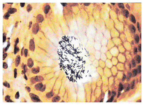 Helicobacter pylori
Stained black
New England Journal of Medicine
{15440} Helicobacter
{19492} type B, antrum
{19491} type B, antrum
{26903} type A
{26906} type A
{26909} type A, some atypia
{19487} Menetrier's, histology
 Menetrier's disease
Pittsburgh Pathology Cases
{18711} "stress ulcers", gross
{19350} "gastric erosion", histology
{39426} hemorrhagic gastritis, nasogastric tube erosions
{07184} hemorrhagic gastritis, nasty case
{15438} benign stomach peptic ulcer
{15439} benign stomach ulcer, with clot
{15560} histology of ulcer, arrow marks bleeding point
{19372} peptic ulcer
{26888} peptic ulcer, stomach
{38656} hyperplastic polyps of stomach
* Patients with familial adenomatous polyposis of the colon are
especially likely to get these (morphology and genes Am. J. Path. 161:
1735, 2002).
{09312} adenomatous polyp
{09331} adenomatous polyp
Gastrointestinal stromal tumors are the common spindle-cell neoplasms
of the stomach, bearing a trademark c-kit mutation and stainable antigen,
and responding to Gleevic (imatinib). They range from totally benign to highly
malignant (South. Med. J. 96: 512, 2003).
Most "leiomyomas", "leiomyoblastomas", and "leiomyosarcomas" are probably "GIST"'s.
Gastric carcinoid is worth mentioning here because
of the big medicolegal flap about long-term gastric acid
suppression
perhaps causing gastric carcinoids. High levels of gastrin
causes enterochromaffin cell hyperplasia, and most people who get gastric
carcinoid have high gastrin levels (atrophic gastritis or Zollinger-Ellison).
And rats actually get such tumors
after lifetime omeprazole.
Stay tuned.
* Gastric carcinoinds that produce ghrelin: J. Clin. Endo. Metab. 86: 5052, 2001).
It
exhibits small glands made of polyhedral cells with round, tame-looking nuclei,
or cells infiltrating singly.
The most common subtype is the signet-ring cancer.
It features large glands made of tall cells with rod-shaped nuclei (as in
the more common, more familiar primary carcinomas of the colon). This is the common type of
stomach cancer in the high-risk countries.
{10472} Stomach cancer, ulcerated
{15509} Stomach cancer, linitis plastica, gross
{15562} Stomach cancer, linitis plastica, histology
{08818} Stomach adenocarcinoma, intestinal type
{17517} Stomach adenocarcinoma, intestinal type
{10473} Stomach adenocarcinoma, diffuse type
{39380} Stomach adenocarcinoma, diffuse type
{39392} Stomach adenocarcinoma, diffuse type
{15510} Signet ring stomach cancer
{19346} Stomach cancer, superficial
{35258} Stomach adenocarcinoma, Pap smear
{15444} non-metaplastic dysplasia
Small Intestine Exhibit
Virtual Pathology Museum
University of Connecticut
Small Bowel Transplant Pictures
Great site
Transplant Pathology Internet Services
Meckel's diverticulum
Small bowel obstruction
Virtual Hospital
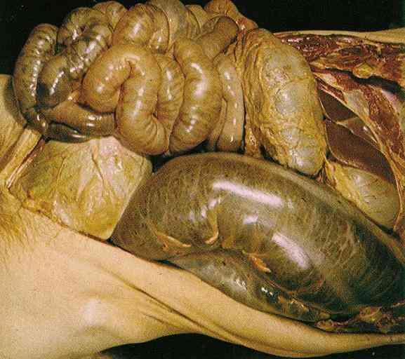 Bowel obstruction -- Autopsy
Source unknown
Not for young or sensitive visitors.
{11111} diverticulum, duodenum
 Ampulla
"Pathology Outlines"
Nat Pernick MD
{10459} Meckel's
{15563} Meckel's
{38674} Meckel's
You remember that the watershed zones of the large intestine
are at the splenic flexure and at the rectosigmoid junction.
* To be sure you're not just looking at livor mortis (this fooled me once), pull on the
bowel. Livor mortis will be stripey, real necrosis will be solid.
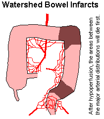
{15538} ischemic enteritis, histology
{19519} ischemic colitis, gross
{49211} ischemic colitis, gross
 Crohn's disease ("regional enteritis", "terminal ileitis"): Review of the new stuff on
etiology Lancet 359:
62, 2002; update NEJM 346: 614, 2002
Crohn's disease ("regional enteritis", "terminal ileitis"): Review of the new stuff on
etiology Lancet 359:
62, 2002; update NEJM 346: 614, 2002
* How fat "creeps" is just now being studied; exotic cytokines and all
Gastroenterology 117: 73, 1999.
Patients suffer for years from ill-defined
abdominal complaints. Eventually, bleeding, malabsorption, nutritional deficiency (remember you
absorb folate, vitamin B12 and bile acids in the terminal ileum), fistula formation, or bowel
obstruction make the disease impossible to overlook.
Tissue necrosis factor alpha antibody
(infliximab) is very effective in eliminating the inflammation
(Gastroenterology 116: 22, 1999; update NEJM 350: 876 & 934, 2004; mechanisms
J. Imm. 176: 2617, 2006). On the evidence, so is
thalidomide (Gastroent. 117: 1278, 1999), working as
an anti-TNF drug.
* A group in Iowa finds pig whipworm to be an effective and safe therapy for
Crohn's, probably by bringing in Th2 cells to downregulate the Th1 cells
(Am. J. Gastro. 98: 2034, 2003). Stay tuned.
* A flap over infliximab causing
cancer in Crohn's patients was found to have no basis: Gut 55: 228, 2006.
Nowell's law triumphant.
{18707} Crohn's disease, gross
{19509} Crohn's disease, gross
{10190} Crohn's disease, histology
{10193} Crohn's disease, histology, with a granuloma
{20251} Crohn's disease, string sign
{20286} Crohn's disease, histology
{25464} Crohn's disease, histology
{26804} Crohn's disease, colon
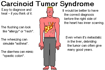
* A brenneroma is a curious hamartoma of the duodenum, composed
of Brenner's glands and fat.
* Today's pathologists distinguish
tame "carcinoids", nastier "well-differentiated neuroendocrine carcinoma", and the
nastiest "poorly-differentiated neuroendocrine carcinoma" based on mitotic counts
and cellular atypia.
{19522} carcinoid, appendix, gross
{19508} carcinoid, small bowel, histology
{49187} carcinoids, small bowel, gross
{24678} carcinoid, small intestine
{26348} carcinoid, ileum
{09175} carcinoid, electron micrograph
{09176} carcinoid, electron micrograph
Here's an outline of the causes:
 Giardia (the usual cause of "malabsorption secondary to hypogammaglobulinemia")
To screen for malabsorption, check for fecal fat using a smear.
If this is present, you have malabsorption. To distinguish
small-intestinal from other causes,
proceed to a d-xylose test. The simple sugar d-xylose
is given by mouth. If the cause of the malabsorption
is a problem with the small intestal mucosa,
the sugar will not appear in the urine.
Most of these people have autoantibodies against reticulin, and
against an endomysial transglutaminase (Nat. Med. 3: 797, 1997).
The most sensitive and specific assay is an IgA anti-endomysial antibody,
against the transglutaminase, of course.
{19500} celiac sprue, small bowel; no villi
{19501} celiac sprue, small bowel; no villi
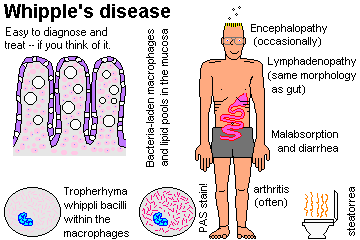
{06103} Whipple's disease, PAS
{24557} incisional hernia
{40681} adhesions
{38677} intussusception, colonic, with gangrene
{39391} intussusception
{46271} volvulus, sigmoid colon ("chronic")
They can also perforate and kill: AJFMP 17: 58, 1996.
Massive fecal impaction
Spectacular x-ray
Brazilian Medical Students
* Adenocarcinoma of the small intestine is thankfully rare. Known risk
factors include Peutz-Jegher's, Crohn's and celiac sprue (Gut 52: 1211, 2003).
Appendix Exhibit
Virtual Pathology Museum
University of Connecticut
)window.location='http://www.mdchoice.com/photo/img/img0103.jpg') Anal pinworms
Anal pinworms
EMBBS
{10196} acute appendicitis, gross
{11445} acute appendicitis, gross
{19526} acute appendicitis, histology
{17558} acute appendicitis, histology
{17562} acute appendicitis, polys
Acute Appendicitis
Australian Pathology Museum
High-tech gross photos
Virtual Pathology Museum
University of Connecticut
* Down's children have an increased rate of Hirschsprung's.
{08086} colonic diverticulum, histology (this is all
mucosa, protruding out into adventitia)
{10514} colonic diverticulum with bleeding point
{19702} diverticulosis coli, x-ray
{24425} diverticulosis coli, x-ray
{39995} diverticulosis; barium fills the "ticks"
Pathology of Inflammatory Bowel Disease
WebPath Tutorial
Ulcerative colitis
Ed Uthman
{26822} ulcerative colitis, gross
{08113} ulcerative colitis
{08809} ulcerative colitis with atypia (worse
on the bottom)
{26825} ulcerative colitis, good pseudopolyps
{10172} ulcerative colitis, histology
{10175} ulcerative colitis, crypt abscess
{10178} ulcerative colitis
{11554} ulcerative colitis, crypt abscess
{49203} ulcerative colitis
{24809} ulcerative colitis, pseudopolyps, gross
{24422} pseudopolyp, histology; there has been substantial healing of the mucosa between
the pseudopolyps,
which in a better case would be ulcerated
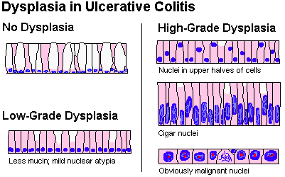
{08105} amebic colitis
Crohn's disease
Ulcerative colitis
Etiology?
Unknown / immune?
Unknown / immune?
Sex?
equal
slightly more among females;
Ethnic group?
more among Jewish
more among Jewish
Hereditary?
Contributes
Contributes
Exacerbate/remit?
Yes
Yes
Stress exacerbates?
Yes
Yes
Smoking?
Makes worse
Quitting increases your risk
Location?
Variable
Rectum and upwards
Bowel?
Small more than Large
Large only
Terminal ileum?
Favorite site
"Backwash"
Colon?
Right more than left, if any
Left more than right
Bile ducts?
May be damaged (Crohn's itself)
May be damaged (sclerosing cholangitis)
Anal troubles?
Common
No
Oral lesions?
Maybe
No
Skip lesions?
Yes
No; continuous
Layers?
All three
Mucosa only
Ulcers?
Linear fissures
Broad / irregular
Pseudopolyps?
Yeah, sort of
Yes unless very mild
Fibrosis?
Severe
No
Fistulas?
Yes
No
Creeping fat?
Yes
No
Granulomas?
Often
No
Bleeding?
Subtle
Heavy-duty
Pain?
Wretched
Not so much
Malabsorption?
Maybe
No
Bowel obstruction?
Maybe
No
B-12 deficiency?
Maybe
No
Lymphs/plasma cells?
Yes
Yes
Arthritis, uveitis?
Yes
Yes
Ankylosing spondylitis?
if (+) for HLA-B27
if (+) for HLA-B27
Diagnosis?
Tough
Easy
Surgery?
Avoid
Cures
Carcinoma risk?
Minor
High
You are already familiar with the very broad, very shallow ulcers.
The smallest lesions feature necrosis of the upper mucosa, with
"mushrooms" or "tufts" of fibrin and neutrophils on the surface
as the initial pseudomembrane.
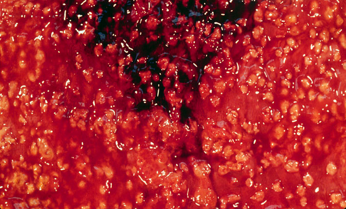 Pseudomembranous colitis
Patches of pseudomembrane
KU Collection
Pseudomembranous colitis
Great photos
Pittsburgh Pathology Cases
Necrotizing entercolitis
Pneumatosis is also present
Loyola Med
{08104} collagenous colitis
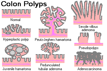
* If very numerous, they indicate increased risk of microsatellite-instability
carcinoma.
However, I cannot agree with "Big Robbins"'s statement that they
are not neoplasms. (1) They tend to occur in the same patients
who get tubular adenomas and there are people who get lots of hyperplastic
polyps for no obvious reason (Am. J. Gastro. 99: 2012, 2004); (2)
the epithelium is obviously abnormal; (3) they dominate the GI pathology
of Cronkhite-Canada and here seem premalignant.
syndrome
{15517} hyperplastic colon polyp, histology
* The genes are the SMAD family (numbers 1-5): Gut 45: 406, 1999.
The proliferation begins in the stroma rather than the epithelium, which probably accounts
for the rather low rate of carcinoma formation.
{15541} juvenile polyp, histology
{46461} pedunculated tubular adenoma
{15499} pedunculated tubular adenoma
{09284} sessile villous adenoma (on the right)
{50560} sessile colon polyp
{15500} sessile colon polyp
{15503} tubulovillous adenoma
Tubular colon adenoma
Photo and mini-review
Brown U.
Tubulovillous colon polyp
Ed Uthman
Gardner's syndrome
WebPath Case of the Week
Colon carcinoma
Photo and mini-review
Brown U.
{09839} adenocarcinoma of the colon
{10540} adenocarcinoma of the colon, cross-section;
note metastases in the lymph nodes
{15456} adenocarcinoma of the colon
{19565} adenocarcinoma of the colon ("napkin ring")
{19567} adenocarcinoma of the colon, histology;
note cribriform "swiss cheese" pattern
{49225} adenocarcinoma of the transverse colon;
anus is at bottom
{20231} adenocarcinoma of the colon, histology
{23887} adenocarcinoma of the colon, stained for CEA
{39598} adenocarcinoma of the colon, perineural invasion
--Shakespearean insult
* Future pathologists: You'll need to see fissures or lacerations
before you diagnose "evidence of sexual abuse" in a child's anus
(AJFMP 17: 289, 1996.)
)window.location='http://www.mdchoice.com/photo/img/img0110.jpg') Foreign body in the rectum
Foreign body in the rectum
X-ray
EMBBS
* Pap smears to detect early lesions here in men at risk: JAMA 281:
1822, 1999.
 Cloacogenic carcinoma
Pittsburgh Pathology Cases
Before we leave the GI tract behind, remember Osler-Weber-Rendu
hereditary hemorrhagic telangiectasia as a cause of hard-to-manage GI bleeding.
Suppurative peritonitis
WebPath Photo
{42229} peritonitis, with lots of fibrin & pus
{49191} peritonitis, after appendicitis
Prothecal peritonitis
Pittsburgh Pathology Cases
Compassion, love for our brothers, for those who love us,
and for those who hate us, love for our enemies --
yes, that is the love which God preached on earth, and which
Princess Marya tried to teach me and I did not understand;
that is what made me loath to relinquish my life;
that is what remained for me had I lived. But now it is too late.
I know it!"
-- Prince Andrei, peritonitis victim, in Tolstoy's "War and Peace"

Peritoneal cyst
Ed Uthman's Pathology Gallery
Vomiting / diarrhea in 2-14 hours. Beware, toxin is heat-stable.

{08790} small intestine normal
{10163} duodenum, normal
{10502} colon, normal mucosa
{11740} stomach, normal
{11741} colon, normal
{11742} colon, normal
{11744} appendix, normal
{11745} appendix, normal
{11804} bowel, normal
{11808} bowel, normal
{11810} ileocecal valve and bowel, normal
{14829} esophagus (esophageal gland), normal
{14830} esophagus (esophageal gland), normal
{14831} esophagus (esophageal gland), normal
{14832} esophagus (esophageal gland), normal
{14833} stomach body, normal
{14834} stomach body, normal
{14836} stomach body (gastric glands), normal
{14837} stomach body (pits & glands), normal

{14838} stomach body (pits & glands), normal
{14839} stomach body (glands), normal
{14840} stomach body (glands), normal
{14841} stomach pylorus (pits & glands), normal
{14842} stomach pylorus (pits & glands), normal
{14843} duodenum (brunner's glands), normal
{14845} duodenum (brunner's glands), normal
{14847} brush border, intestine
{14848} brush border, intestine
{14849} villus, normal intestine
{14850} villus, normal intestine
{14851} crypt of Lieberkuhn, normal
{14852} crypt of Leiberkuhn, normal
{14854} enterochromaffin cells, normal
{14855} villi, jejunum
{14857} Auerbach's plexus
{14859} Meissner's plexus
{14862} Peyer's patches ileum
{14863} appendix, normal
{14865} colon, normal
{14868} colon (crypt), normal
{14869} colon (crypt), normal
{14870} anal rectal junction, normal
{14871} pectinate line, anorectral junction
{15090} ileum, Peyer's patches
{15094} ileum, Peyer's patches
{15095} ileum
{15096} rectum
{15097} rectum
{15100} jejunum, normal
{15101} jejunum, normal
{15102} rectum
{15103} rectum
{15175} esophagus
{15176} esophagus
{15177} esophagus
{15178} esophagus
{15179} esophagus
{15182} stomach, body
{15183} stomach, body and pylorus
{15184} stomach, body and pylorus
{15185} stomach, body and pylorus
{15186} stomach, body and pylorus
{15204} duodenum
{15206} duodenum
{15207} duodenum
{15208} duodenum
{15236} appendix
{15238} esophagus, gland
{15239} esophagus, duct and gland
{15242} esophagus, lymphocyte aggregate
{15243} stomach, cardia
{15244} stomach, cardia
{15245} stomach, cardia
{15246} stomach, body
{15247} stomach, body
{15248} muscularis mucosa, stomach
{15249} auerbach's plexus, stomach
{15250} meissner's plexus, stomach

{15251} lamina propria, stomach
{15253} surface mucus cells, stomach
{15254} stomach, venule
{15255} stomach, pyloric/body junction
{15256} stomach, pyloric/body junction
{15257} jejunum, normal
{15258} jejunum, crypts
{15259} jejunum, terminal bars (tb)
{15260} paneth cell, jejunum
{15261} brunner's gland, duodenum
{15262} brunner's gland, duodenum
{15264} auerbach's plexus, duodenum
{15266} appendix, lymphocyte aggregate
{15267} peyer's patch, ileum
{15268} peyer's patch, ileum
{15269} lamina propria, ileum
{15270} colon, normal
{15271} colon, normal
{15272} colon, lymphocytes
{15273} colon, taenia coli
{15329} ileum, Peyer's patches
{15581} stomach, normal unfixed

{15582} stomach, rugae unfixed
{15584} stomach, normal unfixed
{15586} stomach, normal
{15595} small bowel unfixed, normal
{15596} small bowel unfixed, normal
{15597} small bowel unfixed, normal
{15598} small bowel unfixed, normal
{14835} stomach cardia, normal
{16505} tonsil, normal
{17557} appendix, normal
{19365} jejunum, normal
{19366} jejunum, normal
{19368} jejunum, normal scanning EM
{19450} esophageal gastric junction, normal
{19494} jejunum, normal
{20842} stomach, cardia
{20868} valve of kerckring, duodenum
{20878} taenia coli, colon
{24412} duodenum, normal
{24413} small intestine, normal
{24423} appendix, normal
{24424} appendicitis and normal appendix
{24432} polyp, adenomatous and normal colon
{24810} appendix, normal
{24811} appendix, normal
{25463} esophagus, normal
{49192} appendix, normal


| Visitors to www.pathguy.com reset Jan. 30, 2005: |
Ed says, "This world would be a sorry place if
people like me who call ourselves Christians
didn't try to act as good as
other
good people
."
Prayer Request
 Teaching Pathology
PathMax -- Shawn E. Cowper MD's
pathology education links
Ed's Autopsy Page
Notes for Good Lecturers
Small Group Teaching
Socratic
Teaching
Preventing "F"'s
Classroom Control
"I Hate Histology!"
Ed's Physiology Challenge
Pathology Identification
Keys ("Kansas City Field Guide to Pathology")
Ed's Basic Science
Trivia Quiz -- have a chuckle!
Rudolf
Virchow on Pathology Education -- humor
Curriculum Position Paper -- humor
The Pathology Blues
 Ed's Pathology Review for USMLE I
![]()
![]()
 | Pathological Chess |
 |
Taser Video 83.4 MB 7:26 min |

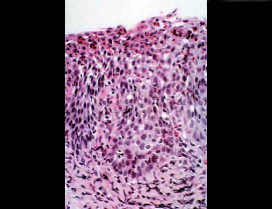 Reflux
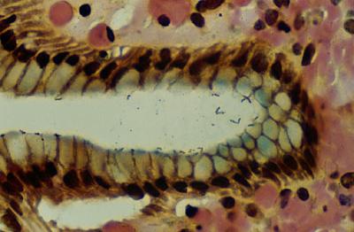 Helicobacter
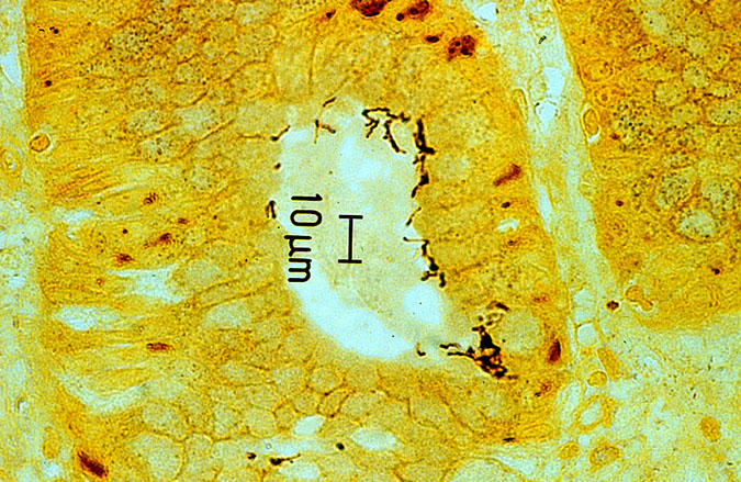 Helicobacter
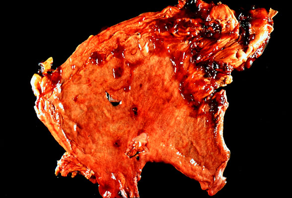 Curling ulcers
 Worm in appendix
 Perforated colon canbmr