Ed Friedlander, M.D., Pathologist
scalpel_blade@yahoo.com


Cyberfriends: The help you're looking for is probably here.
Welcome to Ed's Pathology Notes, placed here originally for the convenience of medical students at my school. You need to check the accuracy of any information, from any source, against other credible sources. I cannot diagnose or treat over the web, I cannot comment on the health care you have already received, and these notes cannot substitute for your own doctor's care. I am good at helping people find resources and answers. If you need me, send me an E-mail at scalpel_blade@yahoo.com Your confidentiality is completely respected.
 DoctorGeorge.com is a larger, full-time service.
There is also a fee site at myphysicians.com,
and another at www.afraidtoask.com.
DoctorGeorge.com is a larger, full-time service.
There is also a fee site at myphysicians.com,
and another at www.afraidtoask.com.
Translate this page automatically
 |
With one of four large boxes of "Pathguy" replies. |
 I'm still doing my best to answer
everybody.
Sometimes I get backlogged,
sometimes my E-mail crashes, and sometimes my
literature search software crashes. If you've not heard
from me in a week, post me again. I send my most
challenging questions to the medical student pathology
interest group, minus the name, but with your E-mail
where you can receive a reply.
I'm still doing my best to answer
everybody.
Sometimes I get backlogged,
sometimes my E-mail crashes, and sometimes my
literature search software crashes. If you've not heard
from me in a week, post me again. I send my most
challenging questions to the medical student pathology
interest group, minus the name, but with your E-mail
where you can receive a reply.
Numbers in {curly braces} are from the magnificent Slice of Life videodisk. No medical student should be without access to this wonderful resource. Someday you may be able to access these pictures directly from this page.
Also:
Medmark Pathology -- massive listing of pathology sites
Freely have you received, freely give. -- Matthew 10:8. My
site receives an enormous amount of traffic, and I'm
handling about 200 requests for information weekly, all
as a public service.
Pathology's modern founder,
Rudolf
Virchow M.D., left a legacy
of realism and social conscience for the discipline. I am
a mainstream Christian, a man of science, and a proponent of
common sense and common kindness. I am an outspoken enemy
of all the make-believe and bunk that interfere with
peoples' health, reasonable freedom, and happiness. I
talk and write straight, and without apology.
Throughout these notes, I am speaking only
for myself, and not for any employer, organization,
or associate.
Special thanks to my friend and colleague,
Charles Wheeler M.D.,
pathologist and former Kansas City mayor. Thanks also
to the real Patch
Adams M.D., who wrote me encouragement when we were both
beginning our unusual medical careers.
If you're a private individual who's
enjoyed this site, and want to say, "Thank you, Ed!", then
what I'd like best is a contribution to the Episcopalian home for
abandoned, neglected, and abused kids in Nevada:
My home page
Especially if you're looking for
information on a disease with a name
that you know, here are a couple of
great places for you to go right now
and use Medline, which will
allow you to find every relevant
current scientific publication.
You owe it to yourself to learn to
use this invaluable internet resource.
Not only will you find some information
immediately, but you'll have references
to journal articles that you can obtain
by interlibrary loan, plus the names of
the world's foremost experts and their
institutions.
Alternative (complementary) medicine has made real progress since my
generally-unfavorable 1983 review linked below. If you are
interested in complementary medicine, then I would urge you
to visit my new
Alternative Medicine page.
If you are looking for something on complementary
medicine, please go first to
the American
Association of Naturopathic Physicians.
And for your enjoyment... here are some of my old pathology
exams
for medical school undergraduates.
I cannot examine every claim that my correspondents
share with me. Sometimes the independent thinkers
prove to be correct, and paradigms shift as a result.
You also know that extraordinary claims require
extraordinary evidence. When a discovery proves to
square with the observable world, scientists make
reputations by confirming it, and corporations
are soon making profits from it. When a
decades-old claim by a "persecuted genius"
finds no acceptance from mainstream science,
it probably failed some basic experimental tests designed
to eliminate self-deception. If you ask me about
something like this, I will simply invite you to
do some tests yourself, perhaps as a high-school
science project. Who knows? Perhaps
it'll be you who makes the next great discovery!
Our world is full of people who have found peace, fulfillment, and friendship
by suspending their own reasoning and
simply accepting a single authority that seems wise and good.
I've learned that they leave the movements when, and only when, they
discover they have been maliciously deceived.
In the meantime, nothing that I can say or do will
convince such people that I am a decent human being. I no longer
answer my crank mail.
This site is my hobby, and I presently have no sponsor.
This page was last updated February 6, 2006.
During the ten years my site has been online, it's proved to be
one of the most popular of all internet sites for undergraduate
physician and allied-health education. It is so well-known
that I'm not worried about borrowers.
I never refuse requests from colleagues for permission to
adapt or duplicate it for their own courses... and many do.
So, fellow-teachers,
help yourselves. Don't sell it for a profit, don't use it for a bad purpose,
and at some time in your course, mention me as author and KCUMB as my institution. Drop me a note about
your successes. And special
thanks to everyone who's helped and encouraged me, and especially the
people at KCUMB
for making it possible, and my teaching assistants over the years.
Whatever you're looking for on the web, I hope you find it,
here or elsewhere. Health and friendship!
QUIZBANK: Liver and biliary (all)
Once again, consider this all "worth knowing".
Review the liver's architecture and function. Describe its capacity to regenerate, and the limits on this
capacity.
Describe the lesions that can produce jaundice. Cite physiology to place them into the appropriate
categories.
Use, and furnish (given the definition), each word in the glossary and elsewhere in the handout.
Tell how alcohol affects the liver.
Give a complete account of the generalized syndrome of liver failure, and the causes of massive hepatic
necrosis.
Describe various conditions that result in ischemia of the liver. Describe the causes and effects of thrombosis
of the hepatic and portal veins.
Describe the pathophysiology and clinical problems seen in portal hypertension.
Describe the viral hepatitis family in substantial detail. Describe the significance of various lab tests and
biopsy findings in various stages of these illnesses. Describe the "lupoid hepatitis" family of illnesses, and
primary biliary cirrhosis.
Define cirrhosis, and describe its pathophysiology in detail. Describe distinguishing features of each of the
many causes of cirrhosis.
Describe cholangitis, and liver abscesses.
Describe the common hepatotoxic agents, and their effects.
Tell how liver failure occurs in children, and what the clinician and pathologist will see.
Describe gallstones and their adverse effects. Describe all the common cancers of the hepatobiliary tree.
Recognize the following gross lesions:
acute yellow atrophy
Recognize and distinguish the following microscopic lesions:
acute cholecystitis
Alpha-1 antitrypsin (alpha-1 protease inhibitor): A useful protein produced by the liver for the bloodstream.
It keeps the body's tissues, notably its elastin, from being totally digested early in life by neutrophil
breakdown products. If its release from cells is damaged, it appears as d-PAS-positive granules of varying
sizes within hepatocytes. (* This can happen in advanced chronic liver disease
from any cause, but is far more likely an unrecognized antitrypsin
abnormality: Am. J. Clin. Path. 107: 692, 1997).
Ballooning degeneration: Hydropic swelling of a hepatocyte (i.e., mild, probably-reversible cell injury)
Bile acids (bile salts): Sterols that help solubilize bile. From your biochemistry course.
Bile ductule: The little bile ducts at the edges of the portal triads. They feed into the interlobular bile duct.
Also called "canals of Hering".
Bile lake: An accumulation of bile that has ruptured a canaliculus
Bile plug: Bile visible in a distended canaliculus
{12220} jaundice
Biloma: A pool of bile in a traumatic (laceration, stab, surgery)
lesion of the liver.
Bridging necrosis: Necrosis linking two portal areas or a portal area and a central area.
Cholestatic jaundice: Jaundice caused primarily by failure of conjugated bilirubin to be sent successfully to
the gut
Chronic hepatitis: Morphologic evidence of inflammation and necrosis
plus labs and/or clinical
evidence of liver disease for six months or more.
* You'll find pathologists who prefer to call it "chronic necroinflammatory injury".
Chronic active hepatitis: This is an out-of-use term
that meant Inflammation + interface hepatitis
+ fibrosis involving the liver for six months or
more. This histologic pattern supposedly meant that the disease
would progress to cirrhosis.
Chronic persistent hepatitis: This is an out-of-use
term for lymphocytes and/or plasma cells in the portal areas, without ongoing necrosis;
symptoms and/or abnormal labs for >6 months. This histologic
pattern supposedly meant that the disease would not progress to cirrhosis.
The tendency nowadays is to group chronic persistent hepatitis And chronic
active hepatitis together as "chronic hepatitis", and not to try hard to distinguish them on
morphologic grounds.
* Future pathologists: Here's a scoring system for chronic hepatitis of
otherwise-obscure etiology!
Inflamed patches in the sinusoids, away from the interface:
To describe the hepatitis, choose whichever is worse.
Cirrhosis: Scarring of the whole liver sufficient to seriously interfere with proper perfusion of hepatocytes.
Instead of the familiar lobules, you'll see fibrous bands dividing the liver into
more-or-less round regenerative nodules.
Conjugated bilirubin: Bilirubin that has been conjugated to glucuronic acid, making it water-soluble
Confluent-lytic necrosis: Death of clusters of hepatocytes (* attributed in the current literature to humoral
immunity)
Councilman (acidophil) body: Single-cell necrosis (apoptosis) of a hepatocyte, typically in hepatitis As a result
of attack by a T-killer cell.
* Cytoplasmic dissociation means edema at the edges of a hepatocyte,
granular cytoplasm around its nucleus. The cell is injured.
Focal necrosis: Death of individual cells, evidenced either by Councilman bodies or lytic necrosis (i.e.,
collapse seen on reticulin stain). Inside the lobule, it's "focal lobular necrosis", as in
smoldering hepatitis from any cause.
* Feathery degeneration: A pattern seen when a liver cell retains both bile salts and water. Ask a physical
chemist how it works. When the bile actually digests a group of liver cells, it's called a
bile infarct.
Giant mitochondria: Monsters seen in hepatocytes in alcoholism. They are d-PAS negative (lets you
distinguish
them from alpha-1 antitrypsin). See J. Clin. Path. 45: 412, 1992.
* These mitochondria may have suffered a characteristic loss of DNA due to alcohol-induced free-oxygen-radical
damage or something; the deletion makes it harder for the liver cell to burn fat, and so forth (Gastroent. 108:
193, 1995.)
Ground glass hepatocytes: Distinctive hepatocytes seen in chronic (not acute) hepatitis B infection.
The "ground glass" cytoplasm is an unusual accumulation of a cytokeratin
(Hepatology 28: 347, 1998).
Hepatocellular jaundice: Jaundice due primarily to failure of hepatocytes to properly take up / conjugate
bilirubin.
Hemolytic jaundice: Jaundice due to excessive destruction of red cells or their precursors at any site
* HELLP syndrome: Hemolysis, elevated liver enzymes, low platelets.
A poorly-understood and very serious
complication of pregnancy. Seizure and hypertension management, glucocorticoids,
and/or exchange transfusions may be required.
Interlobular bile duct: The big bile duct in the portal tract. It runs with the branch of the hepatic artery.
Jaundice: Too much bilirubin (conjugated or not) in the bloodstream, for any reason
{12220} jaundice
Limiting plate: The row of hepatocytes immediately adjacent to the portal tract. It should be smooth and
uniform.
* Liver cell unrest: Increased prominence of Kupffer cells and increased ploidy of many hepatocytes. This is a
totally non-specific finding, common to many (if not most) serious illnesses affecting the entire body.
Lobular disarray: Loss of the normal radial arrangement of liver plates within the lobule, typically with
severe distortion of the sinusoids. The hallmark of acute hepatitis.
Lupoid hepatitis: An unfortunate term for the several kinds of non-viral (?), autoimmune hepatitis in which
the histology is that of chronic hepatitis, usually with a lot more plasma cells than in the viral forms.
Lytic necrosis: The hepatocytes in a region (large or small) are gone, leaving behind collapsed stroma.
Older references call this "dropout necrosis".
Mallory's hyaline: Masses ("rope-like", "cottage cheese") of altered cytokeratin and cell stress proteins
(ubiquitin, others: Arch. Path. Lab. Med. 114: 589, 1990). Usually (but not always) a marker for alcoholism.
Massive necrosis: Most of the hepatocytes on the slide are dead. Due to poisoning, viruses, medication reactions, or ischemia. Submassive necrosis
means that at least some entire lobules are destroyed, but in other lobules, enough cells are alive.
Macrovesicular fat: One large lipid drop in a hepatocyte
Microvesicular fat: Several lipid drops in a hepatocyte
Obstructive jaundice: Cholestatic jaundice caused by mechanical obstruction of the common bile duct or
hepatic ducts. Also called surgical/surgeon's jaundice; all other forms of jaundice are medical/internist's
jaundice.
* Oncocytic hepatocytes (oxyphilic hepatocytes,
i.e., mitochondrion-packed) are common
in many livers, especially
where there's been a lot of regeneration, i.e., cirrhosis, which has
let mutant mitochondria overgrow (Virch. Arch. 432:
349, 1998). Fibrolamellar hepatocellular
carcinomas are also mitochondrion-packed.
Piecemeal necrosis: Necrosis of groups of hepatocytes within the limiting plate.
Today the term interface hepatitis is preferred, to prevent confusion with
focal necrosis deeper within the lobule. Often the only evidence of "necrosis" that you see
is a little area with collapsed architecture; if you're lucky, you may spot a councilman body.
INTRODUCTION
Is life worth living? It depends on the liver! The liver is usually our heaviest internal organ, and the most durable. Unlike lungs, kidneys, heart, and
brain, the livers of most 100-year-olds are morphologically and functionally normal.
Liver pathology includes only a few common diseases. The terminology and morphology of these lesions
are notoriously confusing for beginners. Further, you'll have to know them, because you'll frequently look at
livers.
It would be best for you to start by learning the definitions in the "Glossary", and making note of the material
under "For Future Liver Pathologists".
You already know that the liver is the great chemical plant of the body. You remember its location, its
anatomic relationships, its blood supply, and its essential architecture.
Worth mentioning: The "Ito" (perisinusoidal) cells sit in the space of Disse, store vitamin A, and turn on to
carry out fibrosis of the hepatic lobule in developing cirrhosis. All about it: J. Path. 170: 105, 1993.
Normal adult livers weigh 1400-1600 gm. Liver weight is widely variable at autopsy. I've autopsied an end-stage cirrhotic
with a 700 gm liver, and an alcoholic with a 7000 gm liver. The liver hurts when, and only
when, its capsule is stretched.
Despite the discussion in "Big Robbins", the normal liver may or may not be palpable, depending on its
shape. Maybe 1% of livers have a "Riedel's lobe" easily felt on the right side. Others have a small right lobe and
a large left lobe, while still others have random grooves across the organ ("hepar lobatum", or one variant).
The hyperinflated lungs of the emphysema patient usually push the liver downward and make the edge
palpable, but again, this is not reliable; "rib marks" (really from muscle pressure) in emphysema produce the
familiar costal grooves. Remember that a newborn's liver edge is usually easily palpable 1-2 cm below the
costal arch.
The histology of the liver is worth reviewing. Remember that the metabolic lobule ("acinus") is centered on
the portal areas, and the classical lobule is centered on the central vein. Whichever system you use, zone 1 is
the hepatocytes near the portal areas, zone 2 is the hepatocytes midway between the portal areas and central
veins, and zone 3 is the hepatocytes around the central veins.
The familiar polyhedral, pink-staining hepatocytes are often (maybe 10%) binucleate or tetraploid /
octoploid. This is normal. You remember the architecture of the liver plates and sinusoids, the passage of
bile from canaliculi to canals of Hering to bile ducts, and the appearance and function of the hepatic
endothelium and Kupffer cells.
* Prominent Kupffer cells and increased hepatocyte polyploidy is liver cell unrest, common in people who
are sick for a variety of reasons. Its diagnostic significance is nil.
If individual hepatocytes are destroyed but the architecture of the lobule is not destroyed, the remaining
hepatocytes will totally regenerate the liver parenchyma.
If whole lobules are destroyed, the remaining lobules will expand. They will function normally, though bile
may not be drained quite so well.
Of course, if scar tissue alters the flow of blood through the liver (i.e., cirrhosis has occurred), regeneration
will only produce less-than-fully-perfused nodules of liver cells. (This will disappoint well-read problem
drinkers who understood that their hepatocytes had unlimited capacity to regenerate....)
* Liver biopsies are not always easy to read, especially if the community
hospital pathologist isn't focused on liver. The value of a second
opinion: Arch. Path. Lab. Med. 125: 736, 2001.
* Incredible as it may seem, your lecturer got his first exposure to pathology in 1970 with the dean of
experimental liver pathology, Brown's Nelson Fausto, whose focus was and is liver regeneration.
After years of bragging about this, I was delighted to see him as third author of the new "Big Robbins".
Increased bilirubin in the bloodstream is jaundice.
There's no reason to review bilirubin production and metabolism here. You can check "Big Robbins" if you
need refreshing.
Here's a simple review, similar to the one in "Big Robbins", of the various causes of jaundice:
Too much bilirubin being produced ("hemolytic jaundice")
"Ineffective hematopoiesis", i.e., normoblasts dying in the bone marrow
Thalassemias
Megaloblastic anemias
Intravascular hemolysis (many, many kinds)
Extravascular hemolysis
Big hematomas
GI bleeding
Red infarcts
Liver fails to take up and/or conjugate bilirubin ("hepatocellular jaundice")
Newborns
Hypoperfusion
Bad alcoholism
Hepatitis (many causes)
Cirrhosis (many causes)
Gilbert's non-disease, the Crigler-Najjar syndromes
NOTE: From "Biochemistry". Gilbert's is the forme fruste of Crigler-Najjar (Lancet 346: 314, 1995; Lancet
345: 958, 1995). Usually a non-problem, Gilbert's may be exacerbated by other illnesses.
Liver doesn't send bilirubin to the right place ("cholestatic jaundice")
Problems with the liver cells
Drugs (estrogen, anabolic steroids)
Dubin-Johnson (pigmented) non-disease
* A specialist can diagnose Dubin-Johnson without biopsy by its effect
on different urinary coproporphyrin levels. Don't worry about it.
Rotor (non-pigmented) non-disease.
* Byler's disease ("FIC"; familial intrahepatic cholestasis) -- deadly, with mental retardation
and retinitis pigmentosa; thankfully rare, autosomal recessive, the Byler family from which all the
index patients came is Amish and highly inbred;
See Hepatology 26: 155, 1997; gene is ATP8B1.
There is a Byler-like illness
at BSEP, the bile salt export pump, and another at the multidrug-resistance protein 3 site)
"Benign familial recurrent intrahepatic cholestasis", the forme-fruste of Byler.
Patients have intermittent cholestasis and elevated alkaline phosphatase.
Second locus Gastroent. 127: 379, 2004.
Really bad cases of other liver diseases (hepatitis, cirrhosis, alcoholism; i.e., when the liver fails, the picture is
likely to be mixed)
Problems with the bile ducts in the liver
Biliary cirrhosis
Biliary atresia
* Alagille's (dysmorphic child, bile ducts vanish over time; autosomal dominant, gene Jagged1 (Circulation 109: 1354, 2004, the variable liver disease itself Gut 49: 431, 2001)
Problems with the bile ducts beyond the liver (call a surgeon)
Gallstone in the common duct
Cancer (i.e., biliary, pancreatic, ampullary)
Iatrogenic (i.e., the surgeon nicked the common bile duct)
Note that in all but hemolytic jaundice, bile production will be diminished. Stools may become light-colored
(gray if the bile is completely obstructed), and there
will be diminished
intestinal absorption of fat (pee-yew!) and fat-soluble vitamins.
Lab tests are of considerable help in distinguishing these entities.
Obviously, in the first two categories, the serum unconjugated bilirubin will be elevated.
In the third category, the conjugated bilirubin will be elevated until the liver cells themselves are damaged.
Serum bile acids ("bile salts") will also be increased, producing the troublesome
itching seen in these
syndromes. Conjugated (but not unconjugated) bilirubin in the bloodstream spills into the urine. You'll study
other markers for cholestasis in the unit on lab testing.
On biopsy, obstructive jaundice presents the familiar bile plugs, which begin as dilatations of the canaliculi
and end up forming bile lakes when the canaliculi rupture.
* As the liver cells become damaged, they fill with soap bubbles (i.e., bile salts and water), producing feathery
degeneration. You won't need to recognize this. Later, you'll see necrotic cells surrounding bile lakes.
One of the biggest new areas in internal medicine focuses on the treatment (or non-treatment) of the
bewildering array of "chronic hepatitis" states. Most (but not all) result from infection with hepatitis B or C.
Protocols will continue to change.
WHEN THE LIVER FAILS
Regardless of cause, when the liver can no longer function as chemical plant, several unwholesome things
happen.
Jaundice is usual. When the liver is really scrambled, hyperbilirubinemia is mostly the conjugated sort, i.e.,
the cells remember how to conjugate, but not what to do with, the bile. There is usually some unconjugated
hyperbilirubinemia, too.
Hypoalbuminemia is usual, since the liver isn't making albumin. Without albumin in the bloodstream, ascites
and edema develop. By the way, hypocholesterolemia is usual in liver disease too (unless the primary
problem is obstruction of bile flow -- why?), since the liver isn't producing LDL's. (This is part of the reason
for the silly myth that "too low cholesterol is bad for you".)
Coagulopathy of liver disease results from diminished hepatic synthesis of factors II, V, VII (first to go), IX, and X.
(Note that absent vitamin K from malabsorption also prevents synthesis of II, VII, IX, and X.) Monitor all
this by following the prothrombin times.
Further, as the liver fails to clear factors that have become activated in the course of living, low-grade DIC
is likely to develop.
As liver cells fail, detoxification of nasty compounds fails and hyperammonemia and fetor hepaticus (a
distinctive odor to the breath). Other side-effects are reddening of the thenar and hypothenar eminences
("palmar erythema"), spider "angiomas" (you'll learn about these in physical diagnosis), and (in men)
gynecomastia and testicular atrophy. In longstanding liver failure, the parotid glands often enlarge for some
reason.
Thrombocytopenia is due to lack of thrombopoietin: Am. J. Gast. 94:
1918, 1999.
Hepatorenal syndrome is a syndrome of kidney failure.
We used to precipitate this by "lasixing" cirrhotics with ascites.
The pathophysiology, once obscure, is now clear.
First, liver failure interferes with the breakdown of the vasodilator nitric oxide.
Second, portal hypertension itself forces the splanchnic arteries
to open wider at the expense of circulation to the rest of the body.
We now manage all but the worst cases by giving plasma expanders and vasopressin analogues
(Gastroent. 122: 923, 2002)
to constrict the systemic circulation plus dopamine to
open the renal microcirculation
helps (Hepatology 27: 35, 1998; Am. J. Gastroent.
92: 2113, 1997; Clin. Sci. 92: 433, 1997;
Mayo. Clin. Proc. 71: 874, 1996; Lancet 362: 1819, 2003).
Unless the liver disease is reversible (i.e., alcoholic hepatitis
or a drug allergy), this is just buying time while waiting for a liver transplant.
* Hepatopulmonary syndrome is a poorly-understood
phenomenon in which the arteries dilate throughout the lungs
as the liver fails, causing V/Q mismatching. There is no current remedy.
See Gastroent 113: 606, 1997;
Surg. Clin. N.A. 79: 23, 1999.
Hepatic encephalopathy is not a pretty sight, and probably results from a combination of factors, including
nitrogen-containing false neurotransmitters (supposedly including octopamine -- remember that from "Biochemistry"? --
and some others)
produced by the gut flora.
* Fatigue in liver failure may respond to ondansetron: Lancet 354:
397, 1999.
Early in the process, there's a curious distortion of spatial perception. (The stereotype
of accelerated confusion in the problem drinker
is all too familiar -- he pours the whiskey onto his lap,
rather than into the glass in his other hand;
he cannot find his way home even when he sobers up. Whatever the
cause, hepatic encephalopathy makes life far more difficult.) The first
change on physical exam is asterixis, a curious flappy falling-asleep-and-waking-back-up of the fingers-hands-arms-whole body.
Clinicians monitor hepatic encephalopathy by measuring blood ammonia.
{01383} Alzheimer II glia in hepatic encephalopathy (best one is in the center of the field;
it appears as a swollen, pale nucleus)
In acute massive liver failure, cerebral edema is the pathway out of life in about 50% of cases (Lancet
351: 719, 1998).
We're still making educated guesses about the mechanism.
When the liver finally gives up completely, refractory hypotension supervenes from total-body
vascular
relaxation due to some unmetabolized substance.
Nothing you can do will save the patient.
Reminder: Serum liver enzyme (transaminases, lactate dehydrogenase) concentrations become elevated when
liver cells are acutely injured. Note that in burned-out cirrhosis when drinking is stopped, liver enzymes will
be normal.
CIRRHOSIS
Cirrhosis ("roaches of the liver", etc.) is scarring
of
the whole liver sufficient to permanently interfere with circulation of blood to the hepatocytes, no matter what
the cause. You will see
NOTE: Nobody really understands all about how fibrosis supervenes following liver cell death in any disease,
or what we might do to stop it (J. Path. 170: 105, 1993 will leave you confused, but with an appreciation of
the "Ito" cell). Sometimes, you can see layer upon layer of reticulin fibers being laid down as liver cells die in
waves; this is the sign of irreversible (?) damage in chronic
hepatitis, and probably is how scars build up, at least in part.
{00005} cirrhosis
Micronodular cirrhosis: Most of the nodules are smaller than 0.3 cm, and the fibrous-scar bands are relatively
thin.
Think of alcoholism, hemochromatosis (since alcohol and iron will involve all lobules equally), primary-autoimmune biliary
cirrhosis (since portal areas tend to link to adjacent portal areas), or biliary
infection/obstruction (same reason, "secondary biliary cirrhosis"; remember cystic fibrosis).
{08285} micronodular cirrhosis (this happens to have been a case of primary biliary cirrhosis);
liver on left is normal
Macronodular cirrhosis: Most of the nodules are larger than 0.3 cm, and the fibrous-scar bands are relatively
thin.
Think of chronic hepatitis, with its uneven pattern of inflammation,
progressed to cirrhosis (since viral disease is often patchy and will not
involve all lobules equally).
Wilson's disease, galactosemia, and alpha-1 antitrypsin deficiency may produce either pattern. As a
matter of fact, a rehabilitated alcoholic's micronodular liver will, after a few years of sobriety, exhibit enough
large regenerative nodules to qualify as macronodular.
* Pathologists only: "Incomplete septal cirrhosis" is stabilized (regressing?)
macronodular cirrhosis with only thin fibrous
bands. Liver function tests are better, but portal hypertension may be is more severe. Is this a variant of "regressed
cirrhosis"? See
Gastroent. 106: 459, 1994.
{08441} macronodular cirrhosis
Post-necrotic cirrhosis ("end-stage liver"): Macronodular cirrhosis with really big, thick fibrous-scar bands.
Usually results either from submassive necrosis (i.e., whole lobules were destroyed), or (much more often) progression of another type of
cirrhosis to the end stage (and cirrhosis from any cause tends to progress).
{25659} macronodular cirrhosis (some big scars show
progression to postnecrotic cirrhosis)
* Death rates from cirrhosis (age-corrected)
have run a curious pattern over the past 100 years.
Between 1900 and 1934, deaths dropped by about 2/3; this coincided with
the temperance movement and the massive decline in alcohol consumption.
The end of Prohibition and the Great Depression resulted in a tremendous resurgence of alcohol
overindulgence, and the rate of death from cirrhosis skyrocketed, peaking
in 1970. Since then, they've dropped dropped dramatically; I suspect
the explanation is better nutrition and the recovery movement
(Postgrad. Med. 115: 13, Jan 2004.)
CIRCULATORY PROBLEMS
Congestion of the liver receives excessive attention. There's no mystery; if the right side of the heart isn't
pumping well enough, blood pools in the liver.
Except in the most sudden, violent death, the central areas of the liver will be more or less congested. (If
you're at an autopsy and someone asks, "Is that a nutmeg liver?", you can safely guess "Yes!")
Clinicians enjoy showing the hepatojugular reflux of those with
congested livers, especially behind failing right ventricles.
Pathologists enjoy exhibiting their cut nutmegs, which have light-and-dark areas that resemble congested
liver.
{03949} nutmeg liver
If death has been preceded by a few hours of inadequate circulation (heart failure, shock), count on seeing
some hepatocyte necrosis in the centers of lobules. (This is central hemorrhagic necrosis. Why the liver?
Why in the centers? Think about it!)
This isn't "due to the congestion", but merely results from inadequate perfusion with oxygenated blood.
Clinicians may have noted "elevated liver transaminases" ("ischemic hepatitis"), and even mild jaundice.
You can experience the transaminase elevations yourself by running a marathon. Don't worry, the liver will
completely regenerate (since its connective tissue framework is still intact.
If hepatic congestion and underperfusion have been extreme and longstanding, the rare cardiac sclerosis
may supervene. This is substantial fibrosis in the central areas of the
lobule. (Grossly, the liver surface looks like a football, since scar contracts in the centers of the lobules.)
In extreme cases (i.e., tricuspid insufficiency), the fibrous tissue may bridge adjoining lobules (true cardiac
cirrhosis).
* That cardiac cirrhosis is real has recently been demonstrated
by a study of people who have undergone the Fontan procedure for
single-ventricle, a consequence of which is longstanding increased
right-sided venous pressure. See J. Thorac. Card. Surg. 129: 1348, 2005.
Otherwise, cardiac sclerosis is usually just an anatomic pathologist's curiosity.
Liver infarcts
The liver has a dual blood supply and, while hepatocytes are vulnerable to hypoxia, the stroma is very
resistant and hepatocytes regenerate easily. This makes it difficult to truly
arterially infarct the liver.
When a branch of the portal vein is compromised, the worst that usually happens is atrophy of hepatocytes in
a region ("Zahn's infarct"; fresh lesions have much stasis of blood in the sinusoids and thus look blue).
Hepatic vein thrombosis ("Budd-Chiari")
{49262} Budd-Chiari; liver is engorged with blood
and you can see the clots;
Sounds serious, and is. The most common cause is polycythemia vera. Most any other cause of
hypercoagulable blood can produce "Budd-Chiari". Another important cause is invasion of the hepatic veins
by hepatocellular carcinoma.
As you'd expect, in the acute case, the liver swells (ouch!), ascites develops rapidly, and the patient usually
dies of venous infarction of the liver unless surgery or thrombolysis are performed.
In some foreign countries, "chronic Budd-Chiari" is a common problem. Nobody
knows why. At autopsy, look for fibrous "webs"
in the hepatic veins.
* Diabetic micro-angiopathy produces non-cirrhotic fibrosis
of the sinusoids. The entity is newly-named "diabetic hepatosclerosis"
(Arch. Path. Lab. Med. 130: 27, 2006).
Hepatic veno-occlusive disease, clinically a Budd-Chiari mimic
but with no thrombus,
results from intimal thickening of the veins
(onion-skinning, etc.). Think of Jamaican bush-tea (as
in the lung: terrible health problem West. Ind. Med. J. 64: 60, 1997), comfrey,
graft-vs.-host, radiation effect.
* Sickle-cell patients often have chronic venous outflow obstruction (why?).
Be careful about biopsying these people. Blood 101: 101, 2003.
Portal vein thrombosis
Again, this is serious. It results from hypercoagulable blood, invasion by hepatocellular carcinoma,
pancreatitis, or cirrhosis.
The major problems are ascites and venous infarction of the bowel.
Necrosis of the liver
Infections tend to produce random areas of necrosis ("focal", "spotty"), ranging from tiny (viral hepatitis) to
massive (* typhoid).
Poisons and other noxious things, on the other hand, tend to damage distinctive portions of the lobule (why?)
More about this later.
Central necrosis: Ischemia, carbon tetrachloride, chloroform, acetaminophen
{07020} carbon tetrachloride, gross; note the necrosis (yellow, of course)
Mid-zonal necrosis: Yellow fever.
Peripheral necrosis: Acute iron poisoning (J. Tox. 39: 721, 2001),
phosphorus, eclampsia (in the latter, fibrin microthrombi should be visible in the
sinusoids near the portal areas).
{07023} liver showing phosphorus poisoning; note periportal necrosis
Peliosis hepatis ("blood cysts", a misnomer)
Lakes of blood among the hepatocytes. On section, the liver features many easily visible
holes filled with blood.
The pathology has recently been reviewed in depth (For. Sci. Int. 149:
25, 2005.)
Other cases are lined only by hepatocytes ("parenchymal peliosis"); in this case, the lesions
are irregularly-shaped.
Anabolic steroid use is the best-known cause, but many others
are known (oral contraceptives, cachexia) or suspected (hemangiomas,
congestion in people with mild weakness of the veins).
A blow to (or biopsy of) the involved organ may cause these to rupture, with serious hemorrhage.
INFECTIONS
Viral hepatitis: General considerations
The hepatitis family is an alphabet-soup of viruses, several newly-discovered. However, the anatomic
pathology is generally similar. Some viruses are better at producing different patterns than are others.
You can get each of these infections only once.
But B can linger, and C usually does linger, as a minor or major problem.
As with most viral diseases, infectivity peaks just before symptoms appear. Acute hepatitis is heralded by the
blahs. As the immune system gears up, joint pains and rash can occur. Appetite vanishes, and the patient
typically becomes utterly revolted by tobacco. (Smoking cessation is a redeeming feature of the acute
hepatitis family.)
In the acute disease, the liver swells and becomes tender, jaundice often appears (mild cases are "anicteric"),
and (with influx of bile into the bloodstream) the patient starts to itch and to pass brown urine (why?) Serum
transaminases go sky-high, and other lab evidence of liver disease may become apparent.
The best treatment your lecturer knows for the acute phase is masterful inactivity for all but C, intensive therapy for C. Educate the patient, find
out who else needs to be checked for hepatitis or get prophylactic gamma globulin, and give clotting factors if
you must.
Acute viral hepatitis: You will see
* Note that in hepatitis,
the cells may die either by lysis or apoptosis, or both. Perhaps the lysis is due to
the viruses or to antibodies, while the apoptosis is due to the T-cells.
{05961} acute viral hepatitis with Councilman body
Massive necrosis ("fulminant hepatitis"; "acute yellow atrophy") may supervene on any kind of acute
hepatitis, and often kills the patient in short order.
Grossly, as you would expect, the liver is shrunken, red, soft, and flabby, with a wrinkled capsule.
Histologically, the hepatocytes are almost all gone (lytic necrosis and/or apoptosis), leaving a collapsed
fibrous tissue framework. Don't expect to see much inflammation.
Sub-massive necrosis is a little less striking histologically and lasts a little longer, killing the patient in a few
months. (Or the patient may recover after being super-sick for a few months.)
If a patient survives either process, the parenchyma is usually intact, and recovery should be complete,
without cirrhosis. Rarely, the collapsed reticulin meshwork of the liver turns into broad scars (instant
"post-necrotic cirrhosis").
All about massive hepatic necrosis (acute liver failure): Am. J. Med. 96(1A): 3-S, 1994.
{13320} massive necrosis after hepatitis, gross (nothing left but the reticulin and endothelial framework!)
Chronic hepatitis: Inflammation of the liver for more
than six months.
You will see a dense, mostly-lymphocytic inflammatory infiltrate in the portal areas,
with or without spill-over into
the parenchyma.
There may be some smoldering changes resembling acute
hepatitis in the parenchyma.
In mild cases, the limiting plate is intact (i.e., there is no interface hepatitis).
We used to call this "chronic persistent hepatitis".
{12779} mild chronic hepatitis, story
These findings are more ominous:
Drug-induced liver disease (most notably methotrexate; safety protocol Arth. Rheum. 37: 316, 1994; Arth.
Rheum. 38: 1115, 1995), Wilson's, alpha-1 antitrypsin deficiency, and the autoimmune "lupoid"
hepatitis family also typically pass through a "chronic active hepatitis" histopathology stage on their way to
cirrhosis.
{12800} severe chronic hepatitis, piecemeal necrosis
Future pathologists please note: Any and all of these patterns (from acute hepatitis to post-necrotic cirrhosis)
can be mimicked by idiosyncratic reactions to various drugs.
Chronic hepatitis And its sequelae are
often caused by autoimmunity.
hepatitis A ("infectious hepatitis")
This is an unpleasant but almost always self-limited disease caused by a tiny RNA enterovirus (* picornavirus;
"pico-" means "tiny", and "rna" you can figure out).
{0444} hepatitis A virus
hepatitis A is transmitted by the fecal-oral route, i.e., poor sanitation, small kids (i.e., day-care or institutions),
hands (J. Clin. Micro. 30: 757,
1992,
note that there are countries where toilet paper isn't used),
raw oysters (be sure to ask),
some gay male sexual practices (JAMA 267: 1587, 1992), others.
hepatitis A is more common overseas but is no rarity in the U.S.
* hepatitis A is an endemic scourge on some Indian reservations (MMWR 46: 600, 1997;
Am. J. Pub. Health. 80: 1091, 1990).
* The 1998 strawberry outbreak: NEJM 340: 595, 1999.
The incubation period is about two weeks, and this is the time when virus is shed in the feces. The infection in
kids is usually asymptomatic. Adults who get symptoms at all
suffer jaundice and discomfort for a few weeks. Once in a while, the
disease is fatal.
You'll hear different versions of whether the virus itself damages hepatocytes (the other enteroviruses are
cytotoxic), or whether the liver damage is actually wrought by the body's immune response.
Immune response is exactly what you'd expect:
IgM anti-HAV appears in the blood when the symptoms begin, clears the infection, and disappears within 12
months.
IgG anti-HAV appears in the blood during the symptomatic period, and usually stays around for life,
rendering the patient immune.
Occasionally the disease causes acute yellow atrophy and death/transplantation
(Am. J. Gastro. 98: 448, 2003).
hepatitis A very seldom becomes chronic or leads directly to cirrhosis. There is probably no carrier state. At
worst, the disease might be a trigger for autoimmune chronic hepatitis, but the virus won't stay around.
How the vaccine came about: Lancet 343: 321 & 322, 1994; J. Inf. Dis. 169: 996, 1994; JAMA 271: 1328,
1994;
JAMA 273: 906, 1995.
hepatitis B ("serum hepatitis")
The world's most serious DNA-virus-related health problem. The reservoir for the virus ("HBV", "Dane
particle") is the world's 300 million (Proc. Nat. Acad. Sci. 93: 6542, 1996) carriers, most of whom are
asymptomatic and have histologically normal or near-normal livers.
{0445} hepatitis B virus
Even an infinitesimal amount of infected blood, when introduced into another person's tissues, is highly
effective in transmitting the infection.
Routes include
People born uninfected in the poor nations also frequently turn positive during their childhood. This has been
blamed on bedbugs; probably this isn't the main problem (Lancet 343: 761, 1994).
In the U.S. underclass, infection is also rampant, with around 25% of forensic-service death being core-antibody positive
(J. For. Sci. 38: 1075, 1993).
* hepatitis B immunization has resulted in a triumphant reduction in the prevalence of carriage in Taiwan
and the rate of hepatocellular carcinomas
(JAMA 276: 906, 1996). In 1997, I predicted the 1998-9 media hype
that
the vaccine causes multiple sclerosis. The activists don't have the numbers,
the claim is already totally discredited, and many
lawsuits got filed
(Science 281: 630, 1998).
* Catching hepatitis B from the surgeon, even when e-antigen-negative ("low-risk"): NEJM 336: 178, 1997.
Virus antigens:
HBsAg ("Australia antigen"): Surface antigen. Envelope protein. During the productive infection, the liver
cells make considerable excess non-infectious HBsAg, facilitating diagnosis.
HBcAg: Core antigen. Nucleocapsid.
HBeAg: Another nucleocapsid antigen, which means the virus is being replicated.
* Interestingly, entry of the virus into the hepatocyte is by means of binding to polymerized serum albumin.
After a person first meets the virus, the incubation period is usually 1-4 months.
Antigens and antibodies:
HBsAg first appears in the blood shortly before symptoms begin (if they are to begin). It remains in the blood
for the duration of the infection, whether it is acutely symptomatic, slowly-progressive / subclinical, or merely
the carrier state.
HBeAg appears in the blood just after HBsAg, and before symptoms start. It remains as long as there is acute
viral replication (you're very contagious....), and disappears if (and only if) viral replication stops. The
patient is still sick when HBeAg disappears, but can take comfort in the good news.
Anti-HBeAg appears soon after viral replication and HBeAg production stop (if they stop). The patient can
still be sick, but this is another piece of good news.
Don't ask for an assay of
HBcAg, the core antigen in the blood. It's an intranuclear
protein and there's almost none in the blood. However, Anti-HBcAg, in its IgM form, appears in the blood
typically before symptoms begin, and generally remains present for years (IgG anti-HBcAg will eventually
take over, maybe). If a person with clinical hepatitis has cleared his blood of HBsAg, but has not yet
developed detectable anti-HBsAg, the presence of IgM anti-HBcAg confirms that the infection is, indeed,
hepatitis B and is in the core window.
Anti-HBsAg generally appears when the infection is pretty much over, and is a sure sign of recovery.
BEWARE! During the time between disappearance of HBsAg and appearance of anti-HBsAg, the patient
may experience a potentially-lethal type III systemic vasculitis. (Why?)
If your patient is anti-HBsAg positive and anti-HBcAg negative, probably this person has had the hepatitis B
vaccine (why?)
* Well, maybe it's not a sure sign of recovery; the Scripps crew has found viral DNA up to five years after
appearance of the antibody, but the patients don't seem sick or catching (J. Clin. Inv. 93: 230, 1994).
Symptoms begin in hepatitis B infection only when T-cells become angry with HBsAg and HBcAg and start
killing the hepatocytes that produce them. Histopathologists find T-cytotoxic cells where the hepatocytes are
dying. Eventually, the only surviving liver cells are the ones that won't continue making viruses, and these
replenish the liver.
The acute disease may be subclinical, or can cause weeks of jaundice and misery, or can cause fulminant
hepatitis And death, or sub-massive hepatic necrosis with resolution or cirrhosis.
Survivors (and 99% of people survive the acute episode) usually clear themselves of the virus, but maybe
10% fail to do so. These can become healthy carriers, develop chronic hepatitis
that may remit or progress to cirrhosis if untreated.
Rule of thumb: The more severe the initial illness, the less chance of remaining
chronically infected (why?) Terminology: Chronic hepatitis B means HBsAg has been present in the
bloodstream for 6 months or more.
NOTE: Carrying hepatitis B, with or without ongoing liver disease, is an important cause of
cryoglobulinemia and/or "polyarteritis nodosa of hepatitis B" (both immune complex, type III immune injury
problems).
NOTE: People who become carriers are those who mount a poor immune response. Men (weaker immune
response) are more at risk than women; different HLA types differ in susceptibility (Lancet 344: 1194, 1994).
Further, anyone who carries around the virus for a long time is at substantial risk for hepatocellular
carcinoma. (hepatitis B and/or hepatitis C contribute to
most cases of this cancer, which worldwide is one
of the most common fatal diseases. In the case of hepatitis B, the virus may be acting as a mitogen that
allows
Nowell's law
to act, and/or mutating genes at or near its insertion sites: J. Virol. 65: 6761, 1991; there
is no doubt that insertion of the virus can and does scramble chromosomes: Proc. Nat. Acad. Sci. 88: 9248,
1991.)
People who continue to harbor the
virus are probably those that are not especially good at making interferon (the chronically sick, the
immunocompromised, little kids, the unlucky, men much more often than women). Alpha-interferon is now the mainstay of therapy
for
chronic hepatitis B infections, and the results are encouraging, with maybe half of people clearing the
infection. Of course, interferon therapy is expensive and produces 'flu-like symptoms, but it's better than
dying or infecting your spouse. The new trend will probably be intermittent therapy (cheaper, more
acceptable, just as effective: Gastroent. 107: 479, 1994). And thankfully
the risk for hepatocellular carcinoma also drops greatly
(Cancer 66: 2395, 1990 was the first big one).
Future histopathologists: You can stain for HBsAg in the cytoplasm, or core antigen in the nucleus.
"Ground
glass hepatocytes", with altered cytokeratin suggest chronic hepatitis B infection.
We may hope that the hepatitis B vaccine will eventually make this infection, and its dread sequelae, a thing
of the past. Gambia institutes HBV vaccination of its population (Lancet 341: 1129, 1993). Kids in the U.S.
should get immunized, too (Pediatrics 93: 747, 1994). Please be sure you, too, are immune, Doc.
hepatitis D
"Delta hepatitis virus" (HDV) is an incomplete RNA virus that can replicate only while synthesis of
HBsAg is also taking place. Unlike HBV, delta is directly cytopathic to hepatocytes.
Delta may co-infect (i.e., arrive under a person's skin at the same time as the HBV particle) or superinfect
(i.e., arrive under the skin of a person already infected with HBV). Fortunately, delta is relatively hard to
transmit (somewhere between HBV and HIV in infectivity), and hepatitis D is most common
in gay men and IV-drug-abusers.
The results are grim. In co-infections, fulminant disease is common (maybe 5%). In a superinfection,
the victim experiences a second round of acute hepatitis, which tends (maybe 50% of the time) to turn chronic
and progressive. Treating chronic hepatitis D with alpha-IF: NEJM 330: 88, 1994 (it helps around half of
them while being treated; half of these relapse.)
Fortunately, carriers of delta are probably uncommon. Delta kills maybe 1000 people a year.
hepatitis C (the vast majority of the old non-A, non-B hepatitis Cases)
updates Ann. Int. Med. 132:
296, 2000; Mayo Clin. Proc. 73: 355, 1998; Lancet 362: 2095, 2003.
This flavivirus (HCV) and its related disease spectrum are now well-characterized. In the U.S., 1% of
asymptomatic people are positive for HCV (more than this among swingers and MUCH more among IV drug users; maybe 0.3% in those
not in these risk groups; health care workers aren't at increased risk: Lancet 343: 1618, 1994; ear-piercing is
a risk factor: NEJM 334: 1691, 1996; 19% positive for inner-city forensic-pathology service deaths J. For.
Sci. 38: 1075, 1993); in the poor nations, it's around 5%; the highest known prevalence is around 20% in
Egypt (see below). At least 170 million people are infected worldwide (Science 288:
339, 2000), at least 3 million in the USA, with about 10000 deaths yearly.
hepatitis C virus is transmitted by the same routes as hepatitis B, but is probably not nearly so catching. The
best route seems to be blood transfusion or needle-sharing (J. Inf. Dis. 162: 823, 1990; hemophiliacs Blood
84: 1020, 1994).
Needlesticks produce about a 6% rate of infection (Br. Med. J.
315: 333, 1997) and
prophylactic treatment with anti-hepatitis C medicines is now
administered routinely after such events.
Strangely, nobody yet knows the prevalence of perinatally-transmitted hepatitis C,
but it can declare itself later in life as a fulminant illness: Arch. Dis. Child. 88: 160, 2003.
Thankfully, with changing lifestyles (maybe) and surveillance
in the hospital (certainly), the transmission rate of hepatitis C is
only about 1/5 what it was in the 1980's (Sci. Am. 280(3):
17, March 1999.)
The risk from a transfusion is now about 1 in 2 million (Lancet 361: 161, 2003).
* Among several hundred Irish women infected in the '70's
by bad
RhoGam, half still had demonstrable virus,
most of these had some inflammation, many had some fibrosis,
but only two had cirrhosis (NEJM 340: 1228, 1999).
* hepatitis C transmission by acupuncture is now so well-known that you'd do
well to warn your patients to be sure they know who's doing it
(Can. Fam. Phys. 49: 985, 2003).
Sexual transmission seems much less efficient but probably occurs (JAMA 269: 361 and
392, 1993; Gut 45: 7, 1999);
around a quarter of spouses eventually catch it (Ann. Int. Med. 120: 748, 1994). Vertical
transmission from Mom is common, especially if Mom has lots of virus on board: NEJM 330: 744, 1994. In
striking contrast to HIV, hepatitis B, and so forth, around 40% of people who carry the virus haven't got a
clue how they got it. In the US, if you're living clean enough to donate blood, your chance of
coming down with hepatitis C is very low (BMJ 316: 1413, 1998;
NEJM 341: 556, 1999).
If you have the antibody, you have about an 85% chance
that you'll have detectable virus by PCR in your blood, and it's
nearly certain you'll have it in your liver (Gut 42:
570, 1998) -- this is a virus adults are unlikely to get rid of naturally.
A few folks do clear the virus quickly after being infected
(Science 288: 333, 2000).
Egypt, with around 20% of its people infected, has the highest rate.
Probably because of needles (used to administer antimony
in the treatment of schistosomiasis)
not being sterilized between patients (Lancet 355: 887, 2000).
Incubation period is a week to six months. The acute infection is more likely to be subclinical (or cause
minor "belly trouble"), and massive necrosis does not occur.
However, infection usually becomes progressive (Only 15-30% or
so of people shake the bug. Fortunately, progression is slow, and
severe liver failure results in only about 10-30% of people and usually only
after decades.
You can get sick several times if you get
a big dose of the bug several times (Lancet 343: 388, 1994). After acquiring the virus via blood transfusion,
chronic infection with abnormal liver histology happens more often than not (Ann. Int. Med. 137:
961, 2002).
The impact on overall length of life is usually small (NEJM 327: 1906, 1992; Gut 47:
845, 2000) but the
infection is still a serious business.
Around a third of hepatitis C virus carriers have aggressive-looking chronic
hepatitis or cirrhosis (Br. Med. J. 308: 695,
1994). Unlike the other viral diseases, there is often quite a bit of
fatty change (correlates with severity: J. Inf. Dis. 192:
1943, 2005) and/or regenerative change in the bile ducts,
rather few inflammatory cells (maybe just lymphoid aggregates)
in the parenchyma
and a portal infiltrate that's all lymphocytes (no plasma cells or eosinophils)
suggests hepatitis C. The progression to fibrosis usually takes decades; if you're male,
a drinker, and/or older, it may take only a decade or so (Lancet 349:
825, 1997.)
NOTE: As with hepatitis B, carrying hepatitis C, with or without ongoing liver disease, is an important cause
of cryoglobulinemia (Am. J. Med. 96: 124, 1994; NEJM 330: 751, 1994 for the success of alpha-IF therapy).
The cryoglobulins are immune complexes made of the virus and the antibodies.
* How does hepatitis C virus produce fibrosis? There is often remarkably
little inflammation. In one model, the virally-infected hepatocytes produce
huge amounts of transforming growth factor beta, causing stellate cells
to produce collagen. Stay tuned; this may become the basis for a novel
anti-fibrogenic therapy (Gastroenterology 129: 246, 2005).
* NOTE: hepatitis C virus tends to drive out hepatitis B virus over the long-term in patients infected with both
(Gastroent. 106: 1048, 1994, others).
The NIH study -- you're probably recovered if your transaminases are normal and you have no circulating
viral DNA: Ann. Int. Med. 123: 330, 1995. That's about 15% of the asymptomatic-but-antibody-positive
folks.
* Nowadays we monitor disease and therapy using assays for hepatitis C messenger RNA (Am. J. Clin. Path.
107: 362, 1997).
* Strangely, quite a few of these people never have elevated transaminases,
even as they progress to cirrhosis. We have to wonder how these people's infections
were detected (Am. J. Gastro. 98: 1588, 2003).
We now eliminate about half of chronic infections using a combination of pegylated interferon
and ribavirin. This is one of the most important triumphs of medicine in the last few years.
* Future pathologists: The less iron in the sinusoidal cells and portal tracts, the better the response. Nobody
knows why. See Am. J. Clin. Path. 103: 419, 1995.
Like hepatitis B, longstanding infection with hepatitis C places a person at grave risk for hepatocellular
carcinoma (Lancet 345: 413, 1995).
Immunology:
In contrast to hepatitis B, the presence of anti-HCV usually indicates the persistent presence of hepatitis C
virus in the body. Around 60% of people with the antibody have virus detectable by PCR; they're probably
more infectious and more likely to be seriously ill (see, for starters, NEJM 330: 744, 1994).
People with other liver diseases may show false-positives or have persistence of the antibody after elimination
(?) of the virus. PCR has finally made it possible to sort the mess out.
* There's hope that we'll have a hepatitis C vaccine, but it's a long way off: Proc. Nat. Acad. Sci. 91: 1294,
1994 (poxvirus-based). Like HIV, the virus is notorious for mutating rapidly, even in the same patient, and
this isn't good for vaccine-makers. And like HIV, antibodies aren't very protective.
hepatitis G and the hepatitis GC family are hepatitis-C-like flaviviruses.
hepatitis G virus is a relatively common infectious agent
that produces a chronic viremia. It's known to be
transmitted by blood products, sex, needles, and mother-to-child
(Arch. Dis. CHild. 80: F72, 1999). There's an antibody test (Lancet 349: 318, 1997).
Whether these critters make you sick is still under study.
There doesn't seem to be an acute illness (NEJM 336: 741 & 747, 1997).
A link to hepatocellular carcinoma is statistically
possible but unproven (Cancer 86:
936, 1999). More studies failing to show any evidence
that they make you sick: Gut 103: 103, 1998; Arch. Dis. Child.
80: F72, 1999; Ann. Int. Med. 126: 874, 1997.
* People who study these things say that
C, G, and the GC's all evolved from yellow fever or dengue fairly recently.
* TTV ("transfusion transmitted virus") is a DNA virus that's very common
(10% of folks) in Japan, less common in the West. It elevates transaminases
after a transfusion, but nobody's found anyone sick from it yet (Lancet 352:
164, 1998).
hepatitis E: An important, water-borne, epidemic calicivirus infection in the poor nations.
You'll make the diagnosis on the presence of IgM antibodies.
Around 25% of people from the Middle East
have had it, but it is less prevalent in the rest of the world (J. Inf. Dis. 16: 801, 1994). Review from
the CDC in Inf. Dis. Clin. N.A. 14: 669, 2000.
Yellow fever: Councilman bodies, necrosis especially in the mid-zone of the lobule, and a surprising lack of
inflammatory response. Yellow fever today in Bolivia: Lancet 353: 1558, 1999.
Death from yellow fever is probably not so much due to liver failure as to
overactivation of cytokines, much as in sepsis: J. Inf. Dis. 190: 1821, 2004.
CHRONIC HEPATITIS NOT CAUSED BY VIRUSES
Autoimmune ("lupoid"; "plasmacytic") hepatitis: Chronic hepatitis progressing to cirrhosis, without chronic virus
infection but with evidence of immune injury. Review NEJM 354: 54, 2006.
Poorly understood, but fairly common, and deadly. We'll distinguish the different types (which bear little
relationship to real lupus) when we discuss liver function testing. The most common
type features autoantibodies against smooth muscle.
Review: Am. J. Med. 96(1A): 23-S, 1994.
Current thinking is that something first damages the liver (probably one of the viral hepatitis family, or some
drug or poison, or whatever), and patients then get sensitized to their livers and start destroying them over the
long haul. More about this later.
Drugs that trigger "lupoid hepatitis" include some of the older ones, and today minocycline (Br. Med. J. 312:
169, 1996).
Future pathologists: Autoimmune chronic hepatitis usually features a lot more plasma cells than does viral chronic hepatitis.
Unlike in viral infection, the response to immunosuppression (i.e., glucocorticoids) is generally good. The
best protocol, which often cures, is based on azathioprine (NEJM 333: 958 & 1004, 1995).
* Future pathologists! Here's your scoring system (J. Hep. 31: 929, 1999)
Primary biliary cirrhosis: An autoimmune disease caused (we don't know exactly how) by antibodies against
pyruvate dehydrogenase ("anti-mitochondrial antibodies"). Lancet 362:
53, 2003. We'll talk more about this under "Liver Testing".
The bile ducts are selectively attacked by the immune system, eventually resulting in severe obstructive
jaundice.
For some reason, the biliary epithelial cells express pyruvate dehydrogenase, or something very much like it,
on their luminal surfaces (J. Clin. Invest. 91: 2653, 1993).
The histopathology begins with chronic inflammation (mostly
portal, sometimes some interface hepatitis), and progresses through
bile-duct obliteration and collateral formation to micronodular (why?) cirrhosis.
For the details see Mayo Clin. Proc. 73: 179, 1998.
Less easy to explain are the frequent appearance of granulomas and Mallory's hyaline.
{24568} primary biliary cirrhosis, early; cirrhosis has not really developed yet, but portal areas are
inflamed; you could not tell at this magnification that this is primary biliary cirrhosis
Primary biliary cirrhosis is BIG news right now. It is considerably more common than we had once thought,
and it responds to therapy with bile salt analogues (nobody knows why; Br. Med. J. 312: 1181, 1996.)
* The newly-recognized "primary autoimmune cholangitis" looks something like primary biliary cirrhosis, but
has high ANA titers and no anti-mitochondrial antibodies. See Am. J. Surg. Path. 18: 91, 1994;
update on sorting out the autoimmune hepatitis family histologically Am. J. Clin. Path. 114: 705, 2000.
* Idiopathic adulthood ductopenia is disappearance of the interlobular bile ducts; it may be asymptomatic
(elevated GGT prompts its discovery) or progress to cirrhosis (NEJM 336: 835, 1997).
"Secondary biliary cirrhosis" is more likely to be just fibrosis, due to
obstruction of the common bile duct, usually in chronic pancreatitis.
If the stenosis is relieved, the fibrosis often regresses some
(NEJM 344: 452, 2001).
As before, "all poisons are drugs, all drugs are poisons".
To make the call, you want to see one of these:
Especially rough on the liver:
Any of these can produce massive hepatic necrosis. The toadstool and the acetaminophen overdose will
produce massive hepatic necrosis; "Ecstasy" in recreational amounts
is famous for the same (Transplant. Proc. 33: 2743, 2001). The others are more likely to produce a hepatitis-like picture.
Acetaminophen overdose is very common.
The drug is metabolized by two different pathways, one "safe", the other productive of noxious free radicals.
Ordinarily, we use only the "safe" pathway, but when that is overloaded, the drug gets shunted into the bad
pathway.
* We've already seen the "two-pathway" concept in our discussions of atherosclerosis and Alzheimer's disease.
Stay tuned for the discovery of more "two-pathway diseases".
Three or four days after the overdose, the patient gets sick and lapses into hepatic failure. By this time, the
drug itself may be mostly gone.
Future emergency room specialists: You can block the "bad" pathway using good old N-acetyl-cysteine, or
"Mucomist", from the respiratory care department.
CHOLANGITIS
Cholangitis ("ascending cholangitis", etc.) is suppuration involving bile ducts.
The underlying cause is almost always obstruction. Pretty much any gut bacterium can be the opportunist.
E. coli is most common; clostridial gas gangrene of the liver is ultra-deadly.
As you would expect, patients are super-sick with the acute infection. Call a surgeon, since the bile has to be
drained.
The give-away on histologic exam is neutrophils within the bile ducts. Since there's obstruction, look for bile
plugs, too.
Liver abscesses, in the U.S. are usually of bacterial origin, spreading either up the bile ducts ("cholangitis abscess" --
ascending
cholangitis) or via the portal vein ("pyelophlebitic abscess" -- appendicitis, diverticulitis), or from septic emboli (bacterial endocarditis),
or following a dirty wound.
Naturally, patients are super-sick, as with ascending cholangitis.
"Amebic abscesses", a misnomer, are areas of "anchovy paste" necrosis without much inflammation. Hydatid
cysts can become infected, forming real abscesses.
Pericholangitis, broadly defined, is inflammation around the bile ducts. This generally takes the form of
extra lymphocytes, maybe with any other kind of inflammatory cells, in the portal areas.
This is extremely common at autopsy, but its significance is obscure. One could conjecture that the liver
clears the blood of foul products of fatal disease, and that these are excreted in the bile and attract
inflammatory cells.
Patients with ulcerative colitis and Crohn's generally have pericholangitis, which may eventually become
fibrosis (sclerosing cholangitis) and biliary obstruction / biliary cirrhosis. Or one can have idiopathic
primary sclerosing cholangitis, a curious, probably-immune-mediated
entity. The extra-hepatic bile ducts in these diseases come to look like uneven
strings of beads.
* Other infections: Malaria can load the Kupffer cells with pigment, but seldom causes hepatic dysfunction.
Kala-azar (a vicious form of leishmaniasis)
packs Kupffer cells with organisms but does not interfere with liver function. Infectious mono from
any of the usual causes can produce mild hepatocyte failure. Secondary syphilis can give an acute hepatitis,
while tertiary and especially congenital syphilis are noted causes of hepar lobatum, due to scar contraction.
Don't forget leptospira in unexplained jaundice.
Penicillium marneffei is an opportunist in AIDS, especially
in Southeast Asia (Arch. Path. Lab. Med. 128: 191, 2004).
Increased pressure in the portal venous system, for whatever reason (usually increased resistance to flow
and/or increased anastomoses with the arterial circulation).
Pre-hepatic causes
Portal vein obstruction / compression
Thrombus (guess the causes?)
Tumors (guess which ones?)
Really bad pancreatitis
Intra-hepatic causes
Cirrhosis (both fibrosis and AV-shunting contribute)
Central hyaline sclerosis without cirrhosis
Really bad fatty change
Alcoholism / "alcoholic hepatitis", etc.
Reye's
Schistosomiasis (eggs plug portal vein radicles)
* Sarcoid / TB (supposedly, I've never seen this)
* Congenital hepatic fibrosis (a thankfully-rare birth defect, with
very few veins in the expanded portal areas)
Osler-Weber-Rendu telangiectasisa (abnormal vascular communications: NEJM 343:
931, 2000).
"Idiopathic" (dubious)
Post-hepatic causes
Budd-Chiari
Constrictive pericarditis
Tricuspid insufficiency
Really bad right-sided heart failure
Regardless of cause, portal hypertension is troublesome.
Patients get ascites, or large accumulations of fluid in the abdomen. This is troublesome. Mechanisms of
formation include
(1) the obvious increase in hydrostatic pressure in the venules;
(2) the increase (most mechanisms) in hydrostatic pressure within the hepatic sinusoids themselves (the
"increased hepatic lymph formation" of "Big Robbins"; this stuff is likely to be rich in protein, since the
hepatic sinusoidal "endothelium" is discontinuous)
(3) diminished circulatory volume due to low serum albumin, with a tendency of the kidneys to retain sodium
and water. (BEWARE! If you give these patients a diuretic, you can send them into shock, kidney failure,
"hepatorenal syndrome", etc., etc.)
{19381} ascites
Porto-systemic shunting results when blood from the guts finds other routes back to the right side of the heart.
This results in esophageal varices (which can bleed
profusely), hemorrhoids (which can bleed profusely), and the distinctive caput medusae around
the
belly-button. Since this blood isn't detoxified in the liver, hepatic encephalopathy is likely to be exacerbated.
* This also probably is the cause of the usually-mild IgA nephropathy typical of cirrhotics (i.e., asymptomatic
hematuria). IgA from the gut ends up in the kidneys, rather than being cleared by the liver.
Fibrocongestive splenomegaly produces big, firm spleens that often produce clinically significant
hypersplenism (i.e., they make the person anemic, neutropenic, and/or thrombocytopenic). This is bad.
You can treat portal hypertension effectively by doing a porto-caval shunt operation. If the underlying
problem is cirrhosis, this will result in blood flowing directly from the bowel to the systemic circulation,
making hepatic encephalopathy much, much worse. But this beats dying of bleeding varices.
Sclerosing agents save the lives of patients during acute bleeds.
Today, banding ("band ligation") is doing the same (Br. J. Surg. 86:
437, 1999.)
For lasting
control, the patient is likely to have the shunt placed inside
the liver itself, by the radiologist who passes a catheter
down the jugular vein ("transjugular intrahepatic
portosystemic shunt").
* Old-fashioned "prophylactic sclerotherapy" of esophageal varices
actually increased the risk of dying.
Perhaps it just made whatever vein didn't get sclerosed into a bigger varix.
NEJM 324: 1779, 1991.
Of course, portal hypertension isn't the only problem that the cirrhotic has. See "When the liver fails", above.
ALCOHOLIC LIVER DISEASE (Lancet 345: 227, 1995; Mayo Clin. Proc. 76:
1021, 2001)
Woe to those who demand strong drink as soon as they rise in the morning, and linger into the night while
wine inflames them!
How many ship captains does it take to cause the Alaska oil spill? Sir, I have known more old drunkards than old doctors.
 I am presently adding clickable links to
images in these notes. Let me know about good online
sources in addition to these:
I am presently adding clickable links to
images in these notes. Let me know about good online
sources in addition to these:
Pathology Education Instructional Resource -- U. of Alabama; includes a digital library
Houston Pathology -- loads of great pictures for student doctors
Pathopic -- Swiss site; great resource for the truly hard-core
Syracuse -- pathology cases
Walter Reed -- surgical cases
Alabama's Interactive Pathology Lab
"Companion to Big Robbins" -- very little here yet
Alberta
Pathology Images --hard-core!
Cornell
Image Collection -- great site
Bristol Biomedical
Image Archive
EMBBS Clinical
Photo Library
Chilean Image Bank -- General Pathology -- en Español
Chilean Image Bank -- Systemic Pathology -- en Español
Connecticut
Virtual Pathology Museum
Australian
Interactive Pathology Museum
Semmelweis U.,
Budapest -- enormous pathology photo collection
Iowa Skin
Pathology
Loyola
Dermatology
History of Medicine -- National Library of Medicine
KU
Pathology Home
Page -- friends of mine
The Medical Algorithms Project -- not so much pathology, but worth a visit
National Museum of Health & Medicine -- Armed Forces Institute of Pathology
Telmeds -- brilliant site by the medical students of Panama (Spanish language)
U of
Iowa Dermatology Images
U Wash
Cytogenetics Image Gallery
Urbana
Atlas of Pathology -- great site
Visible
Human Project at NLM
WebPath:
Internet Pathology
Laboratory -- great site My team:
Ed Lulo's Pathology Gallery
Bryan Lee's Pathology Museum
Dino Laporte: Pathology Museum
Tom Demark: Pathology Museum
Dan Hammoudi's Site
Claude Roofian's Site
Pathology Handout -- Korean student-generated site; I am pleased to permit their use of my cartoons
Estimating the Time of Death -- computer program right on a webpage
Pathology Field Guide -- recognizing anatomic lesions, no pictures

St.
Jude's Ranch for Children
I've spent time there and they are good. Write "Thanks
Ed" on your check.
PO Box 60100
Boulder City, NV 89006--0100
More of my notes
My medical students
Clinical
Queries -- PubMed from the National Institutes of Health.
Take your questions here first.
HealthWorld
Yahoo! Medline lists other sites that may work well for you
Yahoo! Medline lists other sites that may work well for you

We comply with the
HONcode standard for health trust worthy
information:
verify
here. 

![]()
 Liver, Gall Bladder, Pancreas
Cornell
Class notes with clickable photos
Gastrointestinal Pathology
Virginia Commonwealth U.
Great pictures
Tulane Pathology Course
Great for this unit
Exact links are always changing
 LEARNING OBJECTIVES
cavernous hemangioma
cirrhosis (various types)
congestion ("nutmeg liver")
costal grooves
focal nodular hyperplasia
hepatocellular carcinoma
hepar lobatum
metastases to the liver
Riedel's lobe
acute viral hepatitis
alcoholic hepatitis
alpha1-antitrypsin globules (PAS+)
ascending cholangitis
bridging necrosis
bile plugs and lakes
cavernous hemangioma
cholangiocarcinoma / adenocarcinoma of gallbladder
chronic hepatitis
chronic cholecystitis
cirrhosis (generic, and various etiologies)
congestion / central ischemic necrosis
Councilman body
fatty change (microvesicular, macrovesicular)
giant cell ("neonatal") hepatitis
giant mitochondria (PAS-)
ground glass hepatocytes
hepatocellular carcinoma
interface hepatitis ("piecemeal necrosis")
iron overload (1, 2)
liver cell unrest
lobular disarray
Mallory's hyaline
massive necrosis
primary biliary cirrhosis
Wilson's disease
Hepatitis-Like Lesions
Histopathology and essay
For pathologists
Liver Exhibit
Virtual Pathology Museum
University of Connecticut
Liver Transplant Pictures
Great site
Transplant Pathology Internet Services
PARTIAL GLOSSARY
 Asterixis: "Liver flap". The tremor of early hepatic encephalopathy.
* Francophiles: Cartoon character
"Asterix the Gaul" is ancestor of a wine-loving nation with
an unusually large prevalence of
cirrhosis.
 Intrahepatic lithiasis
Intrahepatic bile duct obstruction
WebPath
* This is obviously an ominous finding in chronic hepatitis, but the old
idea that is was a bad sign in acute hepatitis has been discredited.
Piecemeal necrosis / interface hepatitis:
Hard to see? Mild
Less than 50% of total interface involved? Moderate
50% or more of total interface involved? Severe
Fewer than 5 per 10 high power fields: Mild
5-20 patches per 10 high power fields: Moderate
More than 20 patches per 10 high power fields: Severe
Cirrhosis of the Liver
Australian Pathology Museum
High-tech gross photos
Jaundice
WebPath Photo
-- Anonymous
 The liver's ability to regenerate is legendary. (The Greek titan
Prometheus
had his liver devoured each day by a monster bird, but it always
grew right back.)
The liver's ability to regenerate is legendary. (The Greek titan
Prometheus
had his liver devoured each day by a monster bird, but it always
grew right back.)

* These people lack a pump, which is coded by, of all things,
the hated MRP2 multidrug-resistance protein (Gastroent. 117: 653, 1999)
that pumps cancer chemotherapy agents out of cancer cells.
* Making the call is easy because the liver refuses to take up
the Tc99m-DIPA biliary scan radionuclide.
 Future clinicians: Try grapefruit juice for pruritus of
liver disease (Ann. Int. Med, 126: 920, 1997).
Future clinicians: Try grapefruit juice for pruritus of
liver disease (Ann. Int. Med, 126: 920, 1997).
Gynecomastia in cirrhosis
Patient photo
Brazilian Medical Students
{08846} cirrhosis, trichrome stain (fibrous tissue is blue, of course);
micronodular
{39710} cirrhosis after hepatitis, gross photo showing uneven involvement of the liver lobules.
Just recognize cirrhosis.
The traditional wisdom is that the fibrosis in cirrhosis does not regress.
Only recently have we begun to recognize that there may be regression, though not complete
reversal, if the underlying process goes quiet.
Look
for thin septa with holes in them, lone thick fibers (i.e.,
the surrounding thin stuff is gone), and other stuff that's harder to
see why it means regression. See Arch. Path. Lab. Med. 124:
1599, 2000. Especially in kids cured of thalassemia by marrow transplantation,
extensive reversal of cirrhosis is now known to take place (Ann. Int. Med. 136:
667, 2002); there is also some regression when fibrosis due to
hepatitis C (Dig. Dis. Sci. 43:
2573, 1998) and autoimmune hepatitis (Ann. Int. Med. 127: 981, 1997)
are successfully treated.
 Vascular Liver Disease
Frank Mitros MD
Virtual Hospital
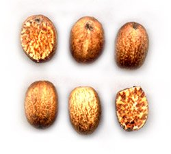
{31889} nutmeg, real
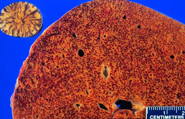 Nutmeg and nutmeg liver
Someone really had fun
making this photo!
* Future pathologists: If you see dilated sinusoids in zone 3, the cause is usually
impairment of venous outflow, but a variety of other illnesses
can cause it as well -- and even obtaining a wedge biopsy during surgery
(Arch. Path. Lab. Med. 128: 901, 2004).
Comfrey is used both topically and as a "holistic" herbal tea;
amazingly, this stuff is still around and still killing people: Pub. Health Nutr. 3: 501, 2000;
the FDA merely sent a "warning to dietary supplement industry leaders" in 2001 and it's still readily
available (2006).
{07022} carbon tetrachloride, microscopic
Sometimes it is due to dilated veins ("phlebectatic peliosis"); in this case, the lesions are round
and lined by endothelium and/or fibrosis, and the liver
looks like swiss cheese.
{08834} acute viral hepatitis; sinusoids are
not visible, lots of inflammatory cells
{11787} acute viral hepatitis, great bile plugging
{12764} acute viral hepatitis
{12758} acute viral hepatitis
{12767} acute viral hepatitis
{12770} acute viral hepatitis (do you see a Councilman body?)
{12773} acute viral hepatitis
{12776} acute viral hepatitis
{13321} massive necrosis after hepatitis, histology
{13322} massive necrosis after hepatitis, histology
{12785} mild chronic hepatitis, H&E (not a very good case, since lymphocytes are very few, and a
portal
area is not even shown; indistinguishable from mild acute hepatitis)
{12788} mild chronic hepatitis, reticulin stain (see the limiting plate intact);
you don't need to tell this isn't normal liver;
{12791} mild chronic hepatitis, trichrome (again, see the limiting plate intact);
again, you can't tell this isn't normal liver;
{12803} severe chronic hepatitis; subtle
{20328} severe chronic hepatitis; almost to cirrhosis (the
nodules are not yet completely separate)
{40279} severe chronic hepatitis, note the necrotic cells
{20183} severe chronic hepatitis with good bridging necrosis;
the hepatocytes are stained orange and the bridge is an area of lytic necrosis
 Chronic viral hepatitis
Photo and mini-review
Brown U.
{08173} hepatitis A virus
{08175} hepatitis B virus
{10532} hepatitis B, * orcein stain (stains the virus)
{11708} hepatitis B, core antigen stained in nuclei
Other anti-viral agents include lamivudine and adefovir dipivoxil
(NEJM 348: 800 & 808, 2003). They are unlikely to eradicate the virus,
but they keep it under control and it is now clear that even the cirrhosis tends to
reverse under this treatment (Lancet 362: 2089, 2003).
A few mutant viruses that can affect the immunized are now
appearing (Epid. & Inf. 124: 295, 2000).
* Maybe from the barber (?! the macho man's horror; see Lancet 345: 658, 1995).
* Children exposed from blood transfusion do much better,
often clearing themselves (NEJM 341: 912, 1999).
The big news in hepatitis C is reports that almost all patients
with the acute illness are apparently cured by interferon alfa-2b (NEJM 345: 1452 & 1495, 2001)
if you get it to them. This is complicated by the discovery that many people
who actually get sick when they meet the virus clear themselves of infection anyway
(Gastroent. 125: 80, 2003); people with anicteric hepatitis C or who
keep the virus on board for twelve weeks are unlikely to self-cure.
What's best to do? Stay tuned.
* Transgenic mice carrying only the core protein develop steatosis,
adenomas, and then hepatocellular carcinomas (Nat. Med. 4:
1065, 1998).
 * Echazabal vs. Chevron. Mario Echazabal had hepatitis C, and Chevron
refused to allow him to work around chemicals that might be hepatotoxic.
He claimed that this violated his rights under the Americans with Disabilities
Act. His supporters accused Chevron of "paternalism", which as you know
is currently anathema in many circles but is (like it or not) the foundation
of occupational health and safety policy.
The Supreme Court decided unanimously for
Chevron in 2002. I agree.
See Am. J. Pub. Health. 93: 540, 2003.
* Echazabal vs. Chevron. Mario Echazabal had hepatitis C, and Chevron
refused to allow him to work around chemicals that might be hepatotoxic.
He claimed that this violated his rights under the Americans with Disabilities
Act. His supporters accused Chevron of "paternalism", which as you know
is currently anathema in many circles but is (like it or not) the foundation
of occupational health and safety policy.
The Supreme Court decided unanimously for
Chevron in 2002. I agree.
See Am. J. Pub. Health. 93: 540, 2003.
For some reason, pregnant women are likely to be severely affected, and may die.
It does not become
chronic. There is no specific treatment.
Doing it:
Interpreting it:
* Pitfall: Sarcoidosis can look exactly like PBC-with-granulomas, but the
AMA is negative.
{24569} primary biliary cirrhosis, histology (i.e., the bile duct is gone)
Primary biliary cirrhosis
Chronic inflammatory infiltrate
WebPath
 Drugs and poisons (a problem easily overlooked;
reviews Gut 44: 731, 1999, NEJM 354: 731, 2006; including how to establish
the relationship and warnings about what will
happen if you leave the patient on the medication that's causing the liver disease)
Drugs and poisons (a problem easily overlooked;
reviews Gut 44: 731, 1999, NEJM 354: 731, 2006; including how to establish
the relationship and warnings about what will
happen if you leave the patient on the medication that's causing the liver disease)
Drug-induced liver disease is a medicolegal nightmare. Since it is
caused by hypersensitivity to medication, and since only a very few people
are vulnerable to a particular drug, it's often missed in clinical trials.
* The most widely-used industrial agent
that causes an idiosyncratic hepatitis in certain exposed
workers is the solvent 1,1,1-trichloroethylene.
"Charcot's triad" is jaundice, right upper quadrant pain, and a bad fever.
* The majority of these patients have positive p-ANCA,
often with curious specificities Am. J. Med. 105: 393, 1998;
Gut 44: 886, 1999.)
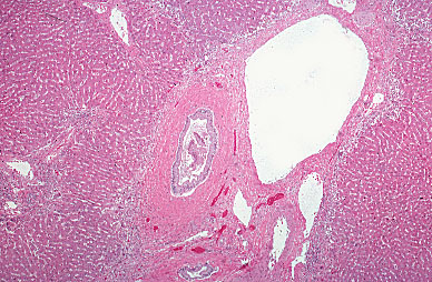 Primary sclerosing cholangitis
Onion-skinning around bile duct
KU Collection
Hepar lobatum
Syphilis
{05952} ascites in a known cirrhotic
{05953} ascites in a known cirrhotic
Massive ascites
Patient photo
Brazilian Medical Students
Splenomegaly with portal hypertension
(Ignore the "hyaline perisplenitis")
WebPath

Alcoholic Liver Disease
Text and pictures
From "Big Robbins"
-- Isaiah 5:11
One and a fifth!
-- Ed

-- Dr. Rabelais
Everybody knows alcohol is bad for the liver, but there is considerable confusion about the various patterns of liver injury and their outcome.
Alcoholic steatosis ("alcoholic fatty liver")
We've already reviewed why fat accumulates in liver cells damaged by alcohol.
To review: The drunken hepatocytes make too much fatty acid, make it into excess triglyceride instead of burning it, then can't complex the triglyceride to apolipoproteins, and can't export the lipoproteins they do make.
Current thinking supports popular wisdom that alcohol itself does the damage (in fatty change and the other faces of "alcoholic liver disease"). Poor nutrition doesn't help, either (i.e., few people make daily trips to the salad bar while on benders.)
If you've ever drunk a case of beer over a great football weekend, I bet you've had fatty liver. Did you notice? Probably not. The "disease" is usually just a pathology finding, unless:
Fatty liver is, by itself, completely reversible once the drinker sobers up. The same applies to fatty liver from other causes i.e., after ileal bypass, in obesity, in ill-controlled adult-onset diabetes, in problem pregnancy, in galactosemia, in methotrexate toxicity, in Wilson's disease, in a few rare disorders of lipid metabolism, and in a few unusual situations.
{49271} fatty cirrhotic liver vs. normal
NOTE: "Microvesicular steatosis" (smaller vacuoles, usually several per cell) is typical of Reye's syndrome, problem pregnancies, mitochondrial problems (Gastroent. 108: 193, 1995), and toxicity from outdated-tetracycline poisoning. It's also reversible; really, "microvesicular" steatosis can occur in any fatty liver.
* In my opinion, the "microvesicular vs. macrovesicular" distinction is of no value. I have seen only one Reye's autopsy, and it was pure macrovesicular fat. I've autopsied many, many problem drinkers, and the pattern is often mixed, and sometimes microvesicular-only. I addressed this question to the pathology on-line group in 1996, and there was no disagreement from a few hundred experienced pathologists.
* NOTE: "Fatty change" confined to the "Ito cells" (stellate cells)is vitamin A overdose. "Stellate cell lipidosis" is often due to vitamin A overdose or Retin-A: Am. J. Clin. Path. 119: 254, 2003.
* Of course, if you continue drinking, you're at serious risk to get cirrhosis, even if "you only had fatty change" on a
biopsy. Bad prognostic indicators include giant mitochondria and mixed microvesicular and macrovesicular
fat. See Lancet 346: 987, 1995..
Non-Alcoholic Steatohepatitis
Frank Mitros MD
Virtual Hospital
Alcoholic hepatitis
Much more serious. Here, we have liver cells dying. The process is worst in the centrilobular regions.
Surviving liver cells often bear Mallory's alcoholic hyaline (not pathognomonic, but suggestive). This is masses of altered prekeratin fibers plus stress proteins. Free Mallory's hyaline is chemotactic for neutrophils.
Also look for giant mitochondria, (* Yokoo bodies, after one of the professors who trained your lecturer), PAS-negative blobs in the cytoplasm.
* Mitochondria (giant or no) in alcoholics' livers often have genetic damage, presumably because alcohol generates toxic free radicals inside them (Gastroent. 108: 193, 1995). This is one mechanism of fatty liver (how)?
Cholestasis is usual because of compromise of bile canaliculi, with bile lakes and bile plugs. At the same time, the bile ducts may proliferate within the portal areas.
As the liver cells die off, look for fibrosis, notably around the central veins ("central hyaline sclerosis"; see Virch. Archiv. 614: 11, 1989). This won't go away, and is undoubtedly a principal route to cirrhosis. (* In some experimental models and some communities, cirrhosis need not be preceded by alcoholic hepatitis. According to others, it is inflammation, rather than fibrosis, that predicts progression. This'll confuse you.)
{39929} alcoholic hepatitis, good Mallory's hyaline
{26702} alcoholic hepatitis, good Mallory's hyaline
{08832} alcoholic hepatitis with fibrosis; there is no
good hyaline; cells are becoming entrapped in the fibrous tissue
{21056} "alcoholic hepatitis", looks like early cirrhosis to me
{26693} "alcoholic hepatitis", looks like early cirrhosis to me
Again, the histology is not pathognomonic for alcoholism. The heart-drug amiodarone in particular is infamous for producing the same histopathology, and post-ileal bypass hepatitis, Wilson's disease (shouldn't miss this one!), NASH, and * East Indian childhood cirrhosis (copper toxicity in the genetically-predisposed; J. Path. 195: 264, 2001) can be dead-ringers.
If you have alcoholic hepatitis, you're in trouble. You may have jaundice, hepatic encephalopathy, and all the signs and symptoms of portal hypertension (not the least being fatal GI bleeding). Further, if you need a general anesthetic, you're likely to die from it.
Some Hippocrates who thinks you're too sick for a biopsy ("His pro-time is prolonged and I don't want to transfuse him") will give you a diagnosis of "cirrhosis", which will stay with you as your chart thickens.
If you've really overdone the alcohol, you can get yellow atrophy and die. This is very unusual.
Fortunately, if you sober up, all that will remain is whatever minor scarring has occurred. The liver cells will regenerate nicely, and your liver will function normally.
Alcoholic cirrhosis ("Laennec's cirrhosis", other names)
Exactly what causes the progression (if it is a progression) from reversible changes (fatty change, Mallory bodies, inflammation) to irreversible (?) disease (i.e., fibrosis-cirrhosis) is obscure.
In order to get cirrhosis, one needs at least 15 pint-years (i.e, a pint of the hard stuff per day for fifteen years, three pints a day for five years, or similar). Many cirrhotics have much more. Yet 2/3 of heavy drinkers die without cirrhosis. Nobody knows why.
At first, the liver is big because of widespread hepatocyte overgrowth and fatty change from ongoing drinking. Later, with advanced scarring and enforced sobriety, the liver becomes rather small.
Microscopically, you'll see fibrosis and nodules instead of the normal architecture and proliferated bile ducts in the scar tissue (a good sign of alcoholism).
Early Laennec's cirrhosis has fine bands and a micronodular pattern. Late, the pattern becomes post-necrotic. When the scar tissue starts seriously obstructing bile flow, clinicians see jaundice and the pathologist sees bile plugs.
{10538} alcoholic cirrhosis, gross
{10844} alcoholic cirrhosis, gross
{18795} alcoholic cirrhosis, gross
{20163} alcoholic cirrhosis, histology; trichrome stain
{08835} alcoholic cirrhosis
{40412} alcoholic cirrhosis
{10535} "alcoholic cirrhosis" closeup
of a lobule; note the Mallory's hyaline and the neutrophils
attacking it. There may be fibrosis
elsewhere, but this merely looks like alcoholic hepatitis.
{39593} alcoholic cirrhosis; lots of bile
duct proliferation (as is usual in alcoholic cirrhosis);
edge of a nodule on each side of the screen
The cirrhotic must decide what he or she wants out of life. Continuing drinking is overall as lethal as untreated AIDS. Sobriety gives maybe a 90% chance of not dying of the cirrhosis within the next five years.
|  |
NON-ALCOHOLIC STEATOHEPATITIS
NASH -- non-alcoholic steatohepatitis, into which fibrosis can creep -- is a poorly-understood but very real condition that is only now getting the attention it deserves.
The anatomic pathology is much like alcoholic liver disease, with fatty change, Mallory hyaline, and sometimes even cirrhosis.
In countries where little alcohol is consumed and there is not much hepatitis or schistosomiasis, this is the most prevalent liver disease. The histology is exactly the same as alcoholic fatty liver, alcoholic hepatitis, or advanced alcoholic cirrhosis.
There is always insulin resistance as well.
The mainstay of therapy is weight loss and exercise
in the overweight, masterful inactivity and perhaps metformin in the non-overweight
(Med. Clin. N.A. 80: 1147, 1996; rationale for metformin Am. J. Gastro. 98: 2093, 2003). Betaine as a remedy: Am. J. Gastro. 96:
2534, 2001; Am. J. Gastro.
* See
also Gastroenterologist 5: 316, 1997; Gastroenterology 120:
1183 & 1281, 2001; the latter shows crystalloids in mitochondria
from these people that are not present in most controls, and suggests
that this is a mitochondrial disease that expresses itself in the
setting of obesity. (More on adult-onset diabetes itself as a likely
mitochondriopathy later.)
Big review emphasizing how little is really known: Gastroenterology 122: 1649, 2002.
NASH as a cause of cryptogenic cirrhosis: JAMA 289: 3000, 2003; 1/3 of patients will
get fibrosis, and 1/9 will get rapid progression to cirrhosis Am. J. Gastro. 98: 2042, 2003.
IRON OVERLOAD
The human body cannot make iron atoms (see Science 226: 922, 1984 for how the Good Lord does it). We must get what we need from outside. The healthy adult has total body iron content of around 4 gm. The red cells contain close to 3 gm total. The cytochromes and other redox enzymes together contain much less than a gram, and most people have some stored iron. An iron storage problem is definitely present when the amount of iron in the body exceeds 10 gm.
A reasonably normal diet contains at least 10 mg of iron daily. This is more than adequate to replace a healthy man's daily losses, and is enough for a non-pregnant woman who does not have heavy menstrual flow. Heme iron is absorbed much more readily than non-heme iron, especially non-heme iron that is complexed with certain small organic molecules. (This explains why what worked for Popeye doesn't work for us.) Dietary iron deficiency anemia can be expected when a girl has been menstruating for about a year while consuming mostly twinkies, french fries, and diet Pepsi. Pregnant women, zealous blood donors, people with diseases in which blood loss (GI, urinary, uterine) cannot be easily controlled, and some growing children are the only people who might need to take iron supplements. (In normal adults, "prophylactic" iron supplementation can only mask serious disease. Health frauds like you-know-what kill by delaying the diagnosis of GI and gynecologic cancers. Fortunately for the public, you-know-what itself uses ferric iron, which makes the stool black, doesn't upset the tummy very much, and isn't absorbed very much. That's nice.) Healthy people absorb and lose around 1.0 mg of iron daily (1.5 mg for menstruating women).
Ferrous (Fe+2), and not ferric (Fe+3) iron, is absorbed across the "mucosal barrier", mostly in the duodenum. It is complexed to a protein called NRAMP-2 that transports much more efficiently when body iron stores are low. So the amount of iron absorbed varies inversely with the amount of ferritin already present in the duodenal epithelial cells, which in turn reflects total body iron stores. Iron absorption by the gut also increases when there is increased normoblastic activity. (The mechanism remains unknown; the latest stuff Blood 96: 4020, 2000.) Absorption is mildly increased in hemolytic anemias or after hemorrhage. Absorption is more markedly increased in "ineffective erythropoiesis", notably in severe sideroblastic anemia and thalassemias. Huge doses of dietary iron can override the regulatory mechanism. And a relative abundance or lack of ascorbic acid (vitamin C) facilitates or inhibits absorption.
Iron atoms are slowly released into the plasma, where they are bound to the globulin transferrin. The amount of transferrin present is also regulated, so that more will be present when more iron is required. The iron is then carried where it is needed. (* Transferrin will only carry ferric iron, while most of the other forms of iron are ferrous.) Likewise, when a red cell (or any other cell) is destroyed, its iron is carried away by transferrin. Extra iron is stored, mostly in the liver and bone marrow. In health, it is available for incorporation into RBC's or for transport by transferrin should a shortage develop.
Storage iron (somewhere in the range of 1 gm in most people) exists in two principal forms. (1) Ferritin is a bit of iron at the center of a protein micelle. The protein shell explains the negative Prussian blue stain. This is the short-term storage form. It is the form found, for example, in bone marrow when it is soon to be incorporated into new RBC's. (2) Hemosiderin is aggregates of ferritin with much of the protein gone. This is "Prussian blue positive" iron. It is a less labile storage form that accumulates when there is excess ferritin. (* "Hemosiderin" is an archaic name chosen by von Recklinghausen. "Exogenous hemosiderin" is an iron-protein complex that forms around sites of iron injection and foreign bodies composed of iron). Hemosiderin is the yellow pigment in the halo surrounding a bruise. Tip: If an injured area is pigmented yellow three months after bruising, the patient probably has iron overload.
Humans have no special mechanism for excreting excess absorbed iron. There is ordinarily a loss of 1 mg/day or so through GI and skin cell turnover and microhemorrhages into the gut and GU tracts. A typical menstrual period results in a loss of 10-20 mg of iron. During the course of a pregnancy, the fetus absorbs 500-1000 mg of iron from the mother's bloodstream. The fetus "gets priority" and often the mother becomes iron-deficient. Donating a 500 mL unit of blood removes approximately 250 mg of iron from the body.
Iron does harm to cells by generating free radicals. Primary iron storage problems are very common, under-diagnosed, easily and inexpensively detected, potentially fatal, and very treatable. And these patients are often considered hypochondriacs for many years before the diagnosis is finally made (Ann. Int. Med. 101: 707, 1984). During the early 1990's, it was finally appreciated that around 1 man in 200 is affected (NEJM 318: 1355, 1988). Hemosiderosis ("systemic siderosis", etc.): increased total body iron (as ferritin and hemosiderin), from any cause. Most of the excess iron is in the reticuloendothelial cells. Spill-over into parenchymal cells is what can cause trouble. It is now known that 13% of people carry an autosomal gene for excessive iron absorption via the gut. (Homozygotes have worse problems.) Depending on diet and iron losses, these people may or may not express their primary hemosiderosis. Secondary hemosiderosis ("acquired hemosiderosis", etc.) occurs in a variety of illnesses. The best-known of these are diseases that require frequent blood transfusions in the absence of bleeding. Except as noted below, these do not usually progress to symptomatic iron overload.
Hemochromatosis is hemosiderosis that has damaged parenchymal cells. Total body iron stores in excess of ten gm are very dangerous. In classic cases, total body iron stores often exceed 100 gm. Primary hemochromatosis (formerly "idiopathic", now "familial", "genetic", "hereditary", or "HLA-linked"): in which the problem is greatly increased absorption of iron from the gut. These are "primary hemosiderosis" people in which the iron overload causes illness. Perhaps 1 man in 200 will be symptomatic with this during life, and thankfully we are diagnosing it more often. Review Am. Fam. Phys. 65: 853, 2002.
Secondary hemochromatosis most often occurs in thalassemia major and in severe sideroblastic anemias. In these conditions, there is greatly increased iron absorption through the gut, and the patient requires many blood transfusions with no way of disposing of the iron load. Obviously you cannot treat these patients by bleeding (why not?) Deferoxamine is life-saving in thalassemia major: NEJM 331: 567, 1994.
Obviously, the distinction between "hemosiderosis" and "hemochromatosis" is not clear-cut. Iron overload is a spectrum of disorders. Thus, it is an extremely common disease. (It is not usually so devastating as better-known genetic diseases).
So, in primary hemosiderosis and hemochromatosis, the problem is that iron is absorbed too easily through the gut. A patient homozygous for primary hemochromatosis who donates blood every eight weeks will remain iron-depleted....) The tendency is encoded at HLA-H (Proc. Nat. Acad. Sci. 94: 2534, 1997 review, also called HFE), discovered in 1996 in the HLA complex on chromosome 6, very closely linked to HLA-A and much like it; mouse hemochromatosis is caused by a bad beta2-microglobulin gene (Proc. Nat. Acad. Sci. 93: 1529, 1996) while the abnormal HLA-H doesn't bind properly to its microglobulin component. (* HLA-A3, B7, and B14 are often present, but the defect is linked with the chromosomes. Almost all B14 owners have the hemochromatosis gene, which is one reason your lecturer is confident that he carries it.) The frequency of the allele is about 14 out of every 100 chromosome 6's. Iron losses due to menstruation and pregnancies prevent expression primary hemochromatosis from ever developing in most women with the gene(s). Primary hemochromatosis is diagnosed nine times more frequently in men.
The classic hemochromatosis triad is liver trouble, diabetes mellitus, and skin discoloration. Today's list of major problems also includes cardiac arrhythmias, cardiac pump failure, and loss of sexuality. The liver may be enlarged on physical exam, or transaminases may be a bit high (South Med. J. 83: 1277, 1990). Around half of identified hemochromatosis patients develop overt diabetes mellitus. (* It is worth screening all adult-onset diabetics for iron overload: Lancet 2: 233, 1989.) It is worth screening everybody for iron overload (Gastroenterology 107: 453, 1994. Most get the peculiar skin discoloration to some degree. The classic form of the disease is usually fully expressed around age 40-60 years. Patients, however, say their ill-health began during their 20's (Am. Fam. Physician 29: 55, March 1984).
Liver disease is the most serious problem in the majority of diagnosed hemochromatosis patients. The hepatocyte lysosomes and mitochondria are packed, and total liver iron stores are often more than one hundred times normal. The liver enlarges even before cirrhosis develops. The radiologist may notice it is unusually radio-dense. Micronodular cirrhosis (scarring that ruins the architecture of each lobule) is usually present by the time the diagnosis is made. When caused by hemochromatosis, this is called "pigment cirrhosis"). For the degree of fibrosis, the liver works surprisingly well, and clinical manifestations of cirrhosis are less severe than in alcoholic cirrhosis. However, cirrhosis is general considered irreversible and will finally kill the patient unless something else does first. (And something else usually does; only 25% of patients diagnosed to have primary hemochromatosis die of cirrhosis.) Once cirrhosis due to hemochromatosis has developed, the patient is at great risk for hepatocellular carcinoma ("hepatoma"; histology Am. J. Clin. Path. 116: 738, 2001). This is fatal and kills another 30% of patients diagnosed to have primary hemochromatosis.
{49253} hemochromatosis causing cirrhosis (Prussian Blue stain, of course)
Although more iron is deposited in the acinar cells than in the islets of Langerhans, around 50% of patients have enough damage to their beta cells to develop symptomatic glucose intolerance.
Cardiac injury is also caused by iron overload. Many hemochromatosis patients have pump failure and/or rhythm disturbances, either of which can be disabling or fatal. This is the other leading cause of death in iron-overloaded people. (How many of these deaths are assumed to be due to some other disease process? No one knows.)
Endocrine injury is an additional problem. Patients are commonly troubled first by loss of libido, and eventually lose their secondary sex characteristics. Testicular atrophy secondary to pituitary failure with deposition in anterior lobe and Leydig cells too. It is quite reversible. Loss of testosterone in both men and women is now considered the explanation for the osteoporosis that develops in hemochromatosis patients (Ann. Int. Med. 110: 430, 1989). In addition, the adrenals, thyroid, and parathyroids are likely to be damaged, with various endocrine insufficiency syndromes. (* The role of "melanocyte stimulating hormone" in "bronze diabetes" is dubious.)
Joint injury caused by iron overload (Am. J. Med. 75: 957, 1983) is yet another major problem. Iron deposition in synovium results in synovial hyperplasia and erosion of bone and cartilage, eventually ruining the joint. In hemochromatosis, this usually affects the fingers, and is a problem for 50% of patients. In addition, the knees (and other weight-bearing joints) of hemochromatosis victims occasionally get accumulations of pyrophosphate crystals ("pseudogout", chondrocalcinosis).
Skin pigmentation in hemochromatosis is primarily due to increased melanin. (* Iron inhibits the enzyme that normally breaks down melanin.) Melanin imparts the "bronze" color; if there is enough hemosiderin in the skin, the combination looks "slate-gray".
Sepsis: For some unknown reason, Vibrio vulnificans (a raw seafood bug), Pasteurella pseudotuberculosis and Yersinia enterocolitica are much more pathogenic in the presence of iron overload. Even E. coli may get a boost in hemochromatosis (Am. J. Med. 87(3N): 40N, 1989).
You'll learn to make the diagnosis soon. In primary iron storage disease, it is important to make the diagnosis early. By the time the patient is obviously sick with hemochromatosis, he is 40 years old, has at least 20 grams of iron stored, has cirrhosis, and will probably be dead within ten years (though you can still help). If you find the tendency to accumulate iron, do this:
* "Why don't you give an iron chelator instead?" Retinal damage from deferoxamine is supposedly reversible.... Some new iron chelator drugs are available now, but I'd still stick with the no-drug approach.
Secondary hemosiderosis and hemochromatosis: Except for the secondary hemochromatosis developing as a result of thalassemia major and severe sideroblastic anemia, these tend not to be severe or result in cirrhosis (though the liver may be enlarged). The abnormal anatomy is similar to primary iron storage disorders, but most of the iron is usually in the Kupffer cells, not the hepatocytes. Hemosiderosis due to red cell transfusions ("transfusional" or "iatrogenic siderosis") is unavoidable in patients with severe anemias of decreased production. These include a variety of bone marrow disorders and some cases of renal failure. Remember, 100 red cell transfusions delivers 25 gm of iron! This is more than enough to produce hemochromatosis. These patients are now being treated with the new iron chelator drugs.
Hemosiderosis due to chronic alcohol abuse results from increased absorption of iron through the gut. The mechanisms are obscure, and this fact makes it hard to distinguish pigment cirrhosis from alcoholic cirrhosis. A few old-timers blame iron-rich wine for hemosiderosis in alcoholics. (A few wines are adulterated with iron, with up to 50 mg/mL. Most have much less). See Arch. Int. Med. 112: 184, 1963. Of course, if a hemochromatosis patient drinks heavily, the liver is doomed (Gastroent. 122: 281, 2002).
Vitamin-and-mineral faddists can occasionally make themselves chronically sick by iron-overloading themselves. (This is difficult to do and probably requires the genetic predisposition. See J. Roy. Soc. Med. 77: 690, 1984.) The Bantu people ingest 100 mg or more of iron daily from beer brewed in "iron pots" (really, steel drums). They tend to get hemosiderosis ("Bantu siderosis"), and a few get pigment cirrhosis. (Again, genes must be important; we now know that a Bantu beer-drinker with one dose of the hemochromatosis gene will probably get hemochromatosis; NEJM 326: 95, 1992.)
You'll screen for common hemochromatosis by checking the transferrin saturation levels, and confirm by serum ferritin levels. The actual iron assays on liver biopsy tissue will probably be replaced soon by MRI (Lancet 363: 341 & 357, 2004).
Hemosiderosis due to increased or ineffective erythropoiesis ("hematopoietic siderosis", typically when there is longstanding hemolysis), mild hemosiderosis is usual because of increased absorption of iron through the gut. In the severe thalassemias and sideroblastic anemias, red cell transfusions are required, erythropoiesis is ineffective, and absorption of iron through the gut is greatly increased. The iron overload progresses to fatal hemochromatosis. These are the patients who are most often treated experimentally with iron chelator drugs.
Porphyria cutanea tarda is a hereditary partial defect of uroporphyrin decarboxylase that is expressed only in the presence of iron overload. You'll encounter this if you keep your eyes open! Update Blood 95: 1565, 2000. * Other unusual genetic diseases include congenital absence of transferrin, in which the liver becomes packed with iron while the marrow remains depleted; no one knows why (NEJM 326: 1705, 1992); and a new not-so-severe non-HLA-linked syndrome with normal transferrin saturation (treacherous: Lancet 349: 95, 1997). You'll diagnose hemochromatosis by labs, and confirm with genetic testing. Liver biopsy is still handy for judging the severity (Am. J. Clin. Path. 118: 73, 2002).
WILSON'S DISEASE ("hepatolenticular degeneration"; Mayo Clin. Proc. 78: 1126, 2003)
Rare but very, very important. Don't miss this diagnosis. Untreated, it is lethal. Treated, it's harmless. You'll never diagnose it unless you think of it. And it always masquerades as something else.
Wilson's disease is an autosomal-recessive problem in which the liver is unable to dispose of excess dietary copper via the bile.
The gene was cloned in 1997, and named ATP7B; it is (no surprise) a copper-transporting ATP-ase (Am. J. Hum. Genet. 61: 317, 1997).
Nobody really understands why serum levels of ceruloplasmin, the copper transport protein, are often (but not always) low in Wilson's. What we do know is that serum copper, not properly carried, spills copiously into the urine in Wilson's disease. This represents the only route of copper excretion, and it is inadequate.
Eventually, the copper accumulates, damaging liver, joints, brain (especially basal ganglia), proximal renal tubule (causing wasting of solutes) and red cells (mild ongoing hemolysis is the rule), and making the distinctive Keyser-Fleischer corneal ring (you probably won't see them without a slit lamp).
The copper itself probably damages the cells in which it accumulates, maybe by free radicals or inhibiting enzymes. * Future pathologists: Stain for copper using rhodanine or rubeanic acid!
The histology in the liver passes through fatty change to chronic hepatitis to micronodular cirrhosis to post-necrotic cirrhosis. At autopsy, you see an unforgettable bluish-coppery colored liver, and the same discoloration in the basal ganglia.
The treatment, of course, is to remove the copper with a metal chelator. Penicillamine works wonders, but it won't cure cirrhosis or restore dead neurons.
{018} Wilson's
HEPATIC AMYLOIDOSIS: You already know about this. Past concerns about biopsying the liver in suspected amyloidosis seem to be groundless (Medicine 82: 291, 2003).
{53548} amyloid in the liver
LIVER POISONS
A host of drugs-poisons produce typical reactions in the liver. These range from predictable ("Take enough
'Tylenol' at once and your liver cells all die") to highly idiosyncratic ("Take 'Halothane' anesthesia a second
time and there's a tiny chance that sensitivity will kill you.") "Inflammation" may mimic acute or chronic
hepatitis.
Acetaminophen ("Tylenol")... Massive hepatic necrosis
Allopurinol... Granulomas
Alpha-methyldopa... Inflammation, granulomas, massive necrosis (idiosyncratic)
Amiodarone... Inflammation, "alcoholic hepatitis" mimic, cirrhosis (idiosyncratic);
kupffer cells loaded with phospholipid ("phospholipidosis")
Anabolic steroids... Cholestasis (at least)
Carbamazepine... Granulomas
Chlorpromazine... Cholestasis (idiosyncratic)
Diltiazem... Granulomas
Estrogens... Cholestasis (idiosyncratic), thrombosis (idiosyncratic)
Ethanol... Fatty change, alcoholic hepatitis, cirrhosis
Fenfluramine... Massive necrosis (idiosyncratic; a dozen Japanese find out what was really
in their Red Chinese holistic-wholesome weight-loss pills beside lotus leaves and chrysanthemum petals: Ann. Int. Med. 139:
488, 2003)
Halothane... Massive hepatic necrosis (idiosyncratic)
{24392} "Halothane hepatitis" case; simply massive necrosis
* Hydralazine... Granulomas
* Hydrazine (a quack cancer remedy)... liver necrosis (Ann. Int. Med. 133: 877, 2000).
Isoniazid... Inflammation (idiosyncratic)
(especially >age 35)
Methotrexate... Inflammation, cirrhosis
(even at "safe" doses)
Oxyphenisatin (laxative)... Inflammation ("lupoid hepatitis")
(who'd abuse that?)
Pennyroyal ("holistic herbal tonic" / amateur abortifacient) ...
simulates viral hepatitis
* Phenylbutazone... Granulomas
(who still uses that?)
* Quinidine... Granulomas
* Sulfa drugs... Granulomas
Tetracycline (outdated)... Microvesicular fatty change
(get rid of yours!)
* Total parenteral nutrition... Fatty change (nobody knows why; this and malnutrition are the major illnesses in which it's likely to be periportal
rather than central; Gastroent. 104: 286, 1993)
Valproic acid: Mixed hepatitis-like and cholestatic
REYE'S SYNDROME (Am. Fam. Phys. 50(7): 1491, 1994;
NEJM 340: 1423, 1999; NEJM 340: 1377, 1999)
This poorly-understood, dread entity is an acute illness that follows another viral illness, usually 'flu or
chicken pox, usually in a child or young teen.
The biphasic clinical story is typical. The pathology is, too. You will see:
The increased intracranial pressure produces vomiting. Later, in severe cases, liver enzymes rise in the
serum, and then the liver itself fails. The patient may die, or be left mildly or moderately brain-damaged.
The etiology of Reye's is obscure.
Several inborn errors of metabolism (notably in urea-genesis, ketogenesis, branched amino-acid metabolism,
and uncoupling of oxidative phosphorylation) produce crises similar to Reye's. Studies of patients with
Reye's have showed a plethora of abnormal compounds around their bodies. See JAMA 260: 3167, 1988;
also Enzymes 45: 209, 1991.
Reye's, and only true Reye's, gives spectacular edema of the mitochondria,
visible by electron microscopy.
In 1986, the public was strongly warned not to give aspirin to children.
As a result, true Reye's has nearly vanished.
"Reye's" cases nowadays are most likely to be
children with decompensated metabolic defects.
Skeptics or no, "the animal model" for Reye's is a rat, which must be given both aspirin and several low doses
of endotoxin (Metabolism 38: 73, 1989).
PEDIATRIC LIVER DISEASE
Extrahepatic biliary atresia ("EHBA")
A catastrophic birth defect in which the common bile duct and/or hepatic ducts ("extrahepatic") and/or many larger
intra-hepatic ducts are without lumens along some or all of their length.
Sometimes this occurs with syndromes. More often, there's only mild inflammation
and replacement of the involved areas with granulation tissue,
perhaps from an intrauterine viral infection. Over time, the lesion progresses, and
eventually the large bile ducts (extrahepatic, then
intrahepatic) become replaced by granulation tissue (Mayo Clin.
Proc. 73: 90, 1998. (* Sounds like a slow virus?)
These children (most often girls) suffer severe neonatal hyperbilirubinemia. Unless surgery can correct the
problem early, the disease will progress to biliary cirrhosis, liver failure, and the need for a transplant.
Neonatal hepatitis ("giant cell hepatitis")
Ongoing liver cell destruction in a baby.
This has a very long differential diagnosis. It includes:
Regardless of cause, the histology tends to be similar in babies.
Look for the lobular disarray, focal cell necrosis, cholestasis, and kupffer cell hyperplasia you'd see in any
kind of hepatitis.
In addition, the infant's liver regenerates well, producing prominent giant hepatocytes with many nuclei.
{38758} neonatal hepatitis, gross
Liver biopsy may show distinctive features (i.e., inclusions in herpes and CMV, smudge nuclei in
adenovirus infection, organisms in toxoplasmosis,
hyaline globs in alpha-1 antitrypsin deficiency.) Don't expect these always to be present.
{46222} herpes simplex hepatitis, gross; trust me; all that
you can appreciate is severe injury
Worth mentioning again here: cirrhosis can result from galactosemia (kids) or alpha-1 antitrypsin (kids
or adults).
{20137} cirrhosis, galactosemia (* future pathologists: Note the "flowers" formed by hepatocytes in this
condition)
As a rule, the prognosis is much more favorable than in biliary atresia. Nevertheless, a few of these children
do die.
BENIGN LIVER MASSES
Hamartomas
Cavernous hemangiomas are little birthmarks, seen in maybe 1% of livers. If you see them at surgery, don't
biopsy them. They give high-resolution scanners fits: Am. J. Roent. 162: 1113, 1994.
* Occasionally a giant hemangioma may need to be resected.
Focal nodular hyperplasia (World J. Surg. 19: 13, 1995)
is a curious lesion that
looks like a chunk of a cirrhotic liver grafted onto a
healthy one. There is usually a central star-shaped large scar.
* Nodular regenerative hyperplasia is a picturesque
lesion in which the entire liver becomes nodular, but without
fibrosis. The cause is damage to the small portal veins, as from
bone marrow or kidney transplantation (nobody really knows why),
thioguanine (famous; Arch. Path. Lab. Med. 128: 2004),
or some other cause of portal hypertension without cirrhosis.
* Mesenchymal hamartoma is a pediatric lesion with a trademark chromosomal marker (Cancer 74: 1237,
1994).
* Bile duct hamartomas are polka-dots, mere
autopsy curiosities ("Von Meyenberg complexes").
Cysts are bounded by biliary epithelium.
In Caroli's disease, some of the bile ducts are dilated (and thus,
more prone to stasis and infection, and for some reason cholangiocarcinoma). Polycystic liver may occur with either mendelian polycystic kidney
disease, or as a distinct autosomal dominant or sporadic disease
(Hepatol. 23: 249, 1996).
* Mouse model Am. J. Path. 150: 2231, 1997. * There is also an autosomal dominant form (gene PRKCSH Nat. Genet. 33: 345, 2003; Am. J. Hum. Genet. 72: 691, 2003;
Another gene SEC63: Nat. Genet. 36: 575, 2004).
Liver cell adenomas may occur sporadically, but are most common in women on birth control pills and men
on anabolic steroids, and these are considered somehow etiologic.
Grossly, adenomas present as soft masses. The histology resembles normal liver, minus bile ducts and good
lobular architecture. There may be cholestasis. Around a quarter are multiple (syndrome: Mayo Clin. Proc.
71: 478, 1996).
Usually liver cell adenomas cause no problems. The lesions are likely to regress when the exogenous
hormones are removed. They seldom turn malignant, though occasionally this happens (Arch. Surg. 129:
712, 1994).
PRIMARY CANCERS OF THE LIVER (i.e., the hepatocytes and their sinusoidal lining cells)
In the poor nations where HBV is endemic and typically transmitted mother-to-child, hepatocellular
carcinoma is easily the most common cancer in men (at extra risk both because of higher HBV carriage and
more body iron), and second only to cervical cancer in women. (Watch lung cancer rise to first for both sexes
in the next decades.)
*
The hepatitis B virus X antigen (HBxAg) integrates into
the host genome to permit chronic infection, and produces
a product that inactivates p53 (Am. J. Path. 150: 1141, 1997).
This is now well-established as causing progression of hepatocellular carcinoma (Cancer Res. 64: 7329, 2004).
We now know that hepatitis C virus is also a very important cause, though again we don't know how (Cancer
73: 2253, 1994). One thing we do know is that hepatitis B and C viruses are much more likely to produce
hepatocellular carcinoma when they cause rapid, prolonged cell turnover, i.e., making
Nowell's law operate
more readily (Cancer 73: 1149, 1994).
Other factors enter into the picture, as well. Iron overload strongly increases risk. Aflatoxin (from aspergillus
in moldy food) is probably responsible for the trademark p53 mutation required for epidemic African
hepatocellular carcinomas; sporadic cancers elsewhere show different mutations in p53 (Cancer 74: 30,
1994).
"Thorotrast" exposure places a person at risk for
hepatocellular carcinoma, and anabolic steroids (from the gym) supposedly do, too. If
alcohol is a risk factor, it is minor.
In the U.S., many (but not all) patients have cirrhosis (from their hepatitis virus and/or iron overload), but this
is less common where most victims are "healthy carriers" of hepatitis B.
Wilson's cirrhosis, lupoid cirrhosis, and primary biliary cirrhosis do not place a person at risk, and alcoholic
cirrhosis itself is a very minor risk factor.
Hepatocellular carcinomas in cirrhotic and non-cirrhotic livers: Am. J. Clin. Path. 105: 3, 1996.
Grossly, the tumor may be a single mass, or arise multicentrically. The better-differentiated the tumor, the
greener it tends to be (since hepatocytes want to make bile). Hepatocellular carcinomas, unlike most
carcinomas, have a great propensity to invade veins, and you will probably see this grossly and
microscopically.
* Worth learning sometime:
The five carcinomas that invade veins in preference to lymphatics (pretty reliable):
The two sarcomas that invade lymphatics in preference to veins (unreliable):
Histologically, the hepatocellular carcinoma may look like nearly-normal liver, or be wildly anaplastic, or
something in between. Appearances vary tremendously. Pathologists look for:
The existence of liver dysplasia is now well-established
(Gastroent. 108: 812, 1995).
It
precedes hepatocellular carcinoma. Today's imaging techniques are
supposedly able to distinguish dysplastic nodules from simple
regenerative nodules in cirrhosis (Br. J. Rad. 77: 911, 2004).
* To tell benign from malignant, proliferating-cell nuclear antigen, a marker for a cell thinking about dividing,
is an excellent immunostain: Cancer 73: 2259, 1994. Pathologists are also counting nucleolar organizer
regions (Cancer 73: 289, 1994; neat pictures of
Nowell's law in action.)
* UCLA got their five favorite international liver pathologists to sit down and draw up some criteria for a
standard nomenclature, which was nice. Am. J. Surg. Path. 17: 1113, 1993.
Not surprisingly for a tumor that arises in this way, hepatocellular carcinomas often appear to arise
multifocally (but not entirely independently; see Hum. Path. 19: 403, 1988).
* The presence of a "capsule" means exactly nothing (Cancer 70: 45, 1992).
The grading system is of little value in predicting the behavior of a particular tumor.
* Future pathologists:
You can use aspiration cytology to tell whether a liver nodule is cancer
(Gut 53: 1356, 2004).
You can't use cytology to grade: Cancer 102: 247, 2004.
* Fibrolamellar hepatocellular carcinomas are rather tame and are almost certainly a different disease.
Grossly they look like focal nodular hyperplasia. Their "pale bodies" contain albumin and fibrinogen.
See Arch. Path. Lab. Med. 128: 222, 2004.
* Spindle cell hepatocellular carcinomas exhibit mesenchymal markers and have an extremely
bad prognosis
(Cancer 77: 51, 1996).
Third-world, hepatitis B-belt hepatocellular carcinoma is usually thoroughly anaplastic and very aggressive:
Cancer 65: 130, 1990.
{18799} hepatocellular carcinoma in macronodular cirrhosis;
hepatocellular carcinomas are white
You can deduce the symptoms from the behavior of the tumor, but by the time the patient knows he or she is
sick, it's usually too late. When a cirrhotic deteriorates rapidly, it's common to find a hepatocellular
carcinoma at autopsy.
Paraneoplastic syndromes include polycythemia (erythropoietin production) and hypoglycemia (insulin-related growth factor
production).
Death occurs through rupture, venous obstruction, liver failure, or (less often) lung metastases.
Clinical behavior of hepatocellular carcinomas: Cancer 66: 2174, 1990. Without treatment, you're dead.
Nowadays, many hepatocellular carcinomas are discovered incidentally,
and especially if there is no cirrhosis, the disease is often curable (Gastroenterology 122:
1609, 2002).
Surgery is often successful since since the tumor
tends to metastasize (to the lungs) only late. The procedure seems to cure about 25% of patients (Arch. Surg.
129: 738, 1994.
More recently, resection of the whole liver with liver transplant is becoming a mainstay of therapy, with about
half of well-chosen patients are alive three years later, and a 30% cure rate claimed (early studies
Am. J. Surg. 167: 317,
1994; Mayo Clin. Proc. 69: 509, 1994; Br. J. Surg. 81: 427, 1994; surgery plus chemotherapy Cancer 73:
2721, 1994; Am. J. Surg. 169: 28, 1995).
Or you can get good palliation by embolizing the hepatic artery, since the cancer's blood supply is mostly
arterial (Cancer 73: 2259, 1994). And nowadays, radiofrequency ablation or even
injecting absolute alcohol under CT guidance
(which gets the patient drunk) can supposedly produce cures.
* Oncologists have a variety of interesting approaches. Recently, small tumors have been treated with
microwaves (Cancer 74: 817, 1994; "residual tumor" may be non-active -- future pathologists will enjoy reading about the picturesque
after-effects in Am. J. Gasto. 98: 2052, 2003) or ethanol injection (Radiology 192: 407, 1994). Both are tributes to
our ability to detect tiny tumors by serum screening and scanning. Big tumors have been treated with protons
(Gastroent. 106: 1032, 1994).
Still, the long-term outlook is grim without major surgery plus a lot of
good luck. As with lung cancer, after non-surgical "cure" of
one hepatocellular carcinoma, it is not unusual for a new clone to arise from the same benign cellular milieu
(Nowell's law triumphant; Cancer 63: 2207, 1989).
* The "fibrolamellar variant" of younger women (mostly) without cirrhosis carries a relatively good prognosis
after surgery and transplantation. It's really a different disease. See Hum. Path. 22: 396, 1991;
Hum. Path. 19: 784, 1988.
Cholangiocarcinoma-type cancers (see below) can occur within the liver, or as a mix with hepatocellular
carcinoma.
They are desmoplastic adenocarcinomas, often mucin-positive. Of course, the cells will not secrete bile.
The risk factors for this lesion are (1) "Thorotrast" exposure;
(2) liver fluke infestation; * (3) idiopathic sclerosing cholangitis; * (4) Caroli's.
Hepatoblastoma is a pediatric solid cancer. Surgical resection (followed by liver transplant, of course) is
often curative.
Ask a pediatric pathologist to show you a sample. As in Wilms' tumor, there's often a variety of different
kinds of epithelial and/or mesenchymal-derived structures. alpha-fetoprotein is high, and FSH/LH often high
too. (What would the latter do?)
The important prognostic factor is stage; histologic type is less important (Ped. Path. 12: 167, 1992).
Histopathologists are now talking about the "cell of origin" (obviously a stem-cell) even predating the
differentiation into endoderm and mesoderm (Am. J. Path. 148: 321, 1996).
Update Cancer 98: 668, 2003.
Hepatic angiosarcoma, a rapidly lethal cancer, is infamous for having been caused by exposure to industrial
poisons (vinyl chloride, arsenic) and the radioactive contrast medium "Thorotrast".
Metastases to the liver need to be mentioned, too.
Liver and lung are the two organs most often involved in fatal cancer.
Around 35% of patients dying of cancer
have "liver mets", and about the same percent have "lung mets". The biggest livers are those bearing
metastatic carcinoma.
Primary cancers of in the liver (hepatocellular carcinoma, angiosarcoma, cholangiocarcinoma) often appear
multiple. But you can distinguish metastatic cancer from primary liver cancer fairly easily:
Metastatic tumor in the liver tends to umbilicate (i.e., undergo central necrosis). Hepatocellular carcinomas
and cholangiocarcinomas seldom do this.
If it's a cirrhotic liver, chances are it's hepatocellular carcinoma.
Here's a rule that usually works: Untreatable liver metastases portend death within twelve months.
* Some folks are living longer now thanks to cryoablation: Am. J. Surg. 171: 27, 1996.
Remember, families of patients dying with liver metastases often believe the patient died of "liver cancer",
unrelated to any known primary.
* Lymphomas and leukemias in the liver cluster on the portal areas. Erythroid cells in the liver cluster in the
sinusoids.
FOR FUTURE LIVER PATHOLOGISTS AND EXAM-TAKERS:
Necrosis in zones of the liver:
Zone 1 (periportal) is hit hardest in phosphorus poisoning and eclampsia
Zone 2 (mid-zonal) is hit hardest in yellow fever
Zone 3 (centrilobular) is hit hardest in hypoxia and shock (i.e., it gets the least oxygen), in acetaminophen
toxicity, and in CCl4 toxicity. NOTE: This is the best place to look for bile plugs.
Other keys to the diagnosis:
Lots of hemosiderin? Is it located predominantly...
Primary hemochromatosis (zone 1 is most heavily involved,
then zone 2, then zone 3). If there's cirrhosis, you'll see iron in the fibrous
tissue
and biliary epithelium.
Hemolysis, transfusional hemochromatosis
longstanding congestion/repetitive ischemia (why?)
Hint: If there is cirrhosis from most any cause, secondary iron overload is common. If the bile ducts and
fibrous stroma of a cirrhotic are iron-poor, the problem is not primary hemochromatosis.
* Chronic hepatitis with a large predominance of plasma cells: Autoimmune (lupoid) hepatitis (chronic
active hepatitis from any cause can look the same)
* Chronic hepatitis with lots of fatty change in a non-drinker:
hepatitis C
Mallory's hyaline in zone 3, plus fat and polys: Alcoholic hepatitis (* less often,
nonalcoholic steatohepatitis,
ileal bypass or amiodarone toxicity)
Random confluent-lytic necrosis: Unusual viral infections, in a baby or immunosuppressed person (* CMV,
herpes simplex or zoster, adenovirus, echovirus; also consider whether the biopsy was obtained during
surgery)
Polys in the lumens of bile ducts: Ascending cholangitis
* Mallory's hyaline in zone 1: Wilson's disease, primary biliary cirrhosis
* Lipofuscin in Kupffer cells: A sign of recent (last few months) hepatocellular necrosis
* d-PAS positive stuff in Kupffer cells: A marker that the patient is on hyperalimentation
Polys in the portal areas, bile duct proliferation: Chronic or intermittent bile duct obstruction
Bile lakes, bile duct proliferation, edema of the portal areas: Acute bile duct obstruction.
* Onion-skinning around the interlobular bile ducts (and eventual
replacement by fibrous onions): Primary sclerosing cholangitis
Lots of eosinophils in the portal areas: Drugs, parasites (remember schistosomes), * primary biliary
cirrhosis,
* primary sclerosing cholangitis
Granulomas in the liver: TB, histoplasmosis (Am. J. Clin. Path. 113:
64, 2000 -- huge array of different lesions, with good granulomas being
the exception), primary biliary cirrhosis, sarcoid, drugs as above, Crohn's
disease, Hodgkin's, remote effect of Hodgkin's, Q-fever, listeria, brucella.
* Fibrosis around individual cells: Congenital syphilis
Star-shaped portal-tract scar containing proliferating bile ducts filled with eosinophilic globs: Cystic fibrosis.
Star-shaped scar around central vein: Central hyaline sclerosis
* Fibrosis with bridging and regeneration, but no nodules: Hepatoportal sclerosis
* Fibrosis with bridging, but no nodules or regeneration: Non-cirrhotic bridging fibrosis
Cholestasis without other abnormalities: Think of drug-effect, post-surgery, sepsis, pregnancy.
Ground-glass hepatocytes: Those caused by hepatitis B virus infection stain positive with * orcein, * Victoria blue,
and the appropriate immunostain.
* No portal veins in the portal areas, but everything else is okay? Hepatoportal sclerosis! Not an
undergraduate medical student's concern.
Update on how to percuss and feel the liver (and whether it's worthwhile; it is): South. Med. J. 87: 182, 1994.
BILE DRAINAGE SYSTEM
{15774} normal gallbladder, from the outside
Worth remembering from previous course work:
Birth Defects
We've already looked at the dread extrahepatic biliary atresia.
Phrygian cap is an anomaly of the gallbladder in which the fundus is folded over on itself. It's a radiologist's
curiosity in which gallstones are more likely to form.
Gallstones (all about them: NEJM 328: 412, 1993; laparoscopic surgeons JAMA 269: 1018, 1993;
lithotripsy Gut 35: 417, 1994)
About 1 adult in 10 has gallstones. Usually these are asymptomatic for life. However, they can cause major
problems. There are 600,000 gallstone surgeries in the U.S. yearly.
Cholesterol stones may be pure or contaminated with calcium, oxalate, and/or bilirubin. They result from
bile becoming super-saturated with cholesterol. They are yellow and range in size from infinitesimal (in
aggregate, these are the potentially-lethal "sludge") to large barrel-shape casts of the gallbladder.
You can read for yourself about "lithogenic bile", etc., etc.
Bile that is merely cholesterol-supersaturated becomes liquid crystals;
excess mucin production from the gall bladder that follows
causes nidus formation and growing crystals (Nat. Med. 10: 1303, 2005).
Decide for yourself whether gallstones are "a
disease caused by the Western diet", or whether paleontologists are correct in exhibiting dinosaurs' gallstones,
or whether there may be truth in both.
Everybody knows "the typical cholesterol stone patient" and the "F's": fair-skinned, fat, female, fertile,
flatulent, forty year old.
* Slow intestinal transit time as a risk factor in those without the usual risk factors: Lancet 341: 8, 1993.
Smoking as a risk for symptomatic stones: Gut 35: 107, 1994.
{00041} faceted, mostly-cholesterol stones; despite the black
bilirubinate coating, the large size and smooth faceted surfaces say "cholesterol"
is their major component
Bilirubin stones ("mulberry stones"; "pigment stones") are calcium bilirubinate. They result from bile
becoming super-saturated with unconjugated bilirubin, i.e., when there is hemolysis (sickle cell,
hemoglobin C, spherocytosis, autoimmune Coombs-positive anemia; also remember intramedullary
hemolysis, especially in the thalassemias).
{18772} bilirubin stones
Regardless of composition, gallstones present problems when they:
{10544} common duct stone
{49181} gallstone ileus
Inflammation of the gallbladder, usually containing gallstones, usually with infection.
The usual bugs are E. coli or enterococci. Less common are clostridia and salmonella (typhoid is carried in
the gallbladder); * Enterocytozoan bienensi is an opportunist in AIDS (NEJM 328: 95, 1993).
No one really understands the pathogenesis of acute cholecystitis, and "Big Robbins" maintains that the
bacteria are secondary invaders. It is inviting to think that a stone may plug the outlet, and the gallbladder
mucosa may then become infarcted due to compression by the muscularis propria's futile efforts to push the
stone out. Read about "poisoning by lysolecithin", etc., etc., on your own time; it is speculative.
In the absence of gallstones ("acalculous" cholecystitis, 10% of cases), there's generally some systemic
disease, and/or perhaps edema of a Reister valve obstructs the outlet.
The pathology is what you'd expect. The gallbladder is red, swollen, and tense. There may be pus on the
inside (even an "empyema"), fibrin all over the outside, or even gangrene of the wall.
{49242} cholecystitis, acute-on-chronic
{08822} chronic cholecystitis, histology
This just means fibrosis of the gallbladder, maybe from repeated bouts of mild or severe acute cholecystitis,
maybe "just from the irritation of having gallstones". There are almost always gallstones. Look for:
The "porcelain gallbladder" has undergone dystrophic calcification.
Non-problems
"Cholesterolosis", or "strawberry gallbladder", is a pathologist's delight seen in maybe 5% of autopsies. Little
clusters of cholesterol-laden macrophages develop just under the epithelium, looking like strawberry seeds.
Its significance to human health is zero.
{13362} strawberry gallbladder ("cholesterolosis"), gross
"Inflammatory polyps" are benign nubbins on the gallbladder wall.
"Mucocele" ("hydrops") is a gallbladder filled with mucus instead of bile, because its cystic duct has become
obstructed. The gallbladder won't visualize on x-ray.
{38827} hydrops ("mucocele") of the gallbladder
Benign tumors of the gallbladder are uncommon, and usually incidental findings.
Carcinoma of the gallbladder
Adenocarcinomas of the gallbladder are uncommon (but more common than cholangiocarcinomas of the bile
ducts). They are usually preceded by (and most people think caused by) gallstones. As you would expect,
women are at increased risk.
* Recently it's been claimed that chronic carrying of typhoid or paratyphoid bacteria increases the risk by
about fifty times (Lancet 343: 83, 1994; somebody please take a look at this study and tell me if it's flawed.)
Usually, these have already invaded the liver by the time they become symptomatic (i.e., have obstructed the
common bile duct).
{26327} adenocarcinoma of the gallbladder, gross
Cancer of the bile ducts
Adenocarcinomas arising from these structures are uncommon. Of course, they produce obstructive jaundice.
Risk factors include ulcerative colitis and infestation with liver flukes or ascaris worms. Unlike for cancer of
the gallbladder, men are at greater risk.
The most common site is at the ampulla of Vater, and this is often considered separately. The "Klatskin
tumor" at the junction of the hepatic and common ducts is also common, but any point on the common bile
duct or a hepatic duct can be the primary.
Cancer of the ampulla tend to be mucin-producing. Cancers arising elsewhere from among the bile ducts tend to
be desmoplastic.
The symptomatology is what you'd expect. Itching makes these patients particularly miserable.
 Drug-induced liver injury
Emilio Orfei
Loyola; great photos
Herpes simplex hepatitis
Please don't let this happen
Loyola Med
{38761} neonatal hepatitis, microscopic; with giant cells and bile stasis
{46223} herpes simples hepatitis, microscopic (note the cell with two nuclei and herpes intranuclear
inclusions)
{13301} cirrhosis, alpha-1 antitrypsin deficiency (trust me)
{13302} cirrhosis, alpha-1 antitrypsin deficiency (trust me)
{13304} cirrhosis, alpha-1 antitrypsin deficiency; PAS stain shows alpha-1 antitrypsin
granules well
Antitrypsin deficiency, liver
Photo and mini-review
Brown U.
Liver Tumors
Frank Mitros MD
Virtual Hospital
Small FNH's are very common and (unfortunately) often get
found on modern imaging studies. Unless it's huge or the radiologist
can't be confident it's FNH, or it hurts, leave it alone.
Dominant polycystic kidney disease
with polycystic liver
WebPath
 Hepatocellular carcinoma ("hepatoma", "Mickey Mantle's disease"): A deadly disease clearly
caused, in the
large majority of cases, by longstanding infestation with hepatitis B and/or C viruses and/or iron overload.
Reviews: Lancet 353: 1253, 1999; Lancet 362: 1907, 2003.
Hepatocellular carcinoma ("hepatoma", "Mickey Mantle's disease"): A deadly disease clearly
caused, in the
large majority of cases, by longstanding infestation with hepatitis B and/or C viruses and/or iron overload.
Reviews: Lancet 353: 1253, 1999; Lancet 362: 1907, 2003.
A dominant mass in a cirrhotic liver is likely to be hepatocellular carcinoma.
Future pathologists: If the histology is quite benign, it's probably just
a large regenerative nodule instead ("macroregenerative nodule");
one has to think there's been at least one genetic hit. If there is slight atypia,
you may be able to diagnose "borderline dysplasia". Don't worry about it now.
{24571} hepatocellular carcinoma in cirrhosis
{39624} hepatocellular carcinoma in cirrhosis
{39708} hepatocellular carcinoma in cirrhosis
{40330} hepatocellular carcinoma in cirrhosis
{40331} hepatocellular carcinoma histology (one of many variants)
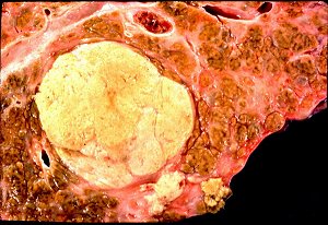 Hepatocellular carcinoma
Arising in post-necrotic cirrhosis
KU Collection
Metastatic adenocarcinoma
Infiltrating ductal carcinoma
WebPath
TB of the liver
Great granulomas
Pittsburgh Pathology Cases
Hepatocellular carcinoma
Chronic alcoholism and viral hepatitis
WebPath
Biliary Tract Disease
Frank Mitros MD
Virtual Hospital
{15775} normal gallbladder, opened up to show mucosa
{08820} "normal gallbladder" with Rokitansky-Aschoff sinuses;
lumen and mucosa on right
{15287} normal gallbladder

* Western civilization fans: A real "phrygian cap" is
the flop-over headgear worn by the French Revolution Lady allegorical figure, and on
the swordpoint on the US Army seal. Smurfs wear phrygian caps.
{08425} gallstone city!
{17591} gallstone city!
{49247} common duct stones;
common bile duct has been opened. Liver at top
{49182} gallstone ileus
Acute-On-Chronic Cholecystitis
Australian Pathology Museum
High-tech gross photos
{08108} strawberry gallbladder, histology; foam
cells (phagocytic macrophages) contain cholesterol
{26330} adenocarcinoma of the gallbladder
{26333} adenocarcinoma of the gallbladder
{49249} adenocarcinoma of the gallbladder
{38821} adenocarcinoma of the gallbladder, with stones
{38824} adenocarcinoma of the gallbladder, no stones
Future surgeons: Courvoisier's law!
In obstructive jaundice, if the gallbladder is palpable, the patient has cancer.
Explanation:
In obstructive jaundice below the level of the cystic duct, and assuming the cystic duct is patent, the pressure in the gallbladder will be high.
If the problem is a common duct stone, the gallbladder will be scarred up due to previous episodes of acute cholecystitis, or chronic cholecystitis. Therefore, it will not expand.
If the problem is cancer of the pancreas, common duct, or ampulla, the gallbladder wall itself is normal, so it will be tremendously expanded and easily palpated.
This works pretty well, but nobody's going to base a decision on it nowadays. Can you think of exceptions? Sure you can!
To date, there is no established role for "alternative and complementary medicines"
in the treatment of any liver disease, though it's possible some of the herbal
remedies might have some effect.
An elaborate protocol by a naturopath for treatment of chronic hepatitis C, which centers around a very special
recipe for breakfast museli, led to drops in transaminase levels but he made no effort
to control for the fact that he also made his patients stop drinking alcohol
This invites an obvious conclusion.
Plus, the risk of hepatotoxicity from herbal
concoctions is well-known. See Liver Int. 23: 213, 2003; Lancet 361: 101, 2003;
Med. J. Aust. 178:
442, 2003; Clin. Liver Dis. 7: 453, 2003.
In Korea, use of "alternative medicine" seems to be the most common
cause of elevated liver enzymes in breast cancer patients (J. Kor. Med. Sci. 19:
397, 2004).
In Oregon,
"complementary herbal remedies" are now THE cause of massive hepatic necrosis requiring
transplantation, exceeding
even acetaminophen overdoses and all the viruses combined.
(Arch. Surg. 138: 852, 2003). Even the much-touted "milk thistle",
though non-toxic, hasn't shown any robust evidence of working (Am. J. Med. 113: 506, 2002).
{08820} gall bladder, normal
I hate liver, liver makes me quiver,
Malignant melanoma
Bile duct primary
Loyola Med
 * SLICE OF LIFE REVIEW
{08826} liver, normal
{11798} liver, normal
{11807} liver, normal
{14874} liver, porcine
{14875} liver human, normal
{14875} liver human, normal
{14876} liver human, normal, central vein
{14876} liver human, normal
{14877} liver (sinusoids), normal
{14877} liver (sinusoids), normal
{14878} liver (sinusoids), normal, space of Disse
{14878} liver (sinusoids), normal
{14879} liver, portal triad
{14880} liver, portal triad; a=vein, b=duct
{14883} gallbladder, normal
{14885} gallbladder (epithelium), normal
{15104} liver
{15105} liver
{15106} liver
{15107} liver, pig
{15281} liver, normal
{15281} liver, normal
{15282} liver, normal
{15282} liver, normal
{15283} liver, normal
{15283} liver, normal; a=vein, b=artery, c=duct
{15284} liver, pig
{15285} liver, pig
{15286} gall bladder, normal
{15286} gall bladder, normal
{15287} gall bladder, normal
{15287} gall bladder, normal
{15288} gall bladder, normal
{15288} gall bladder, normal
{15773} gall bladder, normal
{15774} gall bladder, normal
{15775} gall bladder, normal
{15776} liver, normal
{15778} liver, normal
{16043} liver, normal in fetus
{16074} necrosis, liver junct.; normal
{17592} gallbladder, normal
{19721} gall bladder, normal
{20888} liver, portal triad
{20889} liver, portal vein
{20890} liver, hepatic artery
{20891} liver, bile duce
{20892} liver, hepatocyte plate
{20893} liver, central vein
{20894} liver, central vein
{20895} liver, portal triad
{20896} liver, portal triad and central vein
{20897} gall bladder
{26039} liver, normal
{26042} hepatocytes, normal aspirate
{26045} hepatocytes, normal aspirate
{39498} liver, normal
{39764} liver, normal
liver makes me curl right up and die, makes me cry...
it gives you hives, gives you scurvy, turns my stomach topsy turvy,
liver just simply ain't my bag, makes me gag, makes me want to throw up...
Now liver is neither solid or liquid, but merely an amorphous, viscous colloid of putrid
protein...
It is located between the 7th and the 10th dorsal vertebrae,
JUST south of the diaphragm, "lounging like a worm on a pillow of fat."
Now did you ever look at the word liver?
Is it any coincidence that there are 5 letters in the word liver?
The same number of letters as in the word death? the word drugs? the word hippy?...
Well, maybe you just haven't had it cooked right...
Naaah, I've had that stuff sauteed, I've had it breaded and broiled and broasted and braised,
I've had it diced and sliced and riced and sunnyside up over easy fried, and it still comes out, LIVER....
I hate liver, liver makes me quiver, liver makes me curl right up and die, (makes me cry)...
Why, first time I had it, I didn't like it at all...
Well, what happened?
Well, I was about 9 months..my mammy gave me a hunk of that Gerber's baby food.
Well, I rolled it around, looked at her eyeballs, spit it out and uttered my first words...
At nine months, what'd you say? I said... [repeat Chorus]
-- "Second City", Chicago radio group, 1970's


| Visitors to www.pathguy.com reset Jan. 30, 2005: |
Ed says, "This world would be a sorry place if
people like me who call ourselves Christians
didn't try to act as good as
other
good people
."
Prayer Request
 Teaching Pathology
PathMax -- Shawn E. Cowper MD's
pathology education links
Ed's Autopsy Page
Notes for Good Lecturers
Small Group Teaching
Socratic
Teaching
Preventing "F"'s
Classroom Control
"I Hate Histology!"
Ed's Physiology Challenge
Pathology Identification
Keys ("Kansas City Field Guide to Pathology")
Ed's Basic Science
Trivia Quiz -- have a chuckle!
Rudolf
Virchow on Pathology Education -- humor
Curriculum Position Paper -- humor
The Pathology Blues
 Ed's Pathology Review for USMLE I
![]()
![]()
 | Pathological Chess |
 |
Taser Video 83.4 MB 7:26 min |

 Liver in place
 Hepatic cirrhosis
)window.location='http://edcenter.med.cornell.edu/Pathology_Images/325.gif') Zahn infarct of liver
Zahn infarct of liver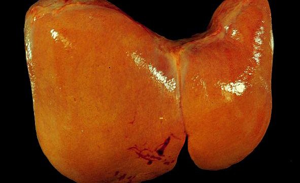 Fatty liver
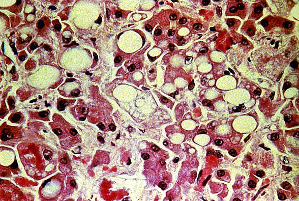 Fatty liver
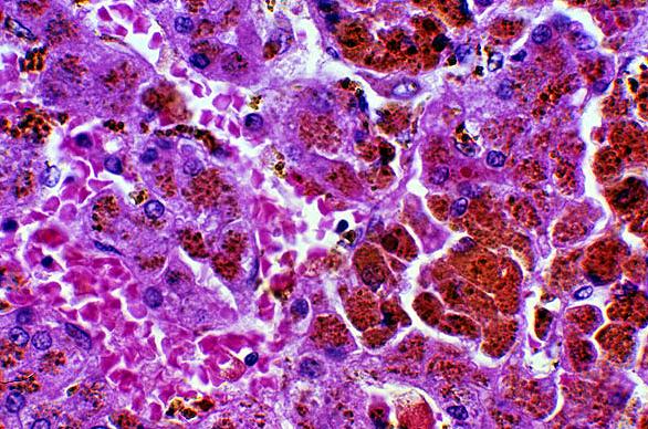 Hemochromatosis
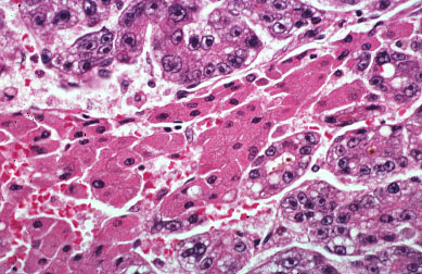 Hepatocellular carcinoma
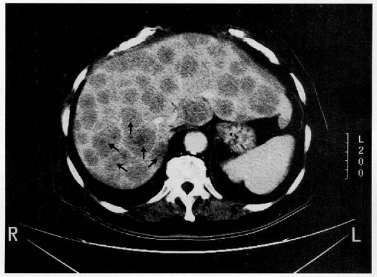 Liver metastases