Ed Friedlander, M.D., Pathologist
scalpel_blade@yahoo.com


Cyberfriends: The help you're looking for is probably here.
Welcome to Ed's Pathology Notes, placed here originally for the convenience of medical students at my school. You need to check the accuracy of any information, from any source, against other credible sources. I cannot diagnose or treat over the web, I cannot comment on the health care you have already received, and these notes cannot substitute for your own doctor's care. I am good at helping people find resources and answers. If you need me, send me an E-mail at scalpel_blade@yahoo.com Your confidentiality is completely respected.
 DoctorGeorge.com is a larger, full-time service.
There is also a fee site at myphysicians.com,
and another at www.afraidtoask.com.
DoctorGeorge.com is a larger, full-time service.
There is also a fee site at myphysicians.com,
and another at www.afraidtoask.com.
Translate this page automatically
 |
With one of four large boxes of "Pathguy" replies. |
 I'm still doing my best to answer
everybody.
Sometimes I get backlogged,
sometimes my E-mail crashes, and sometimes my
literature search software crashes. If you've not heard
from me in a week, post me again. I send my most
challenging questions to the medical student pathology
interest group, minus the name, but with your E-mail
where you can receive a reply.
I'm still doing my best to answer
everybody.
Sometimes I get backlogged,
sometimes my E-mail crashes, and sometimes my
literature search software crashes. If you've not heard
from me in a week, post me again. I send my most
challenging questions to the medical student pathology
interest group, minus the name, but with your E-mail
where you can receive a reply.
Numbers in {curly braces} are from the magnificent Slice of Life videodisk. No medical student should be without access to this wonderful resource. Someday you may be able to access these pictures directly from this page.
Also:
Medmark Pathology -- massive listing of pathology sites
Freely have you received, freely give. -- Matthew 10:8. My
site receives an enormous amount of traffic, and I'm
handling about 200 requests for information weekly, all
as a public service.
Pathology's modern founder,
Rudolf
Virchow M.D., left a legacy
of realism and social conscience for the discipline. I am
a mainstream Christian, a man of science, and a proponent of
common sense and common kindness. I am an outspoken enemy
of all the make-believe and bunk that interfere with
peoples' health, reasonable freedom, and happiness. I
talk and write straight, and without apology.
Throughout these notes, I am speaking only
for myself, and not for any employer, organization,
or associate.
Special thanks to my friend and colleague,
Charles Wheeler M.D.,
pathologist and former Kansas City mayor. Thanks also
to the real Patch
Adams M.D., who wrote me encouragement when we were both
beginning our unusual medical careers.
If you're a private individual who's
enjoyed this site, and want to say, "Thank you, Ed!", then
what I'd like best is a contribution to the Episcopalian home for
abandoned, neglected, and abused kids in Nevada:
My home page
Especially if you're looking for
information on a disease with a name
that you know, here are a couple of
great places for you to go right now
and use Medline, which will
allow you to find every relevant
current scientific publication.
You owe it to yourself to learn to
use this invaluable internet resource.
Not only will you find some information
immediately, but you'll have references
to journal articles that you can obtain
by interlibrary loan, plus the names of
the world's foremost experts and their
institutions.
Alternative (complementary) medicine has made real progress since my
generally-unfavorable 1983 review linked below. If you are
interested in complementary medicine, then I would urge you
to visit my new
8Alternative Medicine page.
If you are looking for something on complementary
medicine, please go first to
the American
Association of Naturopathic Physicians.
And for your enjoyment... here are some of my old pathology
exams
for medical school undergraduates.
I cannot examine every claim that my correspondents
share with me. Sometimes the independent thinkers
prove to be correct, and paradigms shift as a result.
You also know that extraordinary claims require
extraordinary evidence. When a discovery proves to
square with the observable world, scientists make
reputations by confirming it, and corporations
are soon making profits from it. When a
decades-old claim by a "persecuted genius"
finds no acceptance from mainstream science,
it probably failed some basic experimental tests designed
to eliminate self-deception. If you ask me about
something like this, I will simply invite you to
do some tests yourself, perhaps as a high-school
science project. Who knows? Perhaps
it'll be you who makes the next great discovery!
Our world is full of people who have found peace, fulfillment, and friendship
by suspending their own reasoning and
simply accepting a single authority that seems wise and good.
I've learned that they leave the movements when, and only when, they
discover they have been maliciously deceived.
In the meantime, nothing that I can say or do will
convince such people that I am a decent human being. I no longer
answer my crank mail.
This site is my hobby, and I presently have no sponsor.
This page was last updated February 6, 2006.
During the ten years my site has been online, it's proved to be
one of the most popular of all internet sites for undergraduate
physician and allied-health education. It is so well-known
that I'm not worried about borrowers.
I never refuse requests from colleagues for permission to
adapt or duplicate it for their own courses... and many do.
So, fellow-teachers,
help yourselves. Don't sell it for a profit, don't use it for a bad purpose,
and at some time in your course, mention me as author and KCUMB as my institution. Drop me a note about
your successes. And special
thanks to everyone who's helped and encouraged me, and especially the
people at KCUMB
for making it possible, and my teaching assistants over the years.
Whatever you're looking for on the web, I hope you find it,
here or elsewhere. Health and friendship!
 I am presently adding clickable links to
images in these notes. Let me know about good online
sources in addition to these:
I am presently adding clickable links to
images in these notes. Let me know about good online
sources in addition to these:
Pathology Education Instructional Resource -- U. of Alabama; includes a digital library
Houston Pathology -- loads of great pictures for student doctors
Pathopic -- Swiss site; great resource for the truly hard-core
Syracuse -- pathology cases
Walter Reed -- surgical cases
Alabama's Interactive Pathology Lab
"Companion to Big Robbins" -- very little here yet
Alberta
Pathology Images --hard-core!
Cornell
Image Collection -- great site
Bristol Biomedical
Image Archive
EMBBS Clinical
Photo Library
Chilean Image Bank -- General Pathology -- en Español
Chilean Image Bank -- Systemic Pathology -- en Español
Connecticut
Virtual Pathology Museum
Australian
Interactive Pathology Museum
Semmelweis U.,
Budapest -- enormous pathology photo collection
Iowa Skin
Pathology
Loyola
Dermatology
History of Medicine -- National Library of Medicine
KU
Pathology Home
Page -- friends of mine
The Medical Algorithms Project -- not so much pathology, but worth a visit
National Museum of Health & Medicine -- Armed Forces Institute of Pathology
Telmeds -- brilliant site by the medical students of Panama (Spanish language)
U of
Iowa Dermatology Images
U Wash
Cytogenetics Image Gallery
Urbana
Atlas of Pathology -- great site
Visible
Human Project at NLM
WebPath:
Internet Pathology
Laboratory -- great site My team:
Ed Lulo's Pathology Gallery
Bryan Lee's Pathology Museum
Dino Laporte: Pathology Museum
Tom Demark: Pathology Museum
Dan Hammoudi's Site
Claude Roofian's Site
Pathology Handout -- Korean student-generated site; I am pleased to permit their use of my cartoons
Estimating the Time of Death -- computer program right on a webpage
Pathology Field Guide -- recognizing anatomic lesions, no pictures

St.
Jude's Ranch for Children
I've spent time there and they are good. Write "Thanks
Ed" on your check.
PO Box 60100
Boulder City, NV 89006--0100
More of my notes
My medical students
Clinical
Queries -- PubMed from the National Institutes of Health.
Take your questions here first.
HealthWorld
Yahoo! Medline lists other sites that may work well for you

We comply with the
HONcode standard for health trust worthy
information:
verify
here. 


![]()
Define "tumors" and "neoplasms". Tell why they are important. Define "oncology". Tell how tumors are like organs, and how they are different.
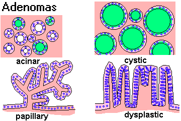
Distinguish "benign tumors" and "malignant tumors" by their gross and microscopic appearances and their behaviors. Tell what "well-differentiated" and "poorly differentiated" mean. Tell how benign tumors can cause problems.
Define "cancer", "malignant", and "metastasis". Tell what any cancer will ultimately do to the patient if it is not cured. Recognize the typical appearances of cancers. Distinguish "carcinomas" and "sarcomas".
Tell how and where various cancers tend to metastasize. Describe patterns for invasion and metastasis by various malignant tumors.
List the three most common cancers in men and in women, and the three most common fatal ones.
Given a tumor name, name the cell of origin and describe the behavior. Given the cell of origin (or a hint) and the behavior (or a hint), name the tumor.
Distinguish "grade" and "stage" of a cancer. Tell why these numbers are important.
Recognize, under the microscope, a squamous cell carcinoma, an adenoma, an adenocarcinoma, a tumor of hematologic origin, and a spindle cell sarcoma. Recognize anaplasia, and generally tell benign from malignant tumors.
Critique the following statement, overheard on a cancer ward: "Cancer cells grow faster than normal cells and are not under the body's control. Therefore, the treatment for cancer involves giving anti-mitotic drugs that kill fast-growing cells."
LEARN FIRST
Tumors (neoplasms) may be considered new, useless organs.
Cancers (malignant neoplasms) invade and spread to remote sites (metastasize). Benign tumors cannot invade or spread to remote sites, but they can cause problems by compressing local structures.
Tumors arise from single cells, and they recapitulate, more or less, the things that cell did in health. The cell of origin gives its name to the tumor.
Benign tumors are usually round like balls. Cancers look like cauliflowers, ulcers, or else simply expand the organ.
Cancers that arise from epithelium are called "carcinomas". Cancers that arise from mesenchyme/mesoderm are called "sarcomas".
Cells and histologic architecture in benign tumors is similar to normal histology. Cells and microscopic architecture in cancers are bizarre. The grade of a cancer is a function of how bizarre the cells look. The higher the grade, the more likely to cancer is to behave aggressively.
The stage of a cancer is how far the clinician knows it has spread.
|
|
|
|
|
|
QUIZBANK: See next handout
One out of about every five persons in the US who die this year will die of tumors (about 500,000 total).
* "Tumor"--literally any swelling. Galen distinguished tumors that are:
"natural"--pregnant uterus
"unnatural"--pus, bony callus
"contrary to nature"--what we now know as neoplasms ("new growths")
The ancient medical writings show an awareness of cancer, and Morgagni remarked that by his era, most physicians were aware that tumors were not treatable except by surgery.
"Oncology" is the study of tumors. In current usage, an oncologist is an internist or surgeon who specializes in the administration of cancer chemotherapy.
In modern usage, a tumor/neoplasm may be thought of as an attempt by the body under some stimulus to make some new sort of organ. (It develops in the wrong shape, in the wrong place, and it persists after the initiating stimulus is removed.)
Tumors are like organs:
All have parenchyma and stroma.
Cells usually look similar to cells in the organ where the tumor arose.
Cells will continue to perform some of the functions of the parent organ.
Tumors are different from organs:
They don't contribute to the homeostasis of the body.
They usually grow more rapidly than surrounding tissues.
Some benign and all malignant tumors never cease to grow.
"Spontaneous regression" of a cancer (i.e., massive shrinkage or disappearance without adequate therapy to explain it) is much-discussed phenomenon that occasionally happens. More often than not, the cancer comes back after a few months. Review, emphasizing the ongoing mystery: In Vivo 14: 773, 2000.
Most tumors show some derangement of histologic architecture.
Malignant tumors are locally invasive and have metastatic potential.
We have already learned a few essential terms:
Neoplasia: A new, useless organ produced by cells bearing mutations.
Dysplasia: Ugly cells in an epithelium, without invasion. Near-synonyms are "carcinoma in situ", "intraepithelial neoplasia", "incipient neoplasia", and "precancer".
Benign: (1) A neoplasm that will compress but not invade the surrounding tissue. (2) Loosely, any non-cancer, non-precancer diagnosis.
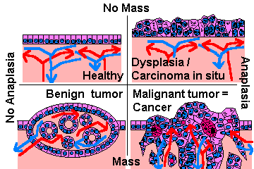
A tumor is either benign or malignant.
Characteristics of benign ("good") tumors:
|
{08800} benign colonic polyp; recapitulates colon
|

{14165} benign ovarian cystic tumor; it weighed approximately 30 lb
{17488} benign fibroadenoma of breast
Characteristics of malignant ("bad") tumors
Malignant tumors generally grow more rapidly than benign tumors.
However, no malignant tumor grows as rapidly as an embryo, nor do cancer cells divide nearly as fast as cells in normal bone marrow or intestinal epithelium. Today, anyone who thinks of cancer cells growing "without any control" is guilty of willful ignorance
Cells differ morphologically and functionally from normal cells, and tumor architecture is less organized than that of parent tissue.
Tumor cells are locally invasive; the tumor grows into the surrounding tissue and destroys it.
 This feature led Dr. Hippocrates to call such tumors karkinoma after karkinos, Greek for "crab."
(Cancer is Latin for "crab".)
This feature led Dr. Hippocrates to call such tumors karkinoma after karkinos, Greek for "crab."
(Cancer is Latin for "crab".)
|
|
The tumor will eventually metastasize, spreading to another site remote from the original tumor
(exceptions: basal cell carcinomas of skin, cancers of glial origin).
Malignant tumor and cancer are synonyms.
Benign or malignant, each tumor has a "cell of origin" from its tissue.
Old studies proved that women heterozygous for G6PD variants express only one form in each
uterine tumor because of lyonization. And we've long known that plasma cell myeloma produces a
monoclonal immunoglobulin product. More about
"Nowell's law" soon.
Tumor cells almost always mimic one cell type of some normal organ, usually the one in which the
tumor arose.
Cells may continue to elaborate keratin, mucus, hormones, immune globulin, etc. And they may
show cross-striations (skeletal muscle cells), microvilli (certain glandular cancers), melanosomes
(melanocyte cancer)., etc., etc.
The resemblance will be better or worse depending on whether the tumor is "well-differentiated" or
"poorly differentiated."
Much of the day-to-day work of a surgical pathologist is figuring out the cell of origin of poorly-differentiated cancers.
The main tools are electron microscopy (now mostly passé) and
immunoperoxidase stains ("the brown revolution").
Worth learning now:
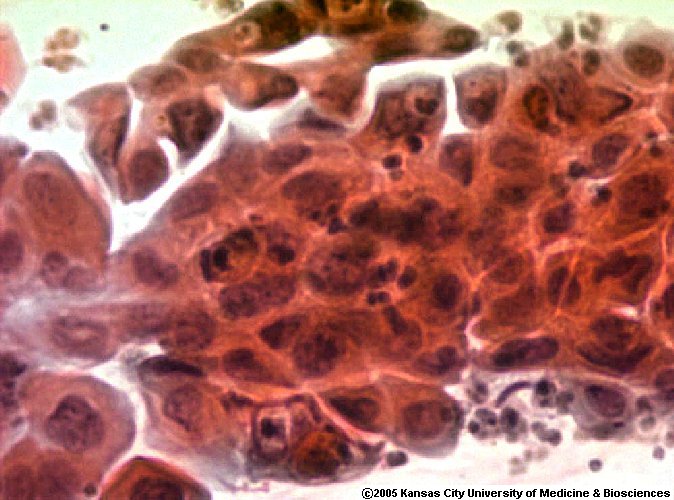
These arise anywhere there is a stratified squamous epithelium, either healthy (skin, esophagus,
mouth, many others) or metaplastic (endocervix, bronchi).
Look for any (or even all) of the following:
The better these things show, the better-differentiated the tumor! (Benign squamous tumors are
uninteresting connective tissue covered with almost-normal-looking stratified squamous epithelium.)
{10085} squamous cell carcinoma
{129} adenocarcinoma, apocrine
Leukemias and lymphomas
Look for cells that resemble blood precursors, not sticking tightly together. (These features
distinguish these common cancers from carcinomas and sarcomas.)
{23776} acute leukemia, basophils predominate
Immunostains:
Benign tumors may cause problems:
The tumor may secrete something in excess (typically a hormone).
The tumor may compress surrounding structures.
{221} meningioma compressing brain
A few benign tumors sometimes transform into malignant tumors. Once this happens, they are no
longer benign (so, benign tumors never metastasize).
In some syndromes, benign tumors may be multiple.
Malignant tumors in the U.S:
The most common cancers:
Males (in descending order): prostate, lung, colon
Females (in descending order): breast, lung, colon
The most commonly cancer killers:
Males (in descending order): lung, prostate, colon
Females (in descending order): lung, breast, colon
NOTE: Worldwide, cancer of the cervix is the great killer of women, especially young women.
NOTE: The other great third-world killer is hepatocellular carcinoma, which is primarily a man's
tumor (because of hepatitis B carrier status and iron overload).
Thanks to the decline in deaths from atherosclerosis, cancer is now the leading cause of death in U.S.
females, and is about tied with
atherosclerosis for US males.
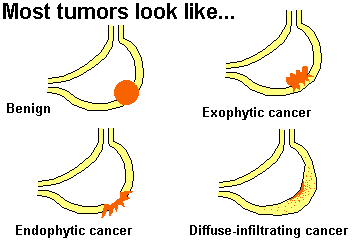
What do cancers look like?
You remember that benign tumors are usually round like a ball.
(The same can be true of some very tame cancers.)
The Gross appearances of cancers usually fits one of three patterns:
Exophytic ("fungating"): tumor grows as a lump, often with a cauliflower-like surface.
(Malignant tumors seldom appear "encapsulated.")
{5801} fibrosarcoma; here and below,
we'll want microscopy for confirmation
Endophytic: tumor grows as an ulcer (i.e., the part that was probably protruding from the surface
sloughed).
{09810} esophageal carcinoma
Infiltrating: tumor cells invade an organ diffusely without changing its shape.
{1573} brain cancer (glioblastoma)
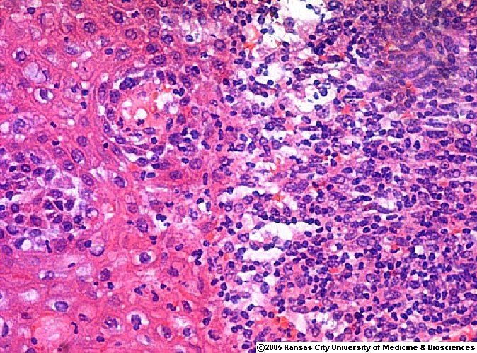
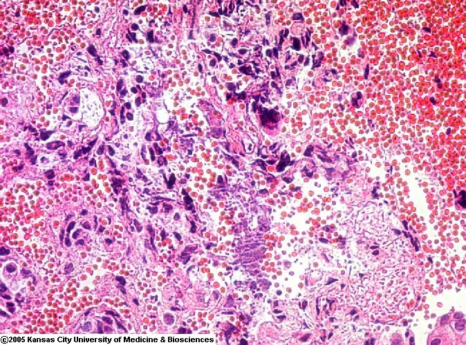
Any cancer is likely to exhibit hemorrhage and/or necrosis, grossly and microscopically. These
result from the cancer cells invading the tumor's own blood vessels.
Microscopic appearance:
Individual cell morphology and tumor architecture may be "well-differentiated" (good prognosis) to
"poorly-differentiated" ("anaplastic", bad prognosis).
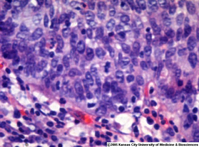
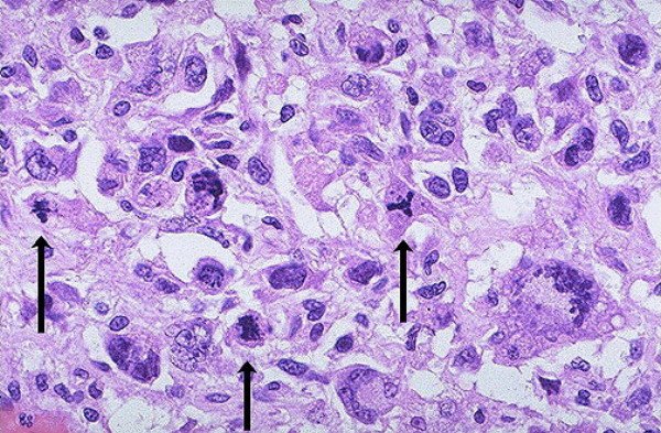
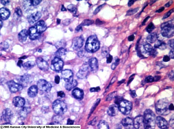
The malignant-looking cell:
{00132} bizarre mitotic figure
*Better measures of cell division, useful in prognosticating tumors, include immunostaining for
PCNA (proliferating cell nuclear antigen) and MIB-1/Ki-67. Many current articles; for example
Am. J. Clin. Path. 107: 229, 1997.
There may also be microscopic visible invasion (tumor cells growing into adjacent normal structures
where they don't belong), and/or indirect evidence (hemorrhage and/or necrosis; either is important
evidence that the tumor in question is malignant).
{10478} squamous cell carcinoma invading; be sure
you can recognize the tentacles ("tumor cords") cut in cross section
Usually, the more abnormal a tumor appears under the microscope, the more rapidly it grows and
the worse the patient's prognosis.
How will I see malignant tumors invading?
Local infiltration
Invasion of surrounding tissues
For some reason, cartilage, tendon, and elastic tissue almost never get invaded.
{08845} cholangiocarcinoma invading around a nerve ("perineural invasion")
"Intraepithelial spread" is possible and may take the form of single cells ("Paget's disease" of the
nipple from an underlying breast cancer, many melanomas) or of dysplasia / carcinoma in situ, in which an
epithelial surface is replaced by a layer of anaplastic cells that has not (yet)
penetrated the basement membrane.
Second idea: "Dysplasia is reversible and carcinoma in situ is not."
I have been hearing this since I entered medical school without anybody
ever showing me any evidence that it is true. How are you going to tell?
Nobody has ever made a study to "just watch" either full-thickness
or partial-thickness intraepithelial anaplasia.
Third idea: "Who cares? Call them both intraepithelial anaplasia.
Mild, moderate, or severe."
This made so much sense that Bethesda decided to call them "intraepithelial
neoplasia" instead.
{08912} carcinoma in situ, endocervix (junction
with normal, i.e., the "carcino-columnar junction")
Metastatic spread:
There are four routes:
(1) Seeding of serosal surfaces (or, in the case of CNS tumors, up and down the neuraxis in the CSF)
(2) Mechanical transplantation (rare, typically iatrogenic; see for example Br. J. Surg. 81: 648, 1994)
(3) Via lymphatics (traditional route for tumors of epithelial origin, i.e., carcinomas)
Tumors spread first to regional lymph nodes, then (because of disruption of directions of lymph
flow) to any lymph nodes or organs
{21051} carcinoma in lymphatics (small intestinal
mesentery)
(4) Via blood vessels (traditional route for tumors of mesenchymal origin, i.e., sarcomas, because the
tumor cells are in direct contact with blood vessels from the beginning)
Regardless of the route of metastatic spread, certain tumors have unexplained preference for certain
metastatic sites.
Why? Even today, there's no clear molecular explanation.
The common sites for metastatic spread for many common cancers include lymph nodes, lung, liver,
bone, and brain.
Most cancers seldom metastasize to the muscles, spleen or gonads.
Requirements for successful metastatic spread:
"Tumor angiogenesis factor" (more about this momentarily...)
Collagen production by local fibroblasts (if dense, tumor is called "desmoplastic")
Metastatic nodules are called "metastases" or (vulgarly) "mets".
{08443} "liver mets"
*There has always been a lot of interest in the basic biology of the metastatic process. It remains an
array of tantalizing clues. See, for example, the discovery that the presence of certain blood group
antigens on the surfaces of tumor cells seems to increase their mobility and their metastatic potential
(NEJM 327: 14, 1992).
Malignant tumors: Grade and Stage:
The grade and stage of a cancer are determined to offer a prognosis and to determine treatment.
Both grade and stage are usually represented by Roman numerals, the best situation by I, the worst
by III, IV, or V depending on the tumor type and determined by rules. Do not confuse grade and
stage!
Tumor grade: assigned by the pathologist to reflect the cancer's degree of differentiation.
Grade I: Well-differentiated, cells look like normal organ (benign = Grade 0)
Flow cytometry can help measure anaplasia by measuring how much
DNA is in each cell.
Leave the arcana of grading up to us in pathology. Rules are constantly changing, and new
prognostic factors emerging (* for example, a current popular
approach is counting argyrophilic nucleolar
organizer regions, AgNOR's: Cancer 74: 1658, 1994; bladder
J. Urol. 162:
63, 1999; thymoma Chest 115: 1115, 1999; prostate Cancer 79:
1956, 1997; sputum cytology Chest 111: 1591, 1997; Cancer 82:
86, 1998; squamous lung carcinoma Cancer 111: 110, 1997).
Tumor stage: assigned by the clinician on the basis of all available information on the extent of
tumor spread.
Stage I might mean the tumor is smaller than 1 cm diameter, without metastases
Stage II might mean the tumor is larger than 1 cm and/or is symptomatic and/or there are metastases
to the regional lymph nodes
Stage III might mean the tumor has infiltrated a non-resectable structure and/or there are distant
metastases
Rules for assigning stage are quite elaborate and different for each type of tumor.
Alternative system: TNM "T" for tumor:
T1 might mean primary tumor is smaller than 1 cm in diameter
T2 might mean primary tumor is larger than 1 cm in diameter
T3 might mean primary tumor is invading something non-resectable
"N" for regional lymph nodes:
N0 would mean no tumor in regional lymph nodes
N1 might mean tumor in a few nearby lymph nodes
N2 might mean many nodes, or some nodes farther downstream, are involved
"M" for metastases:
M0 would mean no distant metastases
M1 would imply distant metastases, etc.
*So the TNM stage for a lung cancer that is invading or encasing the superior vena cava but has
metastases only in two nearby lymph nodes might be T3 N1 M0.
Memorizing tumor staging systems is not an appropriate pathology learning objective, and I will
not test you on it.
Generally tumors of high grade present at high stage, while tumors of low grade present at low
stage.
 Metaphase
Cancer cell in division
Dave Barber MD, KCUMB
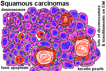
{10088} squamous cell carcinoma, sort-of-good desmosomes
{08977} squamous cell carcinoma, sort-of-good pearls
{09157} squamous cell carcinoma, good desmosomes
{09159} squamous cell carcinoma, electron micrograph,
tonofilaments in the cell on the right
{10987} squamous cell carcinoma, very good pearls
{12596} squamous cell carcinoma, keratin is very pink,
sort-of pearls
{15424} squamous cell carcinoma, good pearls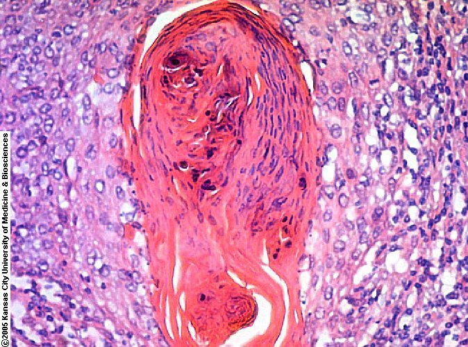 Squamous cell carcinoma
Good pearl
Dave Barber MD, KCUMB
 Squamous cell carcinoma
Right cheek
Pittsburgh Pathology Cases
 Squamous cell carcinoma
Good photos
Pittsburgh Pathology Cases
Squamous cell carcinoma
Not-so-well differentiated
WebPath Photo
Squamous cell carcinoma
Electron micrograph -- desmosomes
WebPath Photo
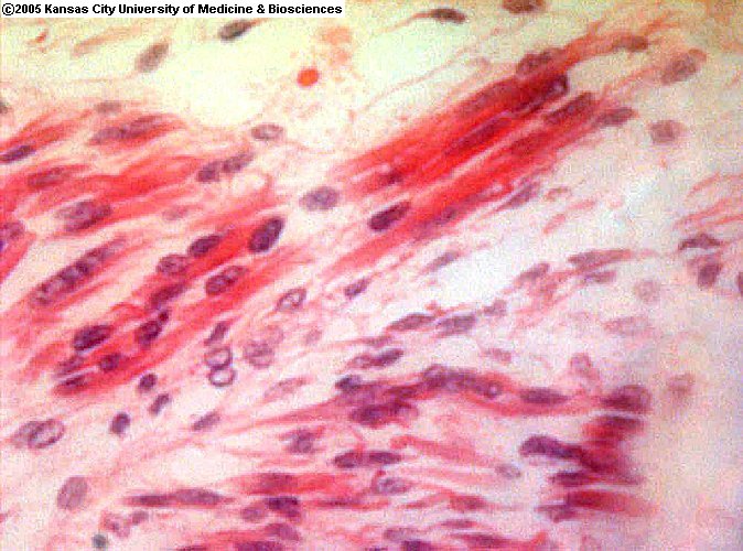
 Invasive squamous carcinoma
Below squamous epithelium
Dave Barber MD, KCUMB
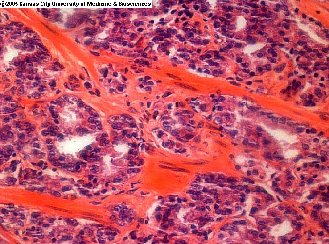
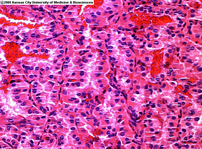
Adenocarcinoma
Anaplastic cells making clear glands
Urbana Atlas of Pathology
Colon adenocarcinoma
Not so well differentiated
Urbana Atlas of Pathology
Colon adenocarcinoma
Fairly well differentiated
Urbana Atlas of Pathology
Adenoid cystic carcinoma
Breast primary
Pittsburgh Pathology Cases
 Adenocarcinoma
Prostate
ERF/KCUMB
 Adenocarcinoma
Prostate
ERF/KCUMB
 Adenocarcinoma
Prostate
ERF/KCUMB
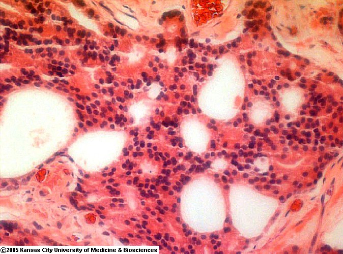
{08806} adenocarcinoma of colon; low-power
shot shows it arising from the mucosa and pushing toward the
muscularis propria
{08852} adenocarcinoma of the pancreas;
find some odd glands and some signet-ring cells
{08865} prostate adenocarcinoma; some swiss cheese
{08866} prostate adenocarcinoma; some swiss cheese
{16671} leiomyosarcoma (smooth muscle
cells, slightly high N/C ratio)
 Abnormal pap smear
High NC ratio
Dave Barber MD, KCUMB
Lymph node with cancer metastasis
Photo and mini-review
Brown U.
{1855} craniopharyngioma
{5831} fibrosarcoma
{5834} liposarcoma
{5958} chondrosarcoma
{08437} uterine adenocarcinoma ("cottage cheese
in the uterine cavity")
{08916} uterine adenocarcinoma
{10208} colon adenocarcinoma
{10211} colon adenocarcinoma, in section
{10435} bronchogenic carcinoma
{17511} gastric adenocarcinoma
{11087} squamous cell carcinoma of larynx
{10436} lip cancer
{12169} lip cancer
{1596} same; microscopy
{1599} same; microscopy
 Hemorrhage in a high-
grade colon cancer
Dave Barber MD, KCUMB
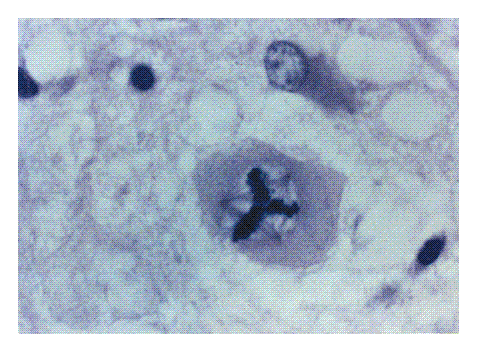
 Bizarre mitotic figures
One is a classic "tripolar"
KU Collection
 Tripolar mitosis
Photomicrograph, nice spindles
New England Journal of Medicine
Today's pathologists have stains to show the laminin in the basement
membrane, if there is any doubt as to whether the tumor is "through".
What is the difference between dysplasia and carcinoma in situ?
First idea: "Carcinoma in situ is full-thickness dysplasia. Turn
the epithelium upside-down. If it looks the same, it is carcinoma in situ.
The cells have completely forgotten how to mature." This merely
reminds us that going from the mildest dysplasia to the meanest
carcinoma in situ is a continuum. The only obvious dividing line is
the moment of invasion -- when the first malignant cell penetrates
the basement membrane.
Carcinoma in situ
Urbana Atlas of Pathology
{21052} carcinoma in lymphatics
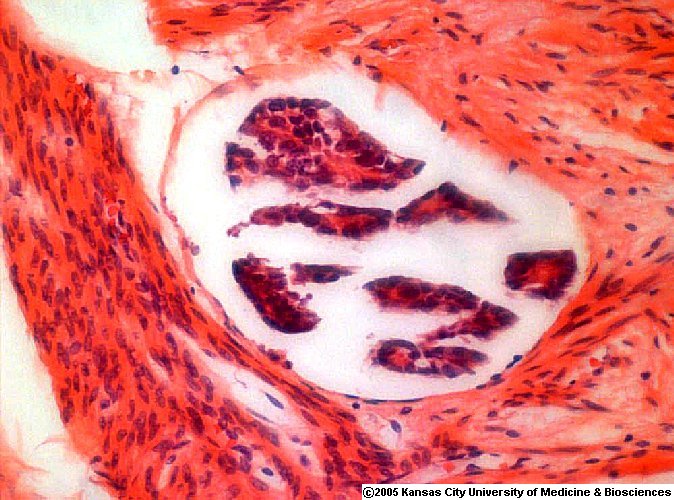 Lymphatic with carcinoma
Uterus
ERF/KCUMB
{08444} "liver mets"
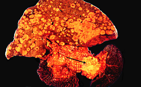 Cancer of the pancreas
Primary and liver metastases
KU Collection
Grade II: Not so well-differentiated
Grade III: Worse than that
Grade IV: Even worse
Grade V: Worst of all (most tumor types are graded I-III or I-IV)
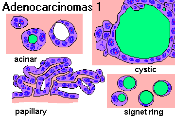
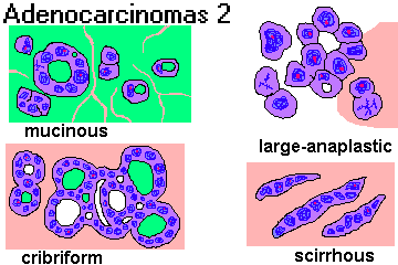
I. To assign a name to a tumor that you have examined, begin by writing the suffix -oma. Most tumor names end in this way. (Unfortunately, the suffix simply means "swelling", and some non-neoplasms also use the suffix, i.e., granuloma, hematoma, xanthoma, traumatic neuroma).
II. If the tumor is malignant, write the root carcin- ("crab") if the tumor is of epithelial origin, or sarc- ("flesh") if the tumor is of mesenchymal origin, before -oma. If the tumor is benign, do not write anything.
III. Now choose one or more roots to describe the cell of origin.
If the tumor originated in glandular epithelium, use the root adeno-. (It probably makes little glands and/or mucin.)
If the tumor originated in squamous or transitional epithelium, is benign, and protrudes above the epithelial surface, use the root papillo-.
If the tumor originated in non-glandular epithelium and is malignant, name it for the cell of origin.
If the tumor originated from a non-epithelial cell, look for a root in the following list. (We do not consider endothelium and mesothelium to be epithelium for this purpose.)
There are a few epithelial roots you will have to learn. For example:
chorio- placenta
pheochromocyto-: adrenal medulla
If the neoplastic cell types are mixed, use a compound, for example, fibroadenoma.
| Some tumors arise in "totipotential cells" and contain a variety of different mature and/or immature tissues from different germ layers, and these are given names with the root terato- ("monster"). |
 |
 Ovarian Teratoma
Australian Pathology Museum High-tech gross photos
|
IV. You can add adjectives as appropriate.
papillary
V. A handful of tumors that are thoroughly malignant have "benign" names. You will just have to
learn these.
VI. A hamartoma is "not a tumor, but is a developmental anomaly" (?) that contains the same
tissues as the organ in which it is found, but in the wrong proportions.
A choristoma ("ectopia") is a mass of normal tissue in an abnormal location.
A tumor that ends in blastoma is composed of cells that resemble those seen in a developing
organ. Most blastomas are malignant (but it depends on the site).
A few tumors of uncertain histogenesis are named eponymously: Ewing's sarcoma, Hodgkin's
disease, Pindborg tumor, Wilms' tumor, Enzinger's sarcoma.
 Immature skeletal muscle
from a teratoma
ERF/KCUMB
well-differentiated
keratinizing
moderately well-differentiated
mucin-producing
poorly differentiated
follicular
pleomorphic
signet-ring cell
cystic (cysto-)
scirrhus
desmoplastic
medullary
comedo-
lymphoma
mesothelioma
myeloma ("multiple", plasma cell)
astrocytoma
carcinoid (a low-grade cancer of APUD cells)
glioma (micro-, oligodendro-)
ependymoma
seminoma
hepatoma (today, "hepatocellular carcinoma")
melanoma
dysgerminoma
leukemia  Mesenchymal hamartoma
Virtual Hospital

| Visitors to www.pathguy.com reset Jan. 30, 2005: |
Ed says, "This world would be a sorry place if
people like me who call ourselves Christians
didn't try to act as good as
other
good people
."
Prayer Request
 Teaching Pathology
PathMax -- Shawn E. Cowper MD's
pathology education links
Ed's Autopsy Page
Notes for Good Lecturers
Small Group Teaching
Socratic
Teaching
Preventing "F"'s
Classroom Control
"I Hate Histology!"
Ed's Physiology Challenge
Pathology Identification
Keys ("Kansas City Field Guide to Pathology")
Ed's Basic Science
Trivia Quiz -- have a chuckle!
Rudolf
Virchow on Pathology Education -- humor
Curriculum Position Paper -- humor
The Pathology Blues
 Ed's Pathology Review for USMLE I
![]()
![]()
 | Pathological Chess |
 |
Taser Video 83.4 MB 7:26 min |

 Neoplasia
 Anaplastic cancer cells
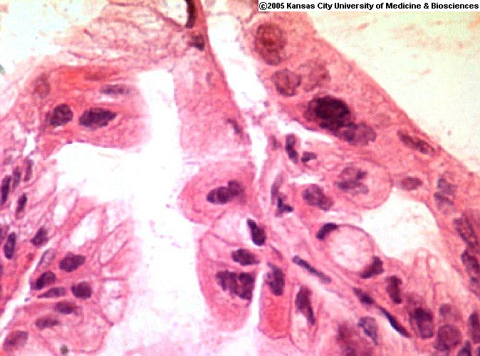 Anaplasia / dysplasia developing
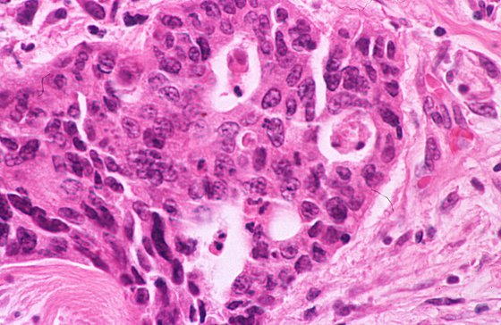 "Glands within glands"
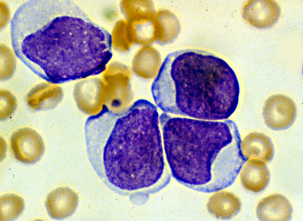 Acute Lymphoblastic Leukemia
 Nasopharyngeal teratoma