Ed Friedlander, M.D., Pathologist
scalpel_blade@yahoo.com


Cyberfriends: The help you're looking for is probably here.
Welcome to Ed's Pathology Notes, placed here originally for the convenience of medical students at my school. You need to check the accuracy of any information, from any source, against other credible sources. I cannot diagnose or treat over the web, I cannot comment on the health care you have already received, and these notes cannot substitute for your own doctor's care. I am good at helping people find resources and answers. If you need me, send me an E-mail at scalpel_blade@yahoo.com Your confidentiality is completely respected.
 DoctorGeorge.com is a larger, full-time service.
There is also a fee site at myphysicians.com,
and another at www.afraidtoask.com.
DoctorGeorge.com is a larger, full-time service.
There is also a fee site at myphysicians.com,
and another at www.afraidtoask.com.
Translate this page automatically
 |
With one of four large boxes of "Pathguy" replies. |
 I'm still doing my best to answer
everybody.
Sometimes I get backlogged,
sometimes my E-mail crashes, and sometimes my
literature search software crashes. If you've not heard
from me in a week, post me again. I send my most
challenging questions to the medical student pathology
interest group, minus the name, but with your E-mail
where you can receive a reply.
I'm still doing my best to answer
everybody.
Sometimes I get backlogged,
sometimes my E-mail crashes, and sometimes my
literature search software crashes. If you've not heard
from me in a week, post me again. I send my most
challenging questions to the medical student pathology
interest group, minus the name, but with your E-mail
where you can receive a reply.
Numbers in {curly braces} are from the magnificent Slice of Life videodisk. No medical student should be without access to this wonderful resource. Someday you may be able to access these pictures directly from this page.
Also:
Medmark Pathology -- massive listing of pathology sites
Freely have you received, freely give. -- Matthew 10:8. My
site receives an enormous amount of traffic, and I'm
handling about 200 requests for information weekly, all
as a public service.
Pathology's modern founder,
Rudolf
Virchow M.D., left a legacy
of realism and social conscience for the discipline. I am
a mainstream Christian, a man of science, and a proponent of
common sense and common kindness. I am an outspoken enemy
of all the make-believe and bunk that interfere with
peoples' health, reasonable freedom, and happiness. I
talk and write straight, and without apology.
Throughout these notes, I am speaking only
for myself, and not for any employer, organization,
or associate.
Special thanks to my friend and colleague,
Charles Wheeler M.D.,
pathologist and former Kansas City mayor. Thanks also
to the real Patch
Adams M.D., who wrote me encouragement when we were both
beginning our unusual medical careers.
If you're a private individual who's
enjoyed this site, and want to say, "Thank you, Ed!", then
what I'd like best is a contribution to the Episcopalian home for
abandoned, neglected, and abused kids in Nevada:
My home page
Especially if you're looking for
information on a disease with a name
that you know, here are a couple of
great places for you to go right now
and use Medline, which will
allow you to find every relevant
current scientific publication.
You owe it to yourself to learn to
use this invaluable internet resource.
Not only will you find some information
immediately, but you'll have references
to journal articles that you can obtain
by interlibrary loan, plus the names of
the world's foremost experts and their
institutions.
Alternative (complementary) medicine has made real progress since my
generally-unfavorable 1983 review linked below. If you are
interested in complementary medicine, then I would urge you
to visit my new
Alternative Medicine page.
If you are looking for something on complementary
medicine, please go first to
the American
Association of Naturopathic Physicians.
And for your enjoyment... here are some of my old pathology
exams
for medical school undergraduates.
I cannot examine every claim that my correspondents
share with me. Sometimes the independent thinkers
prove to be correct, and paradigms shift as a result.
You also know that extraordinary claims require
extraordinary evidence. When a discovery proves to
square with the observable world, scientists make
reputations by confirming it, and corporations
are soon making profits from it. When a
decades-old claim by a "persecuted genius"
finds no acceptance from mainstream science,
it probably failed some basic experimental tests designed
to eliminate self-deception. If you ask me about
something like this, I will simply invite you to
do some tests yourself, perhaps as a high-school
science project. Who knows? Perhaps
it'll be you who makes the next great discovery!
Our world is full of people who have found peace, fulfillment, and friendship
by suspending their own reasoning and
simply accepting a single authority that seems wise and good.
I've learned that they leave the movements when, and only when, they
discover they have been maliciously deceived.
In the meantime, nothing that I can say or do will
convince such people that I am a decent human being. I no longer
answer my crank mail.
This site is my hobby, and I presently have no sponsor.
This page was last updated February 6, 2006.
During the ten years my site has been online, it's proved to be
one of the most popular of all internet sites for undergraduate
physician and allied-health education. It is so well-known
that I'm not worried about borrowers.
I never refuse requests from colleagues for permission to
adapt or duplicate it for their own courses... and many do.
So, fellow-teachers,
help yourselves. Don't sell it for a profit, don't use it for a bad purpose,
and at some time in your course, mention me as author and KCUMB as my institution. Drop me a note about
your successes. And special
thanks to everyone who's helped and encouraged me, and especially the
people at KCUMB
for making it possible, and my teaching assistants over the years.
Whatever you're looking for on the web, I hope you find it,
here or elsewhere. Health and friendship!
If I were asked to what the singular prosperity and growing strength of [the Americans] ought
mainly to be attributed, I should reply, "To the superiority of their women".
-- Alexis de Tocqueville 1789
Never try to impress a woman because if you do, you'll have to keep up that standard for the rest of
your life.
-- W.C. Fields
Not from Adam's brain, to have the same mind as him, nor from Adam's foot, to be subordinate to
him, but from the rib next to Adam's heart, to love and be loved by him.
-- Anonymous
Global views on women's health: Sci. Am. 271(2): Aug., 1994. Good reading, especially for
anyone offering easy, wrong answers to the world's problems.
In sub-Saharan Africa, one woman in 16 dies in childbirth, compared with
one woman in about 5000 in the developed world (Br. Med. J. 326:
567, 2003).
Special thanks to Dr. Tony Racela for the wonderful kodachromes.
You may find them on our intranet at p:\teach\pathol\fem\f101.htm.
QUIZBANK
LEARNING OBJECTIVES:
Cervix:
Uterus:
Give a full account of the etiology, pathogenesis, and clinical correlates of each of the following,
and recognize grossly and/or microscopically as appropriate:
Oviduct:
Ovary:
List each of the three categories of primary ovarian tumors, and for each of these,
the principal tumors and their distinguishing anatomic and clinical features,
risk factors, paraneoplastic syndromes, biological behavior,
and patterns of spread if applicable.
Pregnancy:
Describe how examining the placenta can sometimes help determine
whether twins are identical or fraternal.
 I am presently adding clickable links to
images in these notes. Let me know about good online
sources in addition to these:
I am presently adding clickable links to
images in these notes. Let me know about good online
sources in addition to these:
Pathology Education Instructional Resource -- U. of Alabama; includes a digital library
Houston Pathology -- loads of great pictures for student doctors
Pathopic -- Swiss site; great resource for the truly hard-core
Syracuse -- pathology cases
Walter Reed -- surgical cases
Alabama's Interactive Pathology Lab
"Companion to Big Robbins" -- very little here yet
Alberta
Pathology Images --hard-core!
Cornell
Image Collection -- great site
Bristol Biomedical
Image Archive
EMBBS Clinical
Photo Library
Chilean Image Bank -- General Pathology -- en Español
Chilean Image Bank -- Systemic Pathology -- en Español
Connecticut
Virtual Pathology Museum
Australian
Interactive Pathology Museum
Semmelweis U.,
Budapest -- enormous pathology photo collection
Iowa Skin
Pathology
Loyola
Dermatology
History of Medicine -- National Library of Medicine
KU
Pathology Home
Page -- friends of mine
The Medical Algorithms Project -- not so much pathology, but worth a visit
National Museum of Health & Medicine -- Armed Forces Institute of Pathology
Telmeds -- brilliant site by the medical students of Panama (Spanish language)
U of
Iowa Dermatology Images
U Wash
Cytogenetics Image Gallery
Urbana
Atlas of Pathology -- great site
Visible
Human Project at NLM
WebPath:
Internet Pathology
Laboratory -- great site My team:
Ed Lulo's Pathology Gallery
Bryan Lee's Pathology Museum
Dino Laporte: Pathology Museum
Tom Demark: Pathology Museum
Dan Hammoudi's Site
Claude Roofian's Site
Pathology Handout -- Korean student-generated site; I am pleased to permit their use of my cartoons
Estimating the Time of Death -- computer program right on a webpage
Pathology Field Guide -- recognizing anatomic lesions, no pictures

St.
Jude's Ranch for Children
I've spent time there and they are good. Write "Thanks
Ed" on your check.
PO Box 60100
Boulder City, NV 89006--0100
More of my notes
My medical students
Clinical
Queries -- PubMed from the National Institutes of Health.
Take your questions here first.
HealthWorld
Yahoo! Medline lists other sites that may work well for you

We comply with the
HONcode standard for health trust worthy
information:
verify
here. 

![]()
Let men tremble to win the hand of a woman, unless
they win also with it the utmost passion of her heart.
-- Nathaniel Hawthorne, The Scarlet Letter
Vulva and vagina: Women's problems 13-17, 26-35, 61-62, 67-68, 72
Cervix: Women's problems 80-93
Uterus: Women's problems 1-12, 18, 37-59, 64-66, 69-71, 73-75, 77-79, 94-101
Oviduct: Women's problems 24-25,
Ovary: Women's problems 76, 102-128
Pregnancy: Fetus and pregnancy (all)
 GYN Pathology
Photos by Tony Racela MD (Thanks!)
Notes by Ed
Female I
Introductory Pathology Course
University of Texas, Houston
Female II
Introductory Pathology Course
University of Texas, Houston
Tulane Pathology Course
Great for this unit
Exact links are always changing
Vulva and vagina:
Give a full account of the etiology, pathogenesis, and clinical correlates of each of the following,
and recognize grossly and/or microscopically as appropriate:
Herpes simples
Molluscum
HPV infection
Chlamydia
Garnerella
Candida
Trichomonas
Syphilis
Bartholin abscess
Lichen sclerosus
Condyloma latum
Condyloma acuminatum
Squamous cell carcinoma
Adenocarcinoma of the vagina
Melanoma
Extramammary Paget's
Vaginal adenosis
Gartner duct cysts
Embryonal rhabdomyosarcoma / sarcoma botryoides
Adenocarcinoma of the vagina
Give a full account of the etiology, pathogenesis, and clinical correlates of each of the following,
and recognize grossly, microscopically, and/or cytologically (i.e., on pap smear)
as appropriate:
Herpes simplex
Gonorrhea
Nabothian cyst
Endocervical polyp
Microglandular hyperplasia
Premalignant lesions of the cervix
Squamous carcinoma of the cervix
Adenocarcinoma of the cervix
Remember enough histology to recognize the approximate date of an endometrium,
and especially to spot anovulatory cycles, inadequate luteal phase, and persistent
luteal phase.
Chronic endometritis
Gas gangrene
Endometriosis
Adenomyosis
Endometrial polyps
The various endometrial hyperplasias
Adenocarcinoma of the endometrium and its variants
Mixed mullerian tumors
Leiomyoma and leiomyosarcoma
Give a full account of the etiology, pathogenesis, and clinical correlates of each of the following
and recognize grossly and/or microscopically as appropriate:
Acute and chronic salpingitis
Chlamydia
Gonorrhea
Tuberculosis
Cysts
Actinomycosis
Ectopic pregnancy (also below)
Gestational trophoblastic disease (also below)
Give a full account of the etiology, pathogenesis, and clinical correlates of each of the following
and recognize grossly and/or microscopically as appropriate::
The common simple cysts
Stein-Leventhal syndrome / polycystic ovaries / hyperthecosis
Torsion
Autoimmune disease
Give a full account of the etiology, pathogenesis, and clinical correlates of each of the following
and recognize grossly and/or microscopically as appropriate:
Miscarriage
Ectopic pregnancy
Abruption
Placenta accreta
Placenta previa
Cord problems
Infections
Toxemia of pregnancy
Gestational trophoblastic disease (also above)
{47710} human female
{15787} normal internal female genitalia
We will cover breast in a separate unit.
Primordial germ cells from the yolk sac migrate to the ovarian (or testicular) stroma. (Some of these supposedly go astray and end up in other midline structures, explaining why "germ cell tumors" arise in the retroperitoneum, pineal, and anterior mediastinum.)
In female embryos, the mullerian (paramesonephric) ducts result from infolding of the coelomic lining epithelium. They give rise to the surface epithelium of the ovaries, and the lining of the oviducts and uterus. In male embryos, Mullerian inhibitory substance from the testis makes the mullerian ducts regress.
In female embryos, the wolffian (mesonephric) ducts regress, persisting only as little bits of epithelium along the whole female tract. These give rise to "Gardner's duct cysts" along the cervix and vagina. In male embryos, the wolffian (mesonephric) ducts become the epididymis and vas.
Only the upper two thirds of the endometrium ("the functionalis") cycles. The basal third ("the basalis") does not respond to a woman's steroid hormones and stays in place, giving rise to next month's endometrium. The theca cells surrounding the follicle are ovarian stroma that nurture a particular follicle. You can tell when they have luteinized (i.e., become hormonally active) because they plump-up and become pale-staining (from the lipid used to make steroids). There are a few Leydig-like cells in the hilus, able to make testosterone.
During reproductive life, every month several (not one, as in Big Robbins) follicles mature as graafian follicles. One ovulates and suppresses the others, and becomes a corpus luteum, which will regress when the pregnancy ends or does not occur. It will be recognizable for a few months, gradually being replaced by scar tissue.
The oviduct's mucosa has ciliated cells, secretory cells, and almost-no-cytoplasm "peg cells" ("intercalated cells"). The oviduct's mucosa is thrown up into complicated folds ("plica", or "fimbria" at the end). I've been told that the reason women have orgasms is to make the oviducts wiggle around and be sure to catch the egg if it's just been released. Decide about that for yourself.
Herpes Simplex II
You are already familiar with the classic "herpes cell" seen on Tzanck preparation or pap smears. Cells with multiple gigantic nuclei, usually bearing a central inclusion, are diagnostic.
If herpes is transmitted to the child during birth, severe sickness, brain damage, and even death are likely to occur.
Human Papilloma Virus (HPV)
{11562} chlamydia, pap smear
{25911} chlamydia infection, pap smear
Hemophilus ducreyi
Mycobacterium tuberculosis
Gardnerella
You can make the diagnosis by observing that the vaginal pH
is more alkaline than the usual 4.5.
Or you can confirm the diagnosis by
adding a drop of dilute potassium hydroxide. This will accentuate
the fish smell.
And the bacteria cling to epithelial cells, creating the fuzzy-looking
"clue cell" in pap smear.
Gardnerella is today's "usual suspect" for producing many cases of
premature rupture of the membranes, premature labor, and premature birth
(Hosp. Med. 61: 475, 2000).
Screening for gardnerella during pregnancy cuts prematurity
dramatically: Br. Med. J. 329: 371, 2004.
Candida:
This produces a red, itchy rash that may have fungal colonies visible.
You can scrape them off and see them under the microscope. Biopsy
is unnecessary. The inflammation is superficial and the infection is annoying but not (by itself) dangerous.
Contrary to "Big Robbins", I am not aware of any reason to
believe that some women are more vulnerable because they are "chronic carriers".
The bug is ubiquitous.
Yeast infections are more likely when there is more glucose in the area
(pregnancy, on the oral contraceptive pill, diabetes), or when the normal
bacteria are suppressed with antibiotics.
Trichomonas:
Vulva, vagina, cervix: Trichomonas vulvovaginitis
This produces a bad-smelling, red ("strawberry") inflammation with
a thin discharge. If you are inexperienced and happen
to biopsy it, you'll see
only superficial inflammation.
The protozoan is easily seen in wet mounts, looking like a bouncing pear
moving about with wiggly flagella. Ask a pathologist to show you a pap smear slide
with "Trich"; there is often a second micro-organism, a very long filamentous
bacterium called "leptothrix".
Trichomonas is much less common nowadays thanks to its incidental
discovery on pap smears. As long as both partners are treated, expect
a good result (Lancet 363: 545, 2004).
 TB salpingitis
Photo and mini-review
Brown U.
Calymmatobacterium
Vulva: Donovoniasis, or "granuloma inguinale".
Have a microbiologist show you donovan bodies.
Vagina: Nonspecific vaginitis / "bacterial vaginosis"
The bacterium is normal flora; it grows best in the presence of semen
but no woman is immune.
{08370} Gardnerella vaginalis on Pap smear (these aren't outstanding examples of "clue cells")
{25908} Gardnerella vaginalis infection, good "clue cell"
Clue cell
Tom Demark's Site
Vulva, vagina, cervix: Yeast infection
NON-NEOPLASTIC DISORDERS
Vulvar vestibulitis (various names)
is a pain syndrome in which the vulva becomes tender and intercourse
is painful.
"Acute vulvitis" is nonspecific inflammation of the vulva, by various
surface bacteria.
Distinguishing vulvar cysts:
Skene's glands, on either side of the urethra, can also become inflamed,
especially by gonorrhea.
* Vestibular adenitis is a poorly-understood inflammatory process
at the entry to the vagina. The glands are inflamed
and very painful. They may be excised surgically for a cure.
Skin diseases including psoriasis, lichen planus, vitiligo,
and familial pemphigoid, are very familiar on the vulva. Sometimes
the epidermis simply undergoes hyperplasia, usually without anaplasia. It
thickens ("acanthosis"), and develops extra keratin ("hyperkeratosis").
We call
this vulvar hyperplasia. (This is a cancer risk if an only if there
is some anaplasia.)
Lichen sclerosus ("chronic atrophic vulvitis") is a mysterious
process in which a band of dense, homogeneous, hyaline collagen forms underneath
the epidermis, which is thinned and has a hydropic
basal layer. The skin turns gray and parchment-like
and becomes itchy. It can occur at any age.
Although there is no anaplasia, a few percent turn malignant.
* Of course, lichen sclerosus in a child has gotten parents hauled into
court, where misguided zealots say that it
is "proof of child abuse": Br. Med. J.
320: 331, 2000.
Lichen sclerosus review: Lancet 353: 1777, 1999. Topical
glucocorticoids seem to work better than topical sex steroids.
Today, lasers are being used with good results (Derm. Surg. 30: 1148, 2004).
BENIGN TUMORS
Mucosal polyps are skin tags, fibrous nodules covered
with normal epithelium.
Condyloma acuminatum is the large, usually multiple warts that can
occur on the vulva, perianal region, and (less often) the vagina and cervix.
Remember HPV strains 6 and 11 as causes of condyloma acuminatum (and its
giant variant, "verrucous carcinoma"). Remember
HPV 16 and 18 (also 31, 33, and 35)
as
causes of ordinary, deadly carcinoma; if strains 16 or 18 produces a wart,
it is likely to be flat rather than tree-like. A woman may have several strains.
CARCINOMA OF THE VULVA
Most squamous carcinoma are caused by HPV, and are preceded by
dysplasia and carcinoma in situ ("vulvar intraepithelial neoplasia"),
which is analogous to the lesions in the cervix. A physician may notice
the premalignant lesions and excise them before cancer develops.
Squamous cell carcinomas not caused by HPV usually arise in lichen sclerosus
or idiopathic hyperkeratosis. These are more aggressive.
* Pathologists distinguish a host of subtypes of vulvar squamous carcinomas,
including keratinizing (most common), basaloid, spindle cell, warty,
and verrucous. Don't worry about these for now.
Extramammary Paget's disease is mucin-rich cancer cells
growing within
the epidermis of the vulva or perineum. Local excision should be curative. The pathologist will do
frozen sections to help see if the margins are free.
Even without excision,
the lesion is likely to remain stable for a long time.
Vulva
Nice case photos
Charam M. Ramnani MD
Long considered "mental", it is now pretty clear that this is a fairly
common organic disease. The pathology features (1) too many mast cells, and (2) too
many nerves (Gyn. Ob. Inv. 58: 171, 2004; Ob.Gyn. 91: 572, 1998).
There may also be red speckles visibly grossly, and/or squamous metaplasia of
the vulvar glands and/or T_cells and plasma cells.
Ectopic breast tissue is fairly common on the vulva.
It can enlarge during pregnancy and lactation.
{49364} acute vulvitis
Bartholin glands on either side of the vaginal
introitus are prone to acute infection by ordinary
bacteria, chlamydia, or gonorrhea. They can resolve, leaving the duct obstructed, and a cyst
can form. You'll learn how to marsupialize (i.e., make a pouch) these
on rotations.
{27119} Bartholin gland cyst, vulva
It's now clear that both vulvar hyperplasia and lichen sclerosus are caused
by genetic mutations in the epidermis,
though these have not yet caused anaplasia
(Gyn. Onc. 77: 1717, 2000).
{27110} lichen sclerosis of vulva, histology
* Papillary hidradenoma is an intraductal papilloma of
the breast, only in the vulva along the embryonic milk like.
Microscopically, the pathologist sees a branching fibrous stalk with
a thickened epithelium exhibiting these features of HPV infection:
{27113} condyloma acuminatum of vulva, histology (HPV)
{06026} HPV effect ("koilocytes") in pap smear from cervix
{11470} HPV effect ("koilocytes") in pap smear from cervix
Verrucous Carcinoma
Photo and mini-review
Brown U.
* Leave the diagnosis of such entities as "aggressive angiomyxoma",
"angiomyofibroblastoma", and "angiofibroma" to us.
Most vulvar cancers are squamous cell carcinomas, with adenocarcinomas,
melanomas, and basal cell carcinoma being less common. (The latter are
unlikely to be caused by sunlight; they "just happen").
{25666} melanoma of vulva, gross
{24592} squamous cell carcinoma of vulva, gross
{25664} squamous cell carcinoma of vulva, gross
{25665} squamous cell carcinoma of vulva, gross
{27005} squamous cell carcinoma of vulva, histology
{27008} squamous cell carcinoma of vulva, histology
{25662} carcinoma in situ of vulva, gross
{25663} carcinoma in situ of vulva, gross
It presents as a red,
itchy rash. We don't know exactly where the cancer cells come from.
Unlike in breast, there is seldom an underlying solid cancer.
The pathologist will see tumor cells in the epidermis, as in
the breast. They present clear cytoplasm that will stain for some sort of
mucin.
{11499} Paget's disease of the vulva, gross
{08903} Paget's disease of the vulva, histology
{08906} Paget's disease of the vulva, histology
NON-NEOPLASTIC LESIONS
The only common non-iatrogenic birth defect is a septate vagina, from
failure of the mullerian ducts to fuse. There will also be a double
uterus. The only common
non-infectious, non-neoplastic, acquired lesion of the vagina
is a Gartner duct cyst, from the Wolffian duct remnants.
Melanomas are thankfully uncommon, but do occur sporadically.
Adenocarcinoma of the vagina arises from the glands of girls
exposed to DES, usually in their teens. Fortunately, only one in about 1000 of girls
exposed in this way get cancer, but the impact is devastating. The cells
are glycogen-rich, hence the name "clear cell adenocarcinoma".
Embryonal rhabdomyosarcoma, in its form of "sarcoma botryoides",
is a common cancer of young children.
* There are many other rare tumors. Don't worry about these just now.
Remember the lymphatic drainage. Cancer in the lower two-thirds
of the vagina metastasizes to the inguinal lymph nodes. Cancer of the
upper third metastasizes to the iliac nodes.
 Normal vagina with cervix
WebPath
 Vagina
"Pathology Outlines"
Nat Pernick MD
Girls exposed in utero to diethylstilbestrol (DES) often have
glands in the upper vagina. These appear as red bumps against the
normally-pink mucosa. They may look like endocervical glands with
squamous metaplasia, or like endometrial glands / oviduct without stroma.
These turn cancerous in fewer only about 1 of 700 of affected girls, but when this happens
it is devastating.
{27050} vaginal adenosis (DES exposure in utero), histology
CANCER OF THE VAGINASquamous cell carcinoma is rare, and caused by HPV.
This arises in the setting of intraepithelial neoplasia that may have been
visible.
Melanoma of the vagina
Pittsburgh Pathology Cases
Clear cell carcinoma
Vagina -- DES exposure
WebPath Case of the Week
The sarcoma contains strap- or tadpole-like cross-striated
rhabdomyoblasts, especially
dense in the "cambium layer" beneath the epithelium. They are locally
destructive and can metastasize late. Surgery and chemotherapy usually
bring about a cure.
{08914} normal histology of uterine cervix (endocervix is left, ectocervix is right)
{10271} normal ectocervix histology
{10274} normal endocervix histology
{36059} normal endocervical cells, pap smear
INFLAMMATION
Obviously herpes, gonorrhea, and chlamydia will produce inflammation.
Especially if you see a lot of
lymphocytes with germinal centers, think of chlamydia.
NON-TUMORS Endocervical fibroepithelial polyps are fibrous nubbins covered with epithelium,
hanging out of the cervical os. They act as a wick, drawing bacteria
into the endocervix and endometrial cavity. They are easily cured with curettage.
Microglandular hyperplasia results from progesterone stimulation
of the endocervix (i.e., pregnancy, old-fashioned contraceptive pills).
The glands are abundant and have only a lacy
stroma between them, along with many neutrophils.
{09755} normal cervical pap smear (do you know the cell types?)
The vast majority of cancers of the cervix are squamous cell carcinomas
caused by HPV. In the US, the pap smear technique has greatly reduced
a woman's risk of dying of the disease; as recently as the 1950's,
it was as common a killer as breast cancer is today. Cancer of the cervix remains a major
worldwide killer of women in their reproductive life, with about 190,000 deaths
yearly (Am. J. Ob. Gyn. 189(s4): S37, 2003).
Long before HPV was understood, we knew cancer of the cervix to be a sexually
transmitted disease, with the great risk factors being the number of
male sexual partners, and the number of previous female partners that the
husband had. In the past, putative risk factors have included
smoking (still discussed: JAMA 285: 2995, 2001)
and having an uncircumcised husband (apparently no longer
being investigated); as independent
risk factors, these both seem dubious.
Cancer of the cervix in the U.S. remains more common among the poor,
and more likely to be missed until it is too late (Cancer 101: 1051, 2004).
A virulence factor has now been found in the E6 and E7 oncogenes,
which differ for low-risk and high-risk HPV strains. (See Am. J. Path. 153:
1741, 1998; Cancer 83: 2346, 1998; lots more. These bind p53
and Rb gene products. Finding a virulence
factor is proof of causation -- it has replaced Koch's / Henle's postulates.)
Cervical intraepithelial neoplasia can exist for years or decades
before invasive squamous cancer happens. (And of course, usually it never happens.
But nobody wants to leave these lesions alone.) Or it can progress
very rapidly.
CIN II: Plenty of atypical cells in the lower portions, normal maturation toward
the surface. (The old "moderate dysplasia" and "severe dysplasia").
CIN III: The cells no longer mature as they reach the surface. (The old
"carcinoma in situ").
Invasive cancer arising in from CIN III is usually squamous.
* There are some less common cancers, also:
It is not always clear whether microinvasion has taken place, and today's
hard-core pathologists use double immunostaining for keratin (the cancer cells)
and collagen IV and/or laminin (for basement membrane). See Arch. Path.
Lab. Med. 129: 747, 2005.
Juan Peron's previous wife had also died of cancer of the cervix.
Pap smears were in use in the developed world in the late 1940's, but
had not caught on in Argentina.
In January 1950, Ms. Peron fainted in public and was found to be anemic,
evidently as the result of iron deficiency from blood loss due to her cancer.
It's not clear whether her cancer was found at the time,
but she continued to have heavy vaginal bleeding.
She was taken to surgery and operated by an American "ghost surgeon";
she was never informed of what had been done, who operated her,
or the nature of her illnesss.
How much of this was the "fifties" mentality (concealing unpleasant truths)?
How much was the "VIP syndrome", in which prominent people get their
health problems concealed from the public? You'll have to decide this for yourself.
Ms. Peron was enormously popular with her people, especially for her advocacy
for the poor. She was one of the most beautiful and charismatic women of her era -- perhaps any era.
My reading tells me that most of today's historians consider her a
thoroughly genuine humanitarian. You can read about her final illness in
Lancet 355: 1988, 2000.
Medical school undergraduates do not really need to learn to read pap
smears, but it's enriching. The old-fashioned pap smear (you smear the specimen
on a slide) includes more cells than the newfangled "thin prep" (you put the specimen in
fixative; easier to read Cancer 99: 342, 2003),
and is maybe better for this reason (Br. Med. J. 326:
733, 2003). Computers ("Auto-Pap"/"Focalpoint") now screen pap smears with accuracy about equal
to a human cytotechnologist (Cancer 99: 129, 2003).
And even experienced pathologists do not always make the
right call on either type of test: Arch. Path. Lab. Med. 127: 1413, 2003;
Arch. Path. Lab. Med. 128: 17, 2004).
If a pap smear that you obtained on one of your patients
does not include
any endocervical cells (columnar or squamous-metaplastic, we can tell),
we'll let you know that you probably did not sample the "transformation zone",
where ectocervix joins endocervix and most intraepithelial neoplasia and
invasive
cancers begin.
Your lecturer predicts that in the very near future, routine pap smear
will be replaced (or at least supplemented) by routine DNA probing for the high-risk HPV strains,
with pap smear/biopsy limited to those who are positive.
See Arch. Path. Lab. Med. 127: 940 & 969 & 984 & 991 & 995, 2003;
Arch. Path. Lab. Med. 128: 298, 2004; Postgrad. Med. 118: 37, 2005.
Women with high-grade dysplasia or invasive cancer apparently all
test positive for HPV: Am. J. Ob. Gyn. 189: 118, 2003.
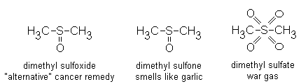
The popular explanation (put forward in For. Sci. Int. 87:
219, 1997) is that she had taken the quack cancer remedy dimethylsulfoxide
(DMSO), and that
it gave rise to the poison gas dimethyl sulfate.
Is this credible?
Yes!
No!
* In the monster movie Godzilla 2000, a photomicrograph of the monster's
skin is examined by a group of scientists. Fascinatingly,
it appears identical to normal
human ectocervix.
Cervix
"Pathology Outlines"
Nat Pernick MD
All adult women have some inflammatory cells in the endocervical canal.
This is to be expected, given the abundant bacteria that thrive on the
glycogen in this area.
Chronic cervicitis
WebPath
Nabothian cysts are endocervical glands that have become plugged
"by the inflammation", and fill with mucus. Most women have a few of these.
{39991} endocervical polyp, gross
* Laminar hyperplasia of the glands is quite common and poorly-understood.
The glands are slender but branch.
* Future pathologists: Don't mistake
the scrambled pattern and enlarged nuclei for adenocarcinoma.
{27137} cervix, micro-glandular hyperplasia, histology
CANCER OF THE CERVIX (Lancet 361: 2217, 2003)
You are already familiar with the concept of dysplasia and intraepithelial
neoplasia. Here are some guidelines for applying the "CIN" system,
from mildest to most severe. A pathologist can tell on pap smear, and confirm
on biopsy; the latter is more precise as long as you get the right spot (which is
made easier by the fact that CIN doesn't stain as well as normal cervix with
iodine ("Schiller test"), and will turn white on application of acetic acid, the acetowhite test).
CIN I: Koilocytes only: Perhaps a condyloma acuminatum or a flat wart. Or perhaps
there is simply squamous metaplasia of the endocervix. Maybe some atypical
cells in the lower third. (the old "mild dysplasia").
{11789} dysplasia of uterine cervix, histology
{11789} cervix, dysplasia, histology
{41963} cervix, dysplasia, histology
{25939} cervix, dysplasia, pap smear
{27101} cervix, dysplasia, pap smear
{27104} cervix, dysplasia, pap smear
{11790} severe dysplasia of uterine cervix, histology
 Glassy cell carcinoma
of the cervix
Pittsburgh Pathology Cases
{08911} uterine cervix, carcinoma in situ, histology
{08912} uterine cervix, carcinoma in situ, histology
{46209} cervical conization specimen. One may cure
CIS by removing the entire ring of abnormal cells.
Pathologists distinguish subtypes of squamous cancer based on their histology
and resemblance to the cells of the normal cervix.
{25962} cervix, carcinoma in situ, pap smear
{34775} carcinoma in situ of cervix, pap smear
{10292} carcinoma of the cervix, gross
{10583} carcinoma of the cervix, gross; bladder is above, rectum below
{10913} carcinoma of the cervix; bladder is right, rectum is left
{46321} carcinoma of cervix, gross
{46322} carcinoma of cervix, gross
Squamous Cell Carcinoma, Cervix
Photo and mini-review
Brown U.
"Microinvasive carcinoma" implies invasion no deeper than 5 mm (Europe)
or 3 mm (US) with no evidence of vascular invasion.
Microinvasive carcinoma will be treated with cervical conization,
preserving fertility; more deeply invasive carcinoma needs hysterectomy.
* Sending the entire conization specimen for frozen section
seems like the best option to assure that as little tissue as possible is taken:
Am. J. Clin. Path. 122: 383, 2004.
* The Death of Eva Peron
Eva Peron ("Evita"), wife of Argentina's left-wing dictator Juan Peron,
died in January 1952 of cervical cancer.

Adenocarcinoma of the cervix is only about 10% as common as squamous
cell carcinoma, but it is less likely to be detected during its in-situ
phase (if it has any in-situ phase; leave the diagnosis of "adenocarcinoma in
situ" to us; it's difficult Arch. Path. Lab. Med. 128: 153, 2004;
Cancer 99: 323, 2003.)
Of course, the two occur together fairly often (Cancer 102: 218, 2004).
Complying with recommendations for routine pap smears
greatly decreases, but does not eliminate, a woman's risk for this
cancer (Cancer 99: 336, 2003).
* "The Toxic Lady!"
Gloria Ramirez died in a California emergency
room as a result of cancer of the cervix causing ureteral obstruction
and kidney failure.
Emergency room personnel who were present for the resuscitation
became acutely sick (lacrimation, fainting).
|
|
INTRODUCTION
Following deliveries, the uterus may prolapse.
Words to know:
Leave the dating of endometrial samples to pathologists. You will usually get one of these diagnoses:
The endometrium is very resistant to bacterial infection.
Infection by common bacterial (strep A, staph) is usually the result of retained products of conception. Surgical removal of the remnants is the mainstay of therapy.
Pyometra is thankfully rare. It is a purulent infection of the uterus, as when products of conception are retained or the os is closed.
Other infections after childbirth or natural or induced abortion include strep, staph, and E. coli. In the Bad Old Days before common-sense hygiene, physicians carried these infections from woman to woman on the delivery unit.
Acute endometritis (i.e., neutrophils) often has no obvious cause;
various mycoplasma are the "usual suspects" and this is now being
confirmed with PCR: Lancet 359: 765, 2002
Chronic endometritis is, by definition, the presence of plasma cells
in the endometrium. Usually this is the result of gonococci or chlamydia
having their home base in the oviducts, or else simply the effect
of compression by a nearby leiomyoma (nobody knows how).
Less often, retained products
of conception are the cause. Obviously an intrauterine contraceptive
device will produce chronic inflammation.
On the Coontagiousness
of Puerperal Fever
Oliver Wendell Holmes MD
Also remember TB, especially in the poor nations.
Thankfully, nobody still uses the magnesium-rich super-absorbent tampons that proved such a good culture medium for the staphylococci that produce toxic shock syndrome.
It's easy to tell this isn't cancer, since the glands are benign and there is stroma with them.
Obviously this can cause discomfort just before and during menstruation. It's supposed to be one of the major causes of menstrual cramps.
* An adenomyoma is a nodule where there is a great deal of adenomyosis.
{14330} adenomyosis, histology
Don't worry about the etiology. The various ideas ("regurgitation", i.e., retrograde menstruation; metaplasia of the coelomic epithelium; metastases via lymphatics) all probably operate at different times.
* Your lecturer is unimpressed with claims that the growths are clonal or bear distinctive mutations; all the recent ones have come from studies of already-established cell cultures.
Being on the oral contraceptive pill seems to prevent endometriosis from forming.
At least one women in 10 will have symptoms of endometriosis during reproductive life. Endometriosis cycles like endometrium does.
The gross appearance of endometriosis depends on how extensive the disease is.
Longstanding ovarian lesions present "chocolate cysts", full of old blood.
Large lesions where the blood has organized present extensive fibrosis. This can obliterate the pouch of Douglas, obstruct the bowel, obstruct the oviduct, and so forth.
Infertility often accompanies endometriosis; exactly how this happens is a minor mystery.
ENDOMETRIAL POLYPS
The histology may seem normal, or show some cystic hyperplasia (see below). The tipoff that curettings contain a polyp is the presence of thick-walled blood vessels (i.e., they've had time to develop and not been shed every month.) Removal by curettage usually is curative.
ENDOMETRIAL HYPERPLASIA
Nobody really knows the "risk of turning into adenocarcinoma",
since the diagnosis is made only on biopsy and this itself affects
the illness (curettage may be curative).
Simple hyperplasia ("cystic hyperplasia", "mild hyperplasias")
features:
ENDOMETRIAL ADENOCARCINOMA
The risk factors are well-known.
Also remember
Patients present with bleeding because of the invasion of the inner wall.
Thankfully, these tumors usually announce themselves early. Only about one woman
in six with cancer of the endometrium will die from it.
Grossly, the lesions look like cottage cheese.
Microscopically, the pathologist sees back-to-back glands. Solid
sheets of cells are more ominous. The grading system:
Increase the grade by one if the nuclei are unusually ugly.
If there is benign-looking squamous metaplasia, the pathologist describes
an "adenoacanthoma". If the squamous areas are anaplastic, the pathologist
describes "adenosquamous carcinoma". This is of little significance.
Metastases eventually can occur, usually via the lymphatics.
Serous adenocarcinoma
of the endometrium (Cancer 101: 2214, 2004)
and clear-cell
carcinoma
of the endometrium are more aggressive,
look like the corresponding ovarian lesions, and is less likely to
be linked to high estrogen or to previous hyperplasia.
* As elsewhere, HER-2/neu amplification is a strong predictor of
bad outcome in the papillary serous lesion (Cancer 104: 1391, 2005).
Watch for herceptin
as an agent to treat these patients.
MIXED MULLERIAN / MESENCHYMAL TUMORS
There is often a history of previous radiation. They tend to be
aggressive and to metastasize
as adenocarcinomas.
Endometrial stromal tumors are of three types. Leave the
diagnosis to us; their histology is not for medical school undergrads.
The etiology is mysterious. They grow in response to estrogen, and shrink (and often vanish)
after menopause.
Usually leiomyomas are asymptomatic, or cause problems by mass effect.
A submucosal leiomyoma can produce bleeding between periods, and interfere
with fertility. Large leiomyomas can cause problems with pregnancy.
The tumors are rubbery white spheres.
Grossly, the "whorled silk" pattern seen on cross-section is famous.
Tumors may calcify, show central necrosis (watershed infarct; when this
becomes infected it's a "pyomyoma"), and/or fatty ingrowth.
The new procedure of embolizing these tumors under fluoroscopy, rather than removing
the uterus, seems safe and effective (Am. J. Ob. Gyn. 190: 1697, 2004;
Ob. Gyn. 106: 52, 2005. AJR 184: 399, 2005).
The most serious risk is infection in the necrotic debris (OB Gyn 104: 1161, 2004
And prior to surgery, leiomyomas may be shrunk using a GNRH antagonist
(BJOG 112: 638, 2005).
Intravascular leiomyomatosis means a bunch of leiomyomas
with a proclivity to grow down the veins. Curiously, this
doesn't metastasize, and regresses after menopause.
Leiomyosarcomas of the uterus are fairly common. If you see
a smooth muscle tumor of the uterus with
ten or more mitotic figures per ten high power fields, or if you see
fewer with anaplasia, it's a leiomyosarcoma. Prognosis depends on the
histology.
Endometrial polyp
WebPath
This is an overgrowth of endometrium, but without the ability to
metastasize (yet). We still haven't sorted out how much is
due to a disturbed hormonal milieu, and how much is due to mutations
(selected-for in a disturbed hormonal milieu).
If a lady has this at the time of her last period,
she will have a cystic endometrium throughout postmenopausal life.
This is quite common at autopsy.
{00096} endometrial hyperplasia, gross
{00099} endometrial hyperplasia, gross
{10907} endometrial hyperplasia, gross
{38986} endometrial hyperplasia, gross
{08918} "cystic hyperplasia" of endometrium, histology
{08919} "cystic hyperplasia" of endometrium, histology
Complex hyperplasia ("complex / adenomatous hyperplasia without atypia")
Atypical hyperplasia ("higher grade hyperplasia")
Leave the details up to the pathologists, including the various metaplasias
that may occur in hyperplastic endometrium. All of these lesions are
prone to regress on administration of progesterone.
{27164} "adenomatous hyperplasia" of endometrium
This is a common cancer in women over age 40.
The primary lesion is likely to be tiny, but to disseminate
over the peritoneal surfaces, probably by reflux out the oviducts.
{05319} uterine carcinoma, radiograph
{08437} endometrial adenocarcinoma, gross
{39635} carcinoma of the endometrium, gross
{18782} adenocarcinoma of the endometrium, gross
{18783} adenocarcinoma of the endometrium, gross
{21075} endometrial adenocarcinoma, gross
{10586} carcinoma of the endometrium; dissection with bladder at bottom, uterus and vagina in
middle, rectum at top
{10589} carcinoma of the endometrium, cross-section of uterus
{27161} adenocarcinoma of endometrium; notice glands-within-glands
{08916} adenocarcinoma of endometrium, low magnification
{08917} adenocarcinoma of endometrium, high magnification
{10694} adenocarcinoma of the endometrium, cytology
Mixed Muellerian tumors arise from the endometrium and contain
both malignant glands and malignant mesenchymal elements.
LEIOMYOMAS (Lancet 357: 293, 2001; Ob. Gyn. 104: 393, 2004)
In addition
to bizarre spindle cells, there may be muscle, bone, fat, and/or cartilage;
nevertheless, these will usually stain with epithelial markers.
These are the banal "fibroids" of the myometrium.
At least 25% of women have these during reproductive life.
They are more common in blacks.
Submucosal leiomyomas can produce bleeding. Subserosal
leiomyomas are visible on the surface but don't mean anything.
Microscopically you will have no trouble recognizing smooth muscle.
Even if you see some odd cells, don't be concerned about malignancy
unless you see mitotic figures.
{08438} leiomyoma of uterus, gross
{09774} leiomyoma of uterus, gross
{10910} leiomyoma of uterus, gross
{24703} leiomyoma of uterus, gross
{39636} leiomyoma of uterus, gross
{49380} leiomyoma of uterus, gross
{08728} leiomyoma, histology
{08729} leiomyoma, histology
{49383} lipoleiomyoma
{20184} calcified uterine leiomyomas, radiograph
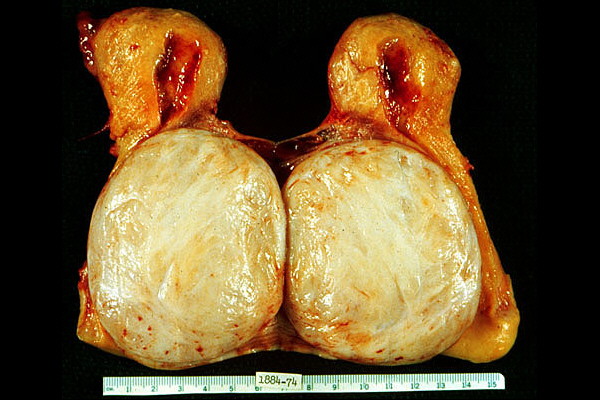 Large uterine leiomyoma
Whorls on cross-section
KU Collection
{09016} leiomyosarcoma of uterus, mitotic figure
{39637} leiomyosarcoma, showing mitotic figure
|
|
PELVIC INFLAMMATORY DISEASE ("salpingitis")
Actinomycosis usually results from the presence of an intrauterine
device, on which the "superglue bug" can build its "sulfur granules"
colonies.
Gonococcal and chlamydial "PID" is a sexually transmitted disease, and unfortunately very common.
It can smolder, with pain being worst during the menstrual periods.
During acute flareups, there is severe pelvic pain, especially when
the cervix is manipulated, with peritoneal signs.
Grossly, the tubes are swollen and inflamed. They may be packed
with pus ("pyosalpinx"). During the acute phase,
the pathologist will see neutrophils and marked edema. In chronic infection,
there is a mix of neutrophils, lymphocytes, and other inflammatory cells.
After everything is over, there is likely to be a lot of scarring,
which will probably interfere with fertility; if the ends of the tubes
are plugged by scar tissue, a "hydrosalpinx" results.
Women still die of PID, either in the acute phase (sepsis)
or from complications
(peritonitis, obstruction).
* Tubal infections that follow abortion or a childbirth infection tend to involve
the mucosal surfaces rather than the lumen.
OTHER LESIONS OF THE OVIDUCT
Cysts arise from embryonic structures. Hyatids of Morgagni / paratubal cysts
arise from mullerian duct remnants and are mere curiosities.
A patient with old pelvic inflammatory disease may be told that her tubes are "cysts".
Adenocarcinoma of the oviduct is very rare and very deadly. Adenomatoid tumors
are hard white spheres that arise from mesothelium.
We will cover ectopic pregnancy below.
The usual infectious agents are the gonococcus and chlamydia.
TB is now
rare in the developed world, but very common in the poor nations.
{39001} intrauterine device (coil type)
{10280} intrauterine device in place
{10904} actinomycosis of the endometrium, histology (note "sulfur granules")
{10901} gonorrheal salpingitis
{27176} acute salpingitis, histology
{27173} chronic salpingitis, histology
Paratubal cyst
WebPath
* Are there perhaps normally more than one ovulation per month? Br. Med. J. 327: 124, 2003.
|
|

{24817} normal ovary in pregnancy
{24695} normal graafian follicle
{24696} normal graafian follicle, higher power
{24698} radiation injury to ovary; note loss of germ cells and radiation change in vessels
* One animal model involves autoimmunization with zona pellucida (ZP3): Mol. Rep. Dev. 48: 140, 1997 -- watch this antigen, as it's being discussed as a reversible contraceptive. Curiously, some mice develop autoimmune oophoritis after thymectomy.
* Treating it in the unborn (!) -- J. Ped. Surg. 32: 1447, 1997.
The mysterious "massive ovarian edema" might be the result of
partial torsion (J. Rep. Med. 41: 359, 1996) or (seems more
likely to me) thrombosis of the ovarian vein.
Ovary with torsion, gross
WebPath
CYSTIC FOLLICLES ("follicular and luteal cysts")
Big ones can cause torsion and/or possess too much luteinized theca cell tissue resulting in hyperestrogenism.
Corpus luteum cysts are simply oversized. They are filled with blood and fatty debris, so when a corpus luteum cyst undergoes torsion or ruptures, it's a bit more of a problem.
STEIN-LEVENTHAL SYNDROME ("polycystic ovarian disease"; "polycystic ovaries")
* Purists: "Polycystic ovaries", i.e., ovaries with numerous cysts, do not always mean Stein-Leventhal.
The failure of ovulation results from no follicle becoming dominant, or reaching the full size for ovulation (J. Clin. End. Metab. 83: 3984, 1998). Since nobody knows how one follicle comes to be selected as dominant, our understanding of Stein-Leventhal is probably a long way off.
Biopsies obtained during laparotomy shows that women with polycystic ovary disease, even when mild and the woman is still ovulating, have much more early-growing follicles than do normal women (Lancet 362: 1017, 2003).
Because androstenedione can turn into estrone, hyperestrogenism can also be a problem, including risk for endometrial adenocarcinoma.
Your lecturer suspects that the principal cause is some hormone awaiting discovery. (* the idea that it was resistin failed, but an important gene is probably nearby: Diabetes 52: 214, 2003) In the meantime, the disease is known to run in families, along with menstrual irregularities, hirsutism (men and women), and insulin resistance (Hum. Repro. 12: 2614, 1997).
* Currently, metformin seems to work well to control Stein-Leventhal (Br. Med. J. 327: 951 & 974, 2003), along with diet and exercise of course. Stay tuned.
The disorder is obviously polygenic; * the best candidate gene so far is VNTR in the insulin gene regulator (Lancet 349: 986, 1997) with other plays being sex-hormone binding globulin (J. Clin. Endo. Metab. 88: 5976, 2003) and a couple of fat-cell-differentiation gene (J. Clin. Endo. Metab. 88: 5529 & 5887, 2003).
* Watch for a link to metabolic syndrome X (Med. J. Aust. 174: 580, 2001).
Stromal hyperthecosis ("cortical hyperthecosis") features hyperplasia and luteinization of the theca cells, making them overproduce androstenedione. The ovaries are big and yellow. Most (but not all) patients are post-menopausal.
 INTRODUCING THE OVARIAN TUMORS
{05318} ovarian carcinoma, radiograph
There are three overriding categories of ovarian tumors.
They are rare before age 30.
Sometimes a benign one will be almost all stroma, and be called an "adenofibroma".
Any of these can also be primary on the peritoneum.
Since the coelomic epithelium is continuous across both ovaries, and since cancer arises in a mutated field, the malignant ones tend to be bilateral when diagnosed; this does not make them incurable.
The malignant ones tend to metastasize over the peritoneum and cause death by obstructing the bowel. These are the common ovarian cancers.
Time on the oral contraceptive pill, or time pregnant, is protective apparently regardless of hereditary risk. Perhaps ovulation gives the coelomic cell the opportunity to divide which allows selection for the mutated clones.
* Recently there was a hoopla about cornstarch and talcum powder applied to the perineum as causing ovarian cancer. This makes no sense biologically, and it sounds like recall bias explains the early reports. Of course it was amply refuted (Am. J. Ob. Gyn. 182: 720, 2000).
They can occur at any age. They are almost always unilateral.
They usually occur in children and young women.
The most common is the benign, banal dermoid tumor ("cystic teratoma") of young women. The other common ones are all cancers.
Metastases to the ovary are common, especially from breast and stomach. The latter especially is a common presentation for stomach cancer, the infamous "Krukenberg tumors" with massive bilateral enlargement of the ovaries, which prove at surgery to be stuffed with signet ring cells.
Most of these are malignant; a majority of the malignant ones arise bilaterally.
This is the single most common ovarian cancer (about 40%).
Benign serous tumors are quite tame-looking and always have a lot of cilia.
Surgery is curative.
Borderline tumors
have piling-up of the cells (to three layers), perhaps with some
anaplasia but with no invasion of the
stroma. Surgery is usually curative; if a borderline has metastasized (usually
over the peritoneal surface), survival is still likely for years or decades.
Fully-developed malignancy is obvious.
Most of these are benign, with well-developed columnar cells and abundant
mucin. They are always multicystic and can be very large. Benign
mucinous tumors are very common. They are usually
unilateral.
Most of the tabloid newspaper tumors that weigh so much are benign mucinous cystadenomas.
Borderline tumors exhibit some stratification of nuclei and/or anaplasia
(much as you'd see in a colon adenoma), but no invasion of the stroma. Again,
surgery is usually curative, and metastatic disease is compatible with long
survival.
Again, real malignancy is obvious. Mucinous carcinomas are bilateral about 20%
of the time.
Mucin in contact with the ovarian stroma can cause it to produce androgens.
These tumors can masculinize.
"Pseudomyxoma peritonei", in which mucin erupts into the peritoneal
cavity and elicits a fibrous response, often results from mucinous
carcinoma of the ovary or appendix.
Endometrioid tumors
Pathologists recognize them by their resemblance to swiss-cheese endometrial
adenocarcinoma.
The risk factors are the same as for endometrial adenocarcinoma; there
is often a history of endometriosis in the ovary as well.
About half are bilateral. Often there is a coexisting endometrial
carcinoma, but this does not imply that these are metastases; the diseases
are still surgically curable.
Be this as it may, this tumor shows lots of
big clear cells in sheets or tubes. These are all malignant.
Brenner tumor
They are almost all benign.
GERM CELL TUMORS (Am. J. Clin. Path. 109(S1): S82, 1998)
Contrary to "Big Robbins", the tissues of a teratoma look like those in the
fetus or young baby, rather than in the adult. The hair is the most likely
stuff to look mature.
About 15% are bilateral. All have the 46XX karyotype.
Rarely a squamous cell carcinoma will arise in one of these, but otherwise
they are thoroughly benign.
Specialized teratoma ("monodermal teratoma", from one germ
layer, actually a contradiction in terms)
Dysgerminoma
Like seminomas, some produce hCG, and they are very sensitive to
radiation and chemotherapy.
Endodermal sinus tumor ("yolk sac tumor")
Like the yolk sac, they are loaded with alpha-1 antitrypsin and
alpha-fetoprotein.
Leave the identification of Schiller-Duval bodies, which
recapitulate the duct of the yolk sac, to the pathologists.
These used to be uniformly lethal, but now most are cured with
chemotherapy.
Miscellany
Choriocarcinoma can be primary in the ovary; since this has none of
Dad's tissue antigens, it's harder to cure than gestational
trophoblastic disease.
* A primary ovarian choriocarcinoma with some of the husband's chromosomes:
Ob. Gyn. 102: 991, 2003.
* A "polyembryoma" contains hundreds of little structures that look like
developing embryos. Puzzle that one out!
This is the new name for tumors that contain abundant
granulosa-type cells (cuboidal steroid-producing cells), often
with some forming Call-Exner bodies (holes for eggs, as in the normal ovary).
There are often theca cells as well, and these may be spindly,
or pink and plump (luteinized).
Many of these produce estrogen. A few produce androgen.
As steroid-producers, expect yellow on the gross.
Those that arise in children ("juvenile granulosa cell tumors") tend to be more anaplastic.
Any granulosa tumor can metastasize.
Thecoma-fibromas Many produce estrogens; a few produce androgens.
They almost never act malignant unless there are a lot
of mitoses. For some reason
they are prone to cause ascites and sometimes even hydrothorax ("Meig's
syndrome). Nobody knows how (* possible serum factors: Am. J. Ob. Gyn. 184:
354, 2001).
Most produce androgens; a few produce estrogens. Androgens from these
tumors (or any other source) will tend to defeminize (i.e., stop the monthly
cycle) and masculinize (=virilize, i.e., enlarge the clitoris,
produce extra body hair, altered hairline, acne, more apocrine sweat, deep voice).
Look for Reinke crystalloids in the Leydig cells.
Miscellany:
"Lipid cell tumors" are benign, full of yellow lipid, and usually virilize.
"Pregnancy luteoma" is a massive corpus luteum.
"Gynandroblastoma" is a mix of male and female sex cord type cells.
"Gonadoblastoma" occurs in intersex people. You'll see both eggs
and a mix of sex-cord and stromal tumors.
Krukenberg tumor
WebPath
SURFACE EPITHELIAL TUMORS
Serous tumors
These recapitulate oviduct, with papillary structures and often cilia.
Often there are psammoma bodies.
{09779} papillary cystadenoma of ovary, gross
Ovarian serous cystadenoma
WebPath
{11515} serous cystadenocarcinoma of ovary, gross
{39560} serous cystadenocarcinoma of ovary, histology
{10382} serous cystadenocarcinoma of ovary, cytology
{10727} serous cystadenocarcinoma of the ovary, psammoma bodies in pap smear
Papillary serious cystadenocarcinoma
Photo and mini-review
Brown U.
Ovarian serous cystadenocarcinoma
Low malignant potential
Ed Uthman
Mucinous tumors
These recapitulate endocervix, with mucin production.
{14165} ovarian mucinous cystadenoma, gross
{14177} ovarian mucinous cystadenoma, gross
{49396} mucinous cystadenoma of ovary, gross; unfortunately there's no ruler, but this might have
weighed 30 lb.
{08938} mucinous cystadenoma of ovary, histology
{08940} mucinous cystadenoma of ovary, histology
{14171} ovarian mucinous cystadenoma, histology
{14180} ovarian mucinous cystadenoma, histology
Mucinous Carcinoma
Photo and mini-review
Brown U.
 Ovarian mucinous cystadenoma
Source unknown
Not for young or sensitive visitors.
For our purposes, all endometrioid tumors of the ovary are malignant.
{21090} endometrioid carcinoma of ovary, gross
{27200} endometrioid carcinoma of the ovary, histology
{25260} endometrioid carcinoma of the ovary
Clear cell adenocarcinoma
Maybe this recapitulates renal tubule, since renal cell carcinoma also
has a lot of clear cells. Or (more likely) it recapitulates the
clear cells seen in endometrial glands during pregnancy.
Clear cell adenocarcinoma of the ovary
Pittsburgh Pathology Cases
These recapitulate the transitional epithelium of the bladder,
as little chunks in a dense fibrous stroma. You need a good eye
to appreciate this.
{27083} Brenner tumor, histology
{39859} Brenner tumor, gross
{40518} Brenner tumor, histology
Brenner tumor
Tom Demark's Site
Mature (benign) teratomas
The most common benign teratoma of the ovary is the dermoid cyst
("cystic teratoma"), with inside-out skin expanding gradually as it
fills with sebum and keratin. There is usually some hair, and
a "Rokitansky" nodule in the wall containing tissues of all three term layers.
{28667} cystic teratoma of ovary, benign, gross ("dermoid cyst")
{28670} cystic teratoma of ovary, benign, gross ("dermoid cyst")
{00111} dermoid cyst of ovary, gross
{11527} dermoid cyst of ovary, gross
{24595} dermoid cyst, gross
{17543} dermoid cyst, gross
{17547} dermoid cyst, brain tissue (white matter)
{17548} dermoid cyst, skin tissue
Ovarian Dermoid
Dino Laporte's PathosWeb
Sometimes a single tissue predominates. The two to remember are the very tame
carcinoids (producing the carcinoid syndrome) and thyroid ("struma ovarii",
rarely with hyperthyroidism.)
Immature teratomas are composed of cells that resemble embryonic
tissue.These are tumors of girls and young women, and are all malignant.
The more neuroepithelium, the worse the prognosis.
{15392} immature ovarian teratoma, histology
{15394} immature ovarian teratoma, histology
{15395} immature ovarian teratoma, histology; the bad section
Struma ovarii
WebPath
This is the counterpart to a man's seminoma of the testis, composed
of large, glycogen-rich, polyhedral "fried egg" cells. They are rare
over age 30.
{24705} dysgerminoma of ovary, gross
{27038} dysgerminoma of ovary, histology
These are aggressive cancers of children or young adults, which
recapitulate the yolk sac.
{27107} endodermal sinus tumor of ovary (Schiller-Duvall bodies)
SEX CORD TUMORS
* Embryonal cell carcinoma can arise in the ovary as in a man's testis.
Granulosa-Theca tumors
{14192} granulosa cell tumor of ovary, gross
{14195} granulosa cell tumor of ovary, gross
{14198} granulosa cell tumor of ovary, histology; notice Call-Exner bodies
These are a mix of variable proportions of inactive fibroblasts
and hormonally-active theca cells. These tumors are solid, hard
balls.
{21084} ovarian fibroma, gross
{24599} ovarian fibroma, gross
{40105} thecoma, gross (good yellow color)
{40127} thecoma, H&E
{40126} thecoma, oil-red O stain for fat
Sertoli-Leydig cell tumors ("androblastomas", "arrhenoblastomas")
These recapitulate the Leydig cells and/or seminiferous tubules.
They are unilateral and solid.
{21086} Sertoli-Leydig cell tumor of ovary (good yellow color)
{20235} Sertoli-Leydig cell tumor of ovary, histology
"Hilus cell tumors" are composed only of Leydig cells. They produce androgen.
|
|
{49415} hydrops fetalis
{49416} fetal death, cord around neck
SPONTANEOUS ABORTION
Today there is some interest in bacterial vaginosis
and chlamydia (Am. J. Ob. Gyn. 183: 431, 2000; others).
After a miscarriage, curettage of the endometrium
yields necrotic tissue, often intensely inflamed with neutrophils.
There will of course always be decidua, and often villi if they
have not been passed.
The usual location is the oviduct, though ovary, peritoneum,
and uterine cornua are other known sites. Rarely an intraperitoneal pregnancy
goes to term.
The best-known cause is old pelvic inflammatory disease, less often
scarring from endometriosis. But about
half of cases in the US happen "for no reason." In countries
where there is much more gonorrhea, there are many more ectopic pregnancies.
No matter where the pregnancy is, the local cells decidualize and
the child develops for a while. In a tubal pregnancy, disaster
strikes at about 6 weeks after the
missed period (about 8 weeks after conception). One of the following
happens:
Treatment is surgical. Or if an ectopic pregnancy is known to be present
but has not ruptured, methotrexate can end it.
PROBLEMS IN LATE PREGNANCY
Many of the problems that develop toward the end are
caused by problems with the umbilical cord. Of course, if the cord
is compressed enough to occlude the veins, the child is in grave
danger. This includes slip knots, cord around neck, or cord being
compressed by the child's head against the cervix.
Infections can pass through a ruptured membrane and produce
inflammation of the amnion (amnionitis) and cord (funisitis).
Common bacteria can infect the placenta, as can syphilis, toxoplasmosis,
and TB. If you see granulomas, think of listeria. Thankfully there's
not much brucellosis in the US any more.
Abruption of the placenta is a huge bleed between the placenta
and the wall. It is among the most dreaded obstetrical complications.
Placenta previa occurs when the placenta covers the lower
uterine segment over the cervical
outlet. This can cause premature labor by affecting the
placenta. As the cervix dilates, bleeding occurs.
{15876} abruption of the placenta, sectioned in situ
You remember the mnemonic for the infections of the unborn child:
* Pathologists are just now starting to examine placenta
changes in children with cerebral palsy, and the results are not
very surprising: clots, partial abruption, inflammation, widespread
infarction (Arch. Path. Lab. Med. 124: 1785, 2000).
* Fun to know: Cesarean section means "to cut". It has nothing to do
with Julius Caesar or laws that he passed. Even in ancient times,
it was routine to take the child by C-section when it was obvious the
mother would die.
Think about it.
One chorion, two amnions: A membrane separates the children, but
it contains only an amniotic layer, not a chorionic layer.
They must be monozygotic twins. Okay, there was one reported exception:
NEJM 349: 154, 2003 (in vitro fertilization, donor oocytes).
Two chorions: A membrane or two separates the children, and contains
both chorion and amnion. They
may be monozygotic or dizygotic (fraternal) twins. Dichorionic
placentas can be separate or fused.
The major hazard is a one-way channel between the twins' umbilical cords.
This causes twin-twin transfusion syndrome. The twin that gets the blood
will be big and can die of circulatory overload. The two that loses the blood
will be small and can die of anemia.
An "acardius" is a very malformed fetus with no heart. It can survive if
it is anastomosed to a normal twin.
TOXEMIA OF PREGNANCY
Somewhere in the world, a women dies every three minutes from
causes related to toxemia of pregnancy (Curr. Op. OB-Gyn, 14: 119, 2002).
Long a major mystery of medicine, the mystery of toxemia of pregnancy
is just now being clarified.
The key molecule is sFlt1, a tyrosine kinase that
binds to VGEF and other factors. This ends up having
a variety of actions on blood vessels, including inhibiting their
growth and causing
them to leak (J. Clin. Inv. 111: 600, 649, & 707, 2003;
NEJM 350: 672, 2004). Ordinarily, this is the "brakes" on
vascular proliferation late in pregnancy. In toxemia of pregnancy,
it appears too soon.
The process begins when the placenta becomes ischemic.
Poor trophoblastic invasion, insufficiency of the uterine arteries, or
goodness-knows-what sets it up.
Once begun, a vicious cycle starts. Something (evidently sFlt1)
is released by the
ischemic placenta that causes endothelial swelling (raising blood
pressure) and leakage (proteinuria and edema),
and damage sufficient to produce DIC. All of this is
pre-eclampsia.
When the woman has a seizure, it's "eclampsia" and the mother and
baby are both at grave risk.
Further, lethal disease in one twin is likely to cause pre-eclampsia
that can be cured by destroying the affected twin to permit safe
continuation of the remaining twin's gestation to term (Am. J. Ob. Gyn. 191:
477, 2004).
* Perhaps the reason that it occurs most often in the first pregnancy
is the finding (awaiting confirmation) that women who have had very little
(<4 months) exposure to semen are at much greater risk (Am. J. Ob. Gyn. 188: 1241, 2003).
Can you think of why this makes sense? Remember that half of the placental antigens
are contributed by Dad.
Pre-eclampsia is considered non-preventable, but restricting
sodium, resting, and maybe prescribing antihypertensive medications can stave off eclampsia.
Don't worry about "the usual suspects" produced
by the ischemic placenta -- thromboxanes,
angiotensin, endothelium, and so forth.
The morphology is distinctive.
Delivery is curative.
HYDATIDIFORM MOLE
About one pregnancy in 1000 in the US is a mole. It's much more common
in China and Southeast Asia.
A hydatidiform mole looks like a mass of grapes, as each villus
swells up. The pathologist will see villi with very poor or absent
blood vessels (i.e., mostly just myxoid stuff; after all, there is no
fetal heart to perfuse the chorionic vessels)
and a lot of edema. The amount of trophoblast on the surfaces
is variable and probably means nothing.
"Partial" moles have unevenly swollen villi, trophoblastic
proliferation is minimal, they may have a non-viable baby
with them, and they are 69,XXY or 69,XXX. The extra set
can come from either parent.
* You can be
a good doctor without knowing the karyotypes of hydatidiform moles, but
it's a triumph of science and a favorite trivia question.
Future pathologists: You can tell a partial mole from a complete mole
because the former stains with a p57 that is only expressed from
the maternal chromosomes (Am. J. Surg. Path. 25: 1225, 2001).
Usually the uterus is larger than it should be,
and bleeding and loss occurs in the fifth month. If the serum hCG levels
have been monitored, they are higher than normal.
Once delivered, the only risk is that an invasive mole
or choriocarcinoma may develop. This will be announced by
persistent elevations of hCG after the mole is gone.
Around 10% of complete moles
go on to cause gestational trophoblastic disease (i.e., invasive mole
or choriocarcinoma), but only about 1% of
partial moles.
Grossly, the tumor is mushy and ultra-bloody (since trophoblast by its
nature invades blood vessels). The tumor has always disseminated widely
by the time it is diagnosed.
Microscopically, there will be no villi. The pathologist will see cytotrophoblast
and syncytiotrophoblast, usually in alternating layers.
Formerly, this was uniformly lethal. Today, the large majority are cured with chemotherapy.
* There are several other lesions involving gestational trophoblast.
Leave their diagnosis to us.
"Exaggerated placental site" is a non-problem.
"Syncytial endometritis" / "placental site nodule"
calls for serial hCG measurements but usually causes no trouble.
"Placental site trophoblastic tumor" is composed of variant
trophoblastic cells and produce human placental lactogen (and maybe
don't produce hCG). Some choriocarcinomas take the forms of other
familiar carcinomas, especially squamous.
* Acupuncture and other "alternative" medicines (with the exception of black cohosh and
phytoestrogens) completely fail for relief of menopausal syndromes (Ann. Int. Med. 137:
805, 2002).
Chorangioma
WebPath Case of the Week
Around 1 known pregnancy in 6 ends with miscarriage (i.e., loss of the child
before 20 weeks). The true number
of lost conceptions is
undoubtedly higher as they are very early (i.e., "the period is late").
About half of miscarriages supposedly have a chromosomal
abnormality. You'll go crazy trying to sort out what's known (and
what's not) about the other causes, which include "failure to implant"
(nobody knows how common this is), disappearance of the corpus luteum
"because of other endocrine disease", "incompetent cervix",
low folic acid/B6 (i.e., poor diet -- does this run with some other
lifestyle-related risk factor? JAMA 288: 1867, 2002; Ob. Gyn.
100: 107, 2002),
and of course antiphospholipid antibody and
(especially later in the course of the pregnancy) congenitally
hypercoagulable blood (NEJM 343: 1015, 2000; Ann. Int. Med. 130:
736, 1999; Factor V-Leiden is now infamous). Of course, donated
ova have a higher rate of miscarriage (about a third) even if the ovum
is known to have implanted.
ECTOPIC PREGNANCY
This occurs whenever the embryo implants someplace other
than the normal intrauterine location. (Contrary to "Big Robbins",
you're not a officially a fetus until you're eight weeks.)
The pathologist can help make the diagnosis in a woman
who may be bleeding, by finding decidualized endometrial tissue with
no villi.
{00102} tubal pregnancy, gross
{40140} ectopic pregnancy, gross
{40365} ectopic pregnancy, gross
You will learn about the problems in advanced pregnancy while you
are on "OB".
These are among the causes of "late decelerations" and so forth
observed on fetal monitoring. If you, the child, are
not getting enough oxygenated blood after
a contraction has been going on for a while, your heart may slow.
Infections (strep, gonorrhea, listeria, others) can involve
the membranes, causing them to rupture early. Sexual intercourse
late in pregnancy can promote these infections. Or a leak may
"just happen", producing oligohydramnios sequence and/or fetal loss
depending on the timing.
Placenta accreta is a portion of placenta that lacks decidua,
adhering instead directly to the myometrium. This is mostly a problem
because after birth, the placenta will separate only with
difficulty and there will be bleeding. One known cause is an
old cesarean scar.
TWINS
Placenta accreta
WebPath
T: Toxoplasmosis
O: Other (syphilis, TB, listeria)
R: Rubella
C: Cytomegalovirus
H: Herpes simplex II
One chorion, one amnion: No membrane separate the children.
They must be monozygotic (identical) twins.
{15651} twin placenta
{39022} twin placenta, gross
{39989} monochorionic monoamniotic twin
{15709} in utero death of a twin
Stillborn twin
Tom Demark's Site
When there is also hemolysis, elevated liver enzymes, and low platelet
count (findings that typically run together), we make the additional diagnosis
of HELLP syndrome.
And how the dread "acute fatty liver of pregnancy" factors into all of this
is equally puzzling.
A large minority of women with HELLP and/or acute fatty liver of pregnancy
are carrying babies with recessively-inherited mitochondrial diseases
(JAMA 288: 2163, 2002). I've predicted that the
placenta produces some toxic factor that remains to be discovered;
there is now a report that it is CD95, the fas ligand (Gastroent. 126:
849, 2004).
Risk factors include hydatidiform mole, twins, diabetes, malnutrition
(selenium deficiency? Am. J. Ob. Gyn. 189: 1343, 2003),
high blood
pressure, and kidney disease. Most often it happens during the first
pregnancy.
The unborn child's physiology
probably contributes something, since if Dad is the product
of a pregnancy with pre-eclampsia, the risk is double (NEJM 355: 867, 2001).
This is the most common of the diseases of trophoblast of pregnancy
("gestational
trophoblastic disease").
"Complete" or "classic" moles have no associated baby, and the
villi are uniformly swollen.
The surrounding trophoblast is hyperplastic, growing in sheets.
They have all the chromosomes from
the father, and are 46XX (usually, two sperms or a duplicated
set from one sperm) or (much less often) 46XY (two sperms).
{27062} hydatidiform mole, histology
{08921} hydatidiform mole, histology
{08922} hydatidiform mole, histology
{18785} hydatidiform mole
INVASIVE MOLE ("chorioadenoma destruens")
This is hypertrophic
trophoblast with at least some villi, penetrating deep into, and maybe through,
the uterine wall, following a hydatidiform mole.
Villi may embolize but not metastasize (i.e., they won't grow into tumors at remote sites).
This lesion is benign and will regress, and is now easily cured
by chemotherapy. The major danger is uterine hemorrhage while the disease
is active.
CHORIOCARCINOMA
This is frank cancer of the trophoblast. The greatest risk is following
a hydatidiform mole, but any pregnancy (term, ectopic, miscarriage, abortion)
can rarely give rise to "chorio".
{25185} choriocarcinoma, gross (looks like ketchup)
{49378} choriocarcinoma with right ovary, gross
{49379} choriocarcinoma, gross
{25186} choriocarcinoma, histology
{25187} choriocarcinoma, histology
{27056} choriocarcinoma, uterus, histology
{27059} choriocarcinoma, uterus, histology
{40660} choriocarcinoma, histology
{00105} cystic follicles in ovary
Choriocarcinoma
WebPath
Choriocarcinoma
Tom Demark's Site
 {47859} pregnant
When politicians get involved, the real losers are usually ordinary, decent folk. As physicians, you are called to bring reason and science into public discussions. Again, the following "women's health issues" unit is non-testable.
ESTROGEN REPLACEMENT: In summer 2002, the first big prospective randomized study of the health effects of estrogen-progesterone replacement was stopped early because the treatment group was having more cardiovascular problems and breast cancer than the controls. Especially the cardiovascular effects were opposite to the "conventional wisdom" that replacement is beneficial. If the conventional wisdom is indeed incorrect, perhaps it reflects that women who go to the doctor more often are both healthier and more likely to receive hormone replacement. Gee whiz. Watch this business. Another study found that replacement increases the risk for venous thrombosis and gallstones (JAMA 288: 99, 2002), without much beneficial effect on fracture risk. The fact that there wasn't an impressive impact on fractures obviously results from the study covering only a few years, which was stupid, and the slightly higher mortality rate in the treated group doesn't look statistically significant. The media parlayed this into estrogens causing tremendous cardiovascular risk, and there will be trouble for the next several years over this.
THE BENDECTIN FIASCO: The United States pays the price for its tort system and ignorance of science.
In 1983, the best drug for severe nausea and vomiting of pregnancy (doxylamine, marketed with pyridoxine as "Bendectin" by Merrell-Dow) was withdrawn because of lawsuits alleging the medicine had caused babies to be born deformed.
The hoopla followed an exposé in The National Enquirer (1979). The principal researcher alleging that Bendectin was a teratogen was denounced by his own co-workers as a blatant fraud (JAMA 263: 1468, 1990). Other animal studies supposedly demonstrating teratogenicity found similar effects for sugar and tap water, but could not show a teratogenic effect for thalidomide (JAMA 264: 569, 1990). The NIH epidemiology found no link whatever (Teratology 40: 151, 1989). I found a single recent abstract (89300349) alleging a link, and it is an sorry effort from the former Soviet Block, where there has been no real biology since nature-mystic Thaddeus Lysenko oversaw Stalin's extermination of his country's honest biologists. However, as late as mid-1994, a jury awarded $19 million to a kid whose mother (Ms. Daubert) took Bendectin; the kid's only problem was common clubfoot. Last case thrown out: Science 267: 167, 1995.
Almost all the other lawsuits were unsuccessful, but had placed a tremendous burden on the corporation. The whole business went to the Supreme Count, which in 1993, in a solid, landmark decision ("Daubert vs. Merrell Dow"), required judges to bar junk science from their courtrooms (JAMA 270: 423 & 2964, 1993; Science 261: 22, 1993, thanks Mr. Justice Scalia). Good luck. Nowadays, nobody is going to market a drug for use by pregnant women, or a vaccine, or anything else that invites silly lawsuits. As usual, it is the people who are really sick who suffer.
THE PAP SMEAR FIASCO: The United States pays the price for its tort system and ignorance of the limits of screening.
The pap smear for cervical cancer was designed as an inexpensive screening test. The key was "inexpensive". The consensus among pathologists is that a 10% false-negative rate is quite acceptable, and if it's that low, probably even good practice. (College of American Pathologists, cited in Path Yearbook 1994 p. 42). The idea was that the test should be repeated frequently, in case the technician misses a few cancer cells on one slide.
Unfortunately, the lawyers and feminist-militants got into the act in the mid-1980's. ("Murderers" missed a few cancer cells on pap smears; this is likely to be fatal only if the women aren't screened as often as they should be.) The price of a "pap smear" rose from $5 to over $50 because of all the regulations; I haven't seen anything to suggest that quality is better, and "to save money", lots of gynecologists switched to a one-slide specimen, which means less sensitivity but much better cover-your-butt. The result, of course, was that the test became unavailable to the underclass women who are at greatest risk, and the rate of invasive cancer of the cervix has increased. See Med. Care. 30: 1067, 1992; Acta Cytol. 36: 461, 1992.
FEMALE GENITAL MUTILATION: ("female circumcision"); Women in the world's poorest nations suffer, while the rich nations ignored a horrible problem for ideological reasons.
The custom is most common in Africa, where some people believe it ensures a woman's chastity and, after she is married, her fidelity by reducing sexual pleasure.
-- Br. Med. J. 313: 1103, 1996.
Until recently, the abominable practice of female circumcision was a taboo subject, thanks to the politics. Suddenly, the medical literature (until recently, utterly silent) is bursting with physicians saying the obvious: female circumcision is horrible.
What is female circumcision? It's a "cultural practice" to which around 100,000,000 living human females have been subjected, mostly in Africa north of the equator. For example, almost every human female in Somalia is circumcised. It involves the surgical amputation of the clitoris, sometimes with the surrounding tissue, part or all of the labia majora, and/or the labia minora. Sometimes the introitus is sewn shut with needle and string, and the new husband cuts the string when he buys the wife. It's usually performed on children, often with no anesthetic. What's the basis for this "cherished traditional cultural practice"? The intent, of course, is to make the girl less interested in sex, and less able to do it, so that she will be a virgin when she is ready to marry and will therefore get more money for her parents.
Complications include huge dermoid cysts arising in the scar, loss of sexual pleasure, inability to menstruate, inability to have intercourse, pelvic and back pain, chronic pelvic infections leading to stones and hydroureters, stitch abscesses, traumatic neuromas, reopening of the incisions, pain on intercourse, vesicovaginal fistulas (dribble continuously), and catastrophic problems with delivery of children. Every one of these is common. Unlike male circumcision, there are no health benefits whatsoever. Opposition to female circumcision comes from mainstream Islam (Saudi Medical Journal 21: 921, 2000, thanks) and Christianity, and even the WHO, which in 1992 (after knowing about the problem since its founding, of course) finally condemned it officially (Eur. J. Ob. Gyn. 45: 153, 1992). Here's a reading list on female circumcision, including a few pieces by Leftists who ask, with a straight face, whether concern for health can ever override "multicultural sensitivity":
Plastic and laser surgeons are able to help these women: Am. J. Ob. Gyn. 187: 1550, 2002.
More changing times: It is finally politically permissible to talk about the endemic of violence against women in sub-Saharan Africa. The problem is unbelievable: JAMA 279: 625, 1998. Ninety-one Sudanese women who had been rendered totally fecal-incontinent from sexual violence: Lancet 354: 2051, 1999. "What are the human rights of a teenager who is the third wife or a man the age of her grandfather who will not allow her to use contraception even though her last confinement nearly killed her?" (Br. Med. J. 321: 570, 2000). It seems to me that "cultural practices" or no, something is wrong if it interferes with people's health. The Taliban's oppression of women, and the resulting impact on women's health, was frightful: JAMA 280: 449, 1998. The extent of Saddam's organized violence against women, especially against Shi'a women between 1991 and 1993, is astonishing (JAMA 291: 1471, 2004).
Attention to women's problems: Right or wrong, it's human for each individual to interact differently with males and females, even when pursuing gender-neutral activities, and in this regard, every human being's behavior is different. Where things go wrong is when fairness is compromised, as in the old days, when medical schools wouldn't take women "because they'll just get pregnant and then never practice." That really happened, and happened frequently. Things are better since the 1950's ended, at least in this regard. Thankfully it's now possible to punish those who really commit sexual harassment, which has historically been the bane of a professional woman's existence, in the health-care setting and otherwise. But there's still a tendency by physicians to thoughtlessly ignore health problems specific to women. In one chart review (at an M.D. institution, mind-you), housestaff (male and female) doing history-and-physicals almost never asked about pap smears, mammograms, or breast self-exam, and were only half as likely to do breast exams (35%) as to do a rectal (70%). (When I was a med student, I was, to my knowledge, the only one who discussed breast self-exam with every adult female patient.) See Academic Medicine 68: 698, 1994. There's plenty of evidence now that women get under-treated, or at least get less treatment for the same things, as do men. See, for example, Am. J. Psych. 150: 1309, 1994 (on being human, as well as disparities in care), JAMA 268: 1872, 1994 (women alcoholics get less care than men), Arch. Int. Med. 152: 972, 1994 (women with myocardial infarcts get treated less aggressively; the latter effect might be due to women being better protoplasm and less sick than men: NEJM 330: 1101, 1994). Hip arthroplasty: NEJM 342: 1016, 2000. Reperfusion in heart attack: NEJM 342: 1094, 2000. Docs: Unless you want Uncle Sam to get involved, you need to do something about this yourselves. And this should come as no surprise: In India, where most families supposedly want sons rather than daughters, three girls die of diarrhea for every boy (Br. Med. J. 327: 126, 2003). South India has by far the highest known suicide rate of anyplace in the world; most often, it is a young woman who commits suicide rather than be forced to marry a man she hates (Lancet 363: 1117, 2004).
In-vitro fertilization: Some people say it's your inalienable right to have Uncle Sam pay for it, while for others, it's anathema on religious-ideological grounds. Averages: To get one born baby when all the factors are favorable, somebody will pay $50,000 if it works the first try; when there's male-factor infertility and five previous failures, the cost is $800,000 per baby (NEJM 331: 239, 1994). I bet third parties (Uncle Sam, managed care) won't pay for this.
Fetal monitoring is fancy-tech, and since it's available, the savvy physician has to use it to cover his butt. However, the benefits to the human race are by no means apparent, and people are finally getting up the courage to say so (Can Med. Assoc. J. 148: 1737, 1994; J. Fla. Med. Assoc. 78: 303, 1994). Even the American College of Obstetricians and Gynecologists has come out with a statement that listening occasionally with the stethoscope is every bit as good as hooking up a monitor (Ob. Gyn. 76: 1130, 1994; this has held up). There are more muddy-the-waters technologies on the horizon; there's no reason yet to think they'll do much to help patients, though they'll surely increase health-care expenditures and incomes (Curr. Op. Ob. Gyn. 5: 647, 1994; Br. J. Ob. Gyn. 100: 733, 1994). One fancy intervention ("home fetal monitoring") didn't seem to do much that simply explaining things to the patient didn't (Am. J. Ob. Gyn. 164: 756, 1994). This trend has continued to the present; in the "managed care" era, the decisions of obstetrician-gynecologists about which technologies to use are a curious subject (Am. J. Ob. Gyn. 188: 162, 2003); a Canadian group points out the obvious fact that "defensive medicine" can itself generate income for physicians (Soc. Sci. Med. 51: 523, 2000), etc., etc. Especially welcome is the loss of the fetal scalp blood monitor as a standard of care.
Androgens for female sexual dysfunction / low libido: Mayo Clin. Proc. 79(4S): S-19, 2004. No joke -- when one partner loses interest in sex, the effect on a marriage is often devastating.
 For more on the vile crime of rape, check the current literature. Examining and treating the woman
who has been raped: NEJM 332: 234, 1995; Ped. Clin. N.A. 46: 809, 1999. Dudes: Forcing yourself on an unwilling woman is the
most un-masculine thing you can do. Most rape is committed by an acquaintance, and (despite
what's below) I suspect most rapes go unreported, which is bad.
"Roofies" is flunitrazepam ("Rohypnol"), which when added
to her drink makes rape easy (review South. Med. J.
93: 558, 2000).
One bizarre article even cites
industry-specific rates for being raped by a co-worker (Am. J. Pub. Health 84: 640, 1994). Under
the common law, a husband may lawfully penetrate his wife without her consent; this is changing.
The serial rapist generally starts shortly after puberty, as a stupid-hurtful way of expressing anger.
The first victim is usually an acquaintance. In most jurisdictions, rape means putting any part of his
penis into any part of her body without her consent. He doesn't have to have an erection, he doesn't
have to ejaculate, it doesn't have to be her vagina, it doesn't matter if she's known to be easy, or if she
consented last night, or if she happily took off her clothes and got in bed with him, or was petting for
a long time, or if she just teased him, or said she would and then changed her mind, etc., etc. A man
can get raped, too. A mentally-retarded or very crazy person cannot lawfully consent, no matter
what her age. Dudes: if she changes her mind later, and says you raped her, it's her word against
yours. Nowadays, the law and public opinion are stacked against you in this (and many other
situations, too) simply because you are a man. For example, a judge isn't even allowed to remind a
jury that it's easy to bring a false accusation or rape, and hard to defend against it, and the defense
may not even be allowed to present evidence that a particular woman had already
made a previous false allegation. Be careful,
dudes, especially around people you don't know aren't "fantasy-prone"
or worse. Until 1994, the subject of "factitious
rape", i.e., a woman making a false accusation, was a forbidden subject in the medical literature for
political reasons, and patently false ideology ("No woman would lie about something so personal")
dominated discussion. Now that we have DNA testing, it's become clear, in retrospect, that plenty of
men serving prison time for a particular rape didn't do it (KC Star March 13, 1993); more will be
forthcoming when more people find the courage to address this forbidden topic. This is in keeping
with my own experience doing exams, which range from godawful cases of genuine rape to women
trying to get out of a non-rape abusive situation to malicious women simply trying to get a particular
man in trouble. The latter works. "Rape counsellors": Before you tell her absolutely to... please be
sure that.... Your lecturer doesn't take anybody's word on anything any more, in the absence of
physical evidence. He also considers himself a macho-man, and hopes that the same science that
should protect a man against a false claim of rape will also bring real perpetrators to justice. For
"Factitious rape", see South. Med. J. 87: 736, 1994; Arch. Sex. Behav. 23: 81, 1994 (the latter
found 41% of police rape accusations to be false; my informal series in 1982-3 was
at least this high). Self-inflicted wounds by a women claiming to be raped:
Am. J. Forens. Med. Path. 20: 374, 1999; Med. Sci. Law. 38: 202, 1998.
Wood's lamp is notoriously nonsensitive and nonspecific in looking for semen:
Pediatrics 104: 1342, 1999.
Examining the clothing can support a claim (saving the woman
a terrible ordeal) or refute a claim (saving the man a terrible ordeal) -- the
authors of this paper talk about the "sensitive" politics:
J. Forens. Sci. 45: 568, 2000. The false memories syndrome
has produced women who accuse both family and physicians of terrible crimes --
for a case of a woman with uterine agenesis who "remembered" incest, a resulting
pregnancy, and a resulting criminal abortion by a particular physician:
J. Ped. Adol. Gyn. 11: 181, 1998. (A few years before, it would have
been extremely difficult to defend against this accusation.)
As I
mentioned, you will find nothing whatsoever on this very important subject in the English-language
medical literature before 1994.
For more on the vile crime of rape, check the current literature. Examining and treating the woman
who has been raped: NEJM 332: 234, 1995; Ped. Clin. N.A. 46: 809, 1999. Dudes: Forcing yourself on an unwilling woman is the
most un-masculine thing you can do. Most rape is committed by an acquaintance, and (despite
what's below) I suspect most rapes go unreported, which is bad.
"Roofies" is flunitrazepam ("Rohypnol"), which when added
to her drink makes rape easy (review South. Med. J.
93: 558, 2000).
One bizarre article even cites
industry-specific rates for being raped by a co-worker (Am. J. Pub. Health 84: 640, 1994). Under
the common law, a husband may lawfully penetrate his wife without her consent; this is changing.
The serial rapist generally starts shortly after puberty, as a stupid-hurtful way of expressing anger.
The first victim is usually an acquaintance. In most jurisdictions, rape means putting any part of his
penis into any part of her body without her consent. He doesn't have to have an erection, he doesn't
have to ejaculate, it doesn't have to be her vagina, it doesn't matter if she's known to be easy, or if she
consented last night, or if she happily took off her clothes and got in bed with him, or was petting for
a long time, or if she just teased him, or said she would and then changed her mind, etc., etc. A man
can get raped, too. A mentally-retarded or very crazy person cannot lawfully consent, no matter
what her age. Dudes: if she changes her mind later, and says you raped her, it's her word against
yours. Nowadays, the law and public opinion are stacked against you in this (and many other
situations, too) simply because you are a man. For example, a judge isn't even allowed to remind a
jury that it's easy to bring a false accusation or rape, and hard to defend against it, and the defense
may not even be allowed to present evidence that a particular woman had already
made a previous false allegation. Be careful,
dudes, especially around people you don't know aren't "fantasy-prone"
or worse. Until 1994, the subject of "factitious
rape", i.e., a woman making a false accusation, was a forbidden subject in the medical literature for
political reasons, and patently false ideology ("No woman would lie about something so personal")
dominated discussion. Now that we have DNA testing, it's become clear, in retrospect, that plenty of
men serving prison time for a particular rape didn't do it (KC Star March 13, 1993); more will be
forthcoming when more people find the courage to address this forbidden topic. This is in keeping
with my own experience doing exams, which range from godawful cases of genuine rape to women
trying to get out of a non-rape abusive situation to malicious women simply trying to get a particular
man in trouble. The latter works. "Rape counsellors": Before you tell her absolutely to... please be
sure that.... Your lecturer doesn't take anybody's word on anything any more, in the absence of
physical evidence. He also considers himself a macho-man, and hopes that the same science that
should protect a man against a false claim of rape will also bring real perpetrators to justice. For
"Factitious rape", see South. Med. J. 87: 736, 1994; Arch. Sex. Behav. 23: 81, 1994 (the latter
found 41% of police rape accusations to be false; my informal series in 1982-3 was
at least this high). Self-inflicted wounds by a women claiming to be raped:
Am. J. Forens. Med. Path. 20: 374, 1999; Med. Sci. Law. 38: 202, 1998.
Wood's lamp is notoriously nonsensitive and nonspecific in looking for semen:
Pediatrics 104: 1342, 1999.
Examining the clothing can support a claim (saving the woman
a terrible ordeal) or refute a claim (saving the man a terrible ordeal) -- the
authors of this paper talk about the "sensitive" politics:
J. Forens. Sci. 45: 568, 2000. The false memories syndrome
has produced women who accuse both family and physicians of terrible crimes --
for a case of a woman with uterine agenesis who "remembered" incest, a resulting
pregnancy, and a resulting criminal abortion by a particular physician:
J. Ped. Adol. Gyn. 11: 181, 1998. (A few years before, it would have
been extremely difficult to defend against this accusation.)
As I
mentioned, you will find nothing whatsoever on this very important subject in the English-language
medical literature before 1994.
Current dogma in "child sexual abuse" circles is that a normal physical exam is consistent with any manner of horrid abuse, including full penetration of a two-year-old by an adult male's erect penis. This ridiculous idea is based on misrepresenting the actual data from an article in Pediatrics 94: 310, 1994, rebutted Pediatrics 97: 148, 1996. I am not making this up, and of course this makes it much more difficult to defend against a coached accusation. The medical profession needs to do something about this, now, despite the political climate.
The term "commercial sex worker", now the politically-correct term for "prostitute", was popularized by the Thai government. Free enterprise brought about "Thailand's economic miracle", which was based largely on sex tourism (Lancet 352: 246, 1998). Girls become CSW's to escape grinding poverty, and the majority pay with their lives when they contract HIV. This seems to me to be a very great evil, and not something we want to honor and dignify by changing our terminology. Thankfully, with the decline in "political correctness", the term is now changing to "trafficked women", re-emphasizing that these women lose their freedom, their dignity, and their health, especially when they are brought from the poor nations to the rich nations as sex slaves (Lancet 363: 564, 565, & 566, 2004).
In the late 1990's, some countries took my advice and made the estrogen-based morning-after pill readily available without a special prescription. It was safe and was not abused. NEJM 339: 1, 1998; a pharmacist is presently allowed to prescribe it in some jurisdictions NEJM 91: 1443, 2001; it almost became available in the U.S. in 2004 but was blocked at the last minute by right-wing politics. Now the big flap is over a pharmacist's right not to full a prescription for the morning-after pill for his/her own religious reasons. Pharmacies may also refuse to dispense out fear of being targeted by militants. Mifepristone (formerly RU486) as a safe, effective morning-after pill without side effects: Lancet 353: 697, 1999. For over a decade, right-wing activists made tremendous political capital off keeping this unavailable. It was legalized in the last days of the Clinton administration. Curiously, we hear nothing from the militants about banning methotrexate, which is also a reliable and convenient way of ending an unwanted pregnancy.
* SLICE OF LIFE REVIEW
{08914} cervix, normal
{09755} cervicovaginal cytology, normal
{10271} cervix, normal
{10274} cervix, normal
{11764} uterus, normal
{11765} cervix, normal
{11766} cervix, normal
{11768} cervix, normal
{11769} breast, normal
{14949} ovary, monkey
{14950} ovary, monkey
{14965} ovary monkey (cortex), normal
{14965} ovary monkey (cortex), normal
{14966} ovary monkey (cortex), normal
{14966} ovary monkey (cortex), normal
{14967} ovary, cortex
{14968} ovary, cortex
{14969} primary follicle, normal
{14969} primary follicle, normal
{14970} primary follicle, normal
{14970} primary follicle, normal
{14971} secondary follicle, normal
{14971} secondary follicle, normal
{14972} secondary follicle, normal
{14972} secondary follicle, normal
{14973} atretic follicle, normal
{14973} atretic follicle, normal
{14974} atretic follicle, normal
{14974} atretic follicle, normal
{14975} corpus hemorrhagicum, normal
{14975} corpus hemorrhagicum, normal
{14976} corpus hemorrhagicum, normal
{14976} corpus hemorrhagicum, normal
{14977} corpus luteum, pig
{14978} corpus luteum, pig
{14979} atretic follicle, with loose pieces of granulosa
{14980} corona radiata, ovary
{14981} oviduct, normal
{14981} oviduct, normal
{14982} oviduct, normal with cilia
{14982} oviduct, normal with cilia
{14983} oviduct (uterine portion)
{14984} oviduct (uterine portion), arrow indicates oviduct
{14985} endometrium secretory, normal
{14985} endometrium secretory, normal
{14986} endometrium secretory, normal
{14986} endometrium secretory, normal
{14987} endometrium secretory (glycogen stain)
{14988} endometrium secretory (glycogen stain) glycogen stain
{14989} vagina, normal
{14989} vagina, normal
{14990} vagina, pap smear
{14991} vagina, pap smear
{15091} vagina, normal
{15091} vagina, normal
{15098} uterus, normal
{15098} uterus, normal
{15099} uterus, normal
{15099} uterus, normal
{15779} female genital organs unfixed, normal
{15780} vagina, normal
{15782} ovary and fallopian tube, normal
{15783} ovary and fallopian tube, normal
{15784} ovary, normal
{15785} ovary, normal
{15786} fallopian tube, normal
{15787} uterus, normal
{15788} uterus, normal
{15789} uterus, normal
{15790} uterus, normal
{15866} placenta, normal
{17495} uterus, normal
{17496} myometrium, normal
{20662} ovary, normal
{20662} ovary, normal
{20663} ovary, normal
{20663} ovary, normal
{20664} ovary, primordial oocyte
{20665} ovary, primordial oocyte
{20666} corpus albicans, ovary
{20667} primary oocyte, ovary
{20668} secondary follicle, ovary
{20669} corpus albicans, ovary
{20670} secondary follicle, ovary
{20671} ovary, blood vessels
{20672} corpus luteum, ovary
{20673} corpus luteum, invading vessels
{20674} granulosa layers, ovary
{20675} oviduct, ampulla
{20676} oviduct, ampulla
{20677} oviduct, ampulla
{20678} oviduct, isthmus
{20679} oviduct, isthmus
{20680} oviduct, isthmus
{20681} uterus, secretory endometrium
{20682} uterus, myometrium
{20683} uterus, glandular epithelium
{20684} uterus, glandular epithelium
{20685} uterus, resting endometrium
{20687} cervix, normal
{20687} cervix, normal
{20689} vagina, stratified squamous epithelium
{20788} endometrium
{20788} endometrium
{20789} endometrium
{20790} uterus, glandular epithelium
{20791} uterus, glandular epithelium
{20952} ovary
{20953} ovary, primary oocyte
{20954} ovary, primary oocyte
{20955} ovary, primordial follicle
{20956} ovary, secondary follicle
{20957} ovary, cumulus oophorus
{20958} ovary, atretic follicle
{20959} ovary, granulosa cells
{20960} ovary, corpus luteum
{20961} oviduct, ampulla
{20962} oviduct, peg cells
{20963} oviduct, isthmus
{20964} uterus, secretory
{20965} uterus, resting
{20966} cervix, squamocolumnar junction
{20967} vagina
{24446} fallopian tube, normal
{24666} placenta, normal
{25778} side-by-side arrangement, normal endocervical cells
{25788} honeycomb arrangement, endocervical cells - normal presentation
{25824} ciliated cells, endocervical cells (normal)
{25873} stratified squamous epithelium, normal cervix
{25875} superficial & intermediate squamous cells; normal pap
{25878} transformation zone, normal cervix
{25879} squamous & endocervical cells, pap smear - normal
{25888} endometrial cells - normal
{25891} endometrial cells - normal, stromal cells
{25893} endometrial cells - normal, stromal cells
{27128} ectocervix, normal glycogenated epithelium
{29291} endometrial cells, normal
{29294} endometrial cells, normal
{39421} trophoblast, normal
{39422} placenta, normal
{39423} trophoblast, normal


| Visitors to www.pathguy.com reset Jan. 30, 2005: |
Ed says, "This world would be a sorry place if
people like me who call ourselves Christians
didn't try to act as good as
other
good people
."
Prayer Request
 Teaching Pathology
PathMax -- Shawn E. Cowper MD's
pathology education links
Ed's Autopsy Page
Notes for Good Lecturers
Small Group Teaching
Socratic
Teaching
Preventing "F"'s
Classroom Control
"I Hate Histology!"
Ed's Physiology Challenge
Pathology Identification
Keys ("Kansas City Field Guide to Pathology")
Ed's Basic Science
Trivia Quiz -- have a chuckle!
Rudolf
Virchow on Pathology Education -- humor
Curriculum Position Paper -- humor
The Pathology Blues
 Ed's Pathology Review for USMLE I
![]()
![]()
 | Pathological Chess |

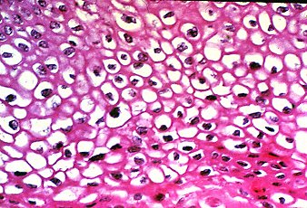 HPV koilocytes
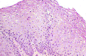 HPV koilocytes
 Squamous cell carcinoma of the cervix
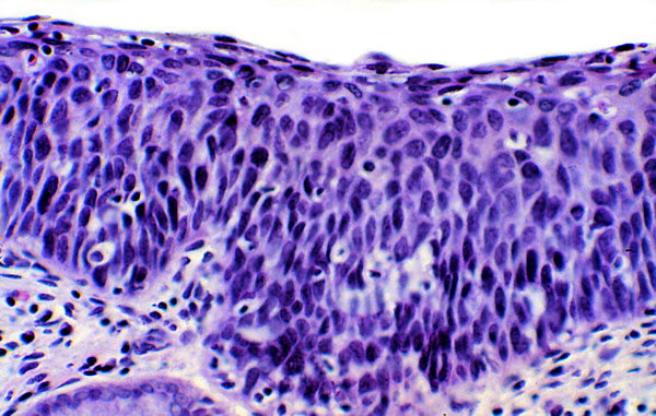 Carcinoma in situ
 Lymphatic malformation
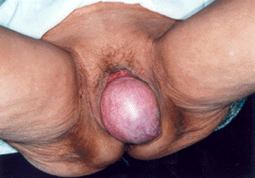 Prolapsed uterus
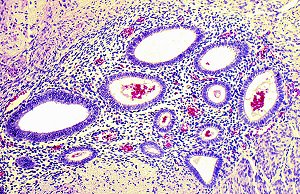 Endometriosis
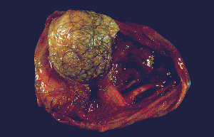 Mature teratoma
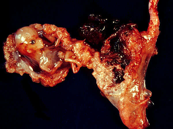 Ectopic pregnancy